Periorbital Hemorrhage Secondary to tPA For Stroke Following Oculoplastic Surgery
Sarah Jacobs1*, Tal Rubinstein2 and Steven Laukaitis2
1Department of Ophthalmology, University of Alabama Birmingham, USA
2Allure Laser and Medispa, USA
Submission: April 30, 2018; Published: May 08, 2018
*Corresponding author: Sarah Jacobs, Department of Ophthalmology, University of Alabama Birmingham & Callahan Eye Hospital, 700 18th St S, Suite 410, Birmingham, AL 35233, USA, Tel: 205-325-8620; Fax: 205-325-8257; Email: jacobs.sarahm@gmail.com
How to cite this article: Sarah J,Tal Rubinstein,Steven L. Periorbital Hemorrhage Secondary to tPA For Stroke Following Oculoplastic Surgery. J Head Neck Spine Surg. 2018; 2(5): 555599. DOI: 10.19080/JHNSS.2018.02.555599
Keywords
Keywords: Tissue plasminogen activator; Hemorrhage; Stroke; Blepharoplasty; Ectropion Ischemic stroke; Complications; Treatment.
Introduction
Tissue plasminogen activator (tPA) is a proven treatment for ischemic strokes when administered within 3-4.5 hours of symptoms. Relative contraindications include minor or spontaneously-improving stroke, seizure at onset, recent gastrointestinal and urinary tract hemorrhage, pregnancy, recent major surgery or serious trauma [1]. As these are relative contraindications, ultimately the provider makes the decision to administer tPA based on analysis of risks and benefits. This is a case of a periorbital hemorrhage in a patient receiving tPA for ischemic stroke within 2 days of oculoplastic surgery. The case highlights
a) The need to identify potential complications of tPA to the eye after oculoplastic surgery;
b) The benefit of understanding how to acutely manage periorbital/orbital hemorrhages provoked by tPA; and
c) The relative contraindication against tPA after oculoplastic surgery when the caretaker is not trained in managing the periorbital complications and when immediate subspecialty care cannot be provided.
Case
A 76-year old male presented to a local emergency room for left hemiparesis, right gaze deviation, and a NIHSS of 19. Less than two days prior, he had undergone uncomplicated outpatient eyelid surgery including bilateral blepharoplasty, lower eyelid ectropion repair, and supraciliary brow lift. His past medical history included hypertension and paroxysmal atrial fibrillation treated with 81mg daily aspirin. His aspirin had been held for 1 week prior to his lid and brow surgery.
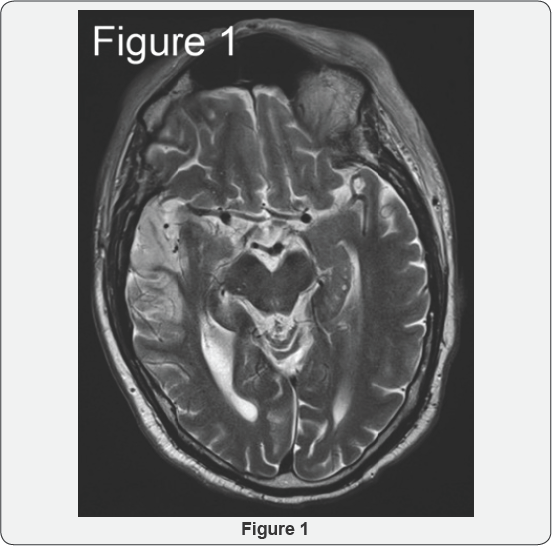
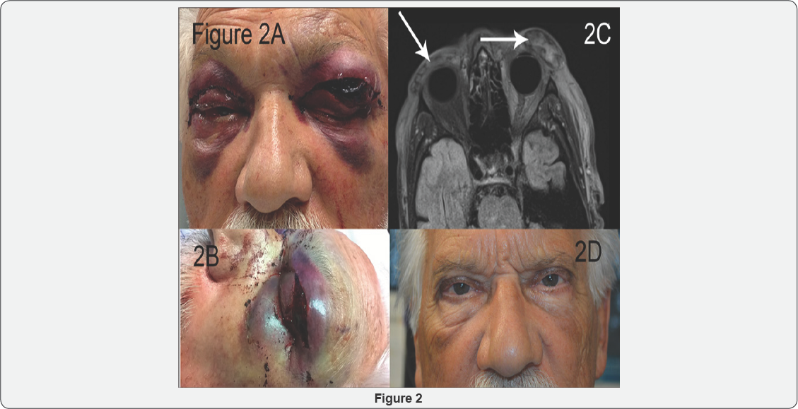
In the emergency department, CT imaging identified a right middle cerebral artery ischemic stroke with a right M1 segment occlusion, confirmed with MRI (Figure 1). He received tPA and mechanical thrombectomy for the stroke, with rapid alleviation of the hemiparesis. However, 3 hours after tPA administration he developed progressively-worsening bilateral periorbital ecchymosis with formation of a tense hematoma on the left. Assessment by an on-call ophthalmologist found that the left eye had no light perception, raising concern for acute hemorrhage within the orbit compressing the optic nerve. The sutures were removed from the left blepharoplasty and ectropion incisions, which allowed egress of an eyelid and orbital hematoma (Figure 2A-2C). By that evening, the patient was noted to have 20/200 vision with normal intraocular pressure on the left side. He received appropriate post-stroke care and was discharged 4 days later.


One week after the initial surgery, the remaining hematoma was surgically evacuated from the left eyelid and the surgical incisions were resutured. At follow up 3 months later, his vision had returned to baseline, with adequate eyelid healing and no significant sequelae (Figure 2D).
Discussion
Few reports identify intraorbital and periorbital hemorrhages after tPA. Thrombolytics after myocardial infarctions have caused orbital hemorrhages [2-4]. Intractable bleeding from a blepharoplasty wound has occurred after thrombolysis for a pulmonary embolism [5]. Sheth & Lee [6] described orbital hemorrhage in a patient who received tPA for stroke after recent orbital trauma [6]. The Americal Society of Aesthetic Plastic Surgery estimates over 165,000 eyelid surgeries are performed yearly in the United States [7]. The periorbital region significantly increases the risk of bleeding. Therefore, despite the rarity of a periorbital hemorrhage due to tPA for stroke after oculoplastic surgery, trainees and physicians caring for such patients need to consider the anatomy and vision-threatening consequences of hemorrhage around the eye, and the appropriate management if they encounter this complication.
In a large survey of ophthalmic plastic surgeons, the risk of postoperative hemorrhage after upper or lower lid blepharoplasty was estimated to be 0.055%, most within the first 24 hours [8]. Hemorrhage that accumulates posterior to the orbital septum can cause orbital compartment syndrome. Orbital compartment syndrome is a potentially blinding condition of increased intraobital pressure, leading to stretching of the optic nerve, compression of the vessels supplying the optic nerve, elevated intraocular pressure, reduced venous outflow from the eye, and orbital ischemia. Symptoms include vision loss, pain, nausea, and vomiting. Signs include proptosis, extensive periorbital edema with ecchymosis, tense eyelids, loss of color vision, ophthalmoplegia, and an afferent pupillary defect [8].
In our case, the large periocular hematoma made bedside exam challenging, even for a trained ophthalmologist. Concerning features included the progressive proptosis, tense eyelids, declining visual acuity, and afferent pupillary defect. Prompt release of the sutures from his incisions allowed external decompression rather than intraorbital progression. This demonstrates the importance of emergent treatment of any significant hemorrhage around the eye after tPA.
While imaging helps diagnose the area of periocular hemorrhage, it is not appropriate to delay prompt treatment of orbital compartment syndrome while awaiting imaging. If an ophthalmologist is not emergently available, treatment should be undertaken by the covering neurologist or emergency department physician. Permanent vision loss can occur within 60-90 minutes after an orbital hemorrhage [9]. Prompt treatment may preserve vision [6]. In the case of tense orbital hematoma formation in the perioperative period, simple removal of the sutures to allow the hemorrhage to drain is crucial. A lateral canthotomy and cantholysis to further decompress the orbit is likely beyond the scope of the neurologist, but these procedures are known to emergency room physicians. Bleeding from the opened incisions is likely to occur, but can then be managed by subspecialty services when they arrive, with topical pro- thrombotics or cautery [5].
This case highlights that thrombolysis with tPA can disrupt the newly formed clots of the upper eyelid and brow within the first 1 week after upper eyelid blepharoplasty and brow ptosis repair. Although these oculoplastic procedures are designated as minor rather than major surgery, tPA-related orbital hemorrhage after such surgery may cause permanent vision loss from compartment syndrome.
The risk of persistent debility or death due to stroke certainly outweighs the risk of orbital or periorbital hemorrhages for cases in which treatment for potential hemorrhage is readily available. However, in settings in which timely treatment of a periorbital or orbital complication cannot be provided, physicians providing tPA should view recent oculoplastic surgery as a relative contraindication. We recommend physicians consider the risk of tPA after oculoplastic surgery, develop the skill set of suture removal to allow basic decompression, and consult ophthalmology early in the tPA administration so that they can provide emergent care if needed.
Key Points
a) Administration of tPA for ischemic stroke can provoke vision-threatening periocular bleeding after even minor oculoplastic surgery.
b) The tPA provider should be vigilant for hematoma formation in patients after periocular surgery, and be prepared to release the eyelid sutures and notify subspecialty services promptly to decompress the wounds.
c) Recent oculoplastics surgery should be considered a relative contraindication for tPA if the provider is unfamiliar with acute periocular hemorrhage management or if subspecialty services for the eye or orbit are unavailable.
References
- Adams HP, Brott TG, Furlan AJ, Gomez CR, Grotta J, et al. (1996) Guidelines for thrombolytic therapy for acute stroke: a suppelement to the guidelines for the management of patients with ischemic stroke. A statement for healthcare professionals from a special writing group of the stroke council, American Heart Association. Stroke 27(9): 17111718.
- Yuen VH, Dolman PJ (1999) Postoperative orbital hemorrhage following thrombolysis for acute myocardial infarction. Can J Ophthalmol 34(1): 33-35.
- Cunneen TS, Morlet N (2007) Retro-orbital hemorrhage after thrombolysis for acute myocardial infarction. N Engl J Med 357(14): 1448-1449.
- Leong JK, Ghabrial R, McCluskey PJ, Mulligan (2003) Orbital haemorrhage complication following postoperative thrombolysis. Br J Ophthalmol 87(5): 655-656.
- Burroughs JR (2009) Management of intractable postoperative blepharoplasty bleeding after thrombolysis for a pulmonary embolism. Ophthal Plast Reconstr Surg 25(4): 314-315.
- Sheth SA, Yee AH (2013) Mystery case: an unexpected complication of IV thrombolysis for acute ischemic stroke. Neurology 81(7): e42-e43.
- Hotta TA (2014) ASAPS (American Society of Aesthetic Plastic Surgery)--2013 Annual Statistics. Plast Surg Nurs 34(2): 47-48.
- Hass AN, Penne RB, Stefanyszyn MA, Flanagan JC (2004) Incidence of postblepharoplasty orbital hemorrhage and associated vision loss. Ophthal Plast Reconstr Surg 20(6): 426-432.
- Ballard SR, Enzenauer RW, O'Donnell T, Fleming JC, Risk G, et al. (2009) Emergency lateral canthotomy and cantholysis: a simple procedure to preserve vision from sight threatening orbital hemorrhage. J Spec Oper Med 9(3): 26-32.


