




 JDVS Home



Abstract
The present study was conducted to investigate the histological and histochemical features of the vomeronasal organ of adult dogs. The organ was surrounded by a J-shaped cartilage, with a gradual caudal reduction in the distance between the two free limbs extending rostro-caudally. Structurally, the tubular organ presented pseudostratified columnar ciliated epithelium with goblet cells towards the lateral as well as dorsal half of the medial wall and olfactory epithelium towards the ventral half of the medial wall at level I. At level II and III, both medial and lateral walls were lined by sensory pseudostratified columnar epithelium. The medial and lateral propria-submucosa contained sero-mucous glands, numerous fine blood capillaries, thin-walled blood vessels of different sizes, venous caverns, and nerve fascicles. Glandular ducts predominantly opened towards the dorsal or ventral-most portions of the organ’s lumen. Histochemically, the glandular acini showed the presence of glycogen, neutral mucopolysaccharides, weakly acidic sulfated mucopolysaccharides, hyaluronic acid, sialomucins, mucins, and acidic mucopolysaccharides, but lacked proteinaceous components. The goblet cells exhibited variable histochemical reactions, indicating diverse carbohydrate moieties and the presence of proteins in their secretions. The structural features indicate that the vomeronasal organ is highly sensory and plays important roles in various olfactory-related activities..
Keywords:Vomeronasal organ, Dog, Histology, Histochemistry, Epithelium
Introduction
Dogs are famous for their incredible sense of smell, primarily mediated by the main olfactory epithelium which is a part of the olfactory sensory system responsible for relaying smell information to the brain. It uses epithelial dendrites to detect odour molecules and sends this information to the main olfactory bulb [1]. The vomeronasal organ (VNO), considered a secondary olfactory structure, is a tube-like organ situated on both sides of the ventro-lateral portion of the nasal septum and is especially sensitive to non-volatile chemical cues found in pheromones. These signals help dogs interpret information that is unavailable through normal smell alone. Signals detected by the VNO are conveyed to the accessory olfactory bulb, initiating certain reproductive behaviours [2]. Together, these systems allow dogs to interpret a highly detailed chemical world. The VNO plays a key role in chemical communication and influences social behavior, mating behavior, emotional understanding, and maternal bonding with neonates. This is particularly important in species whose newborns have underdeveloped visual and auditory systems and rely on particular pheromones to recognize their mothers [3]. Furthermore, the VNO is commonly thought to play a role in flavor perception during feeding through olfaction [4]. In mammals, sexual behaviours of males and females are induced, and their hormonal status may also be changed via the stimulation of the VNO [5-7]. It allows dogs to access a layer of information far beyond ordinary smell, essentially giving them a second, highly specialized “scent system.”
It may play a role in producing Flehmen response to pheromones, especially in numerous felid and ungulate species [8, 9]. The “Flehmen response” is a unique facial action seen in several mammals, used to channel inhaled chemicals toward the VNO [10, 11]. Dogs do not show a dramatic lip-curling response like horses because their upper lips are too rigid and firmly fixed via the frenulum to permit this type of movement [12]. Instead, they show a subtle Flehmen-like reaction: pausing mid-sniff, slight head lift, raising the upper lip, momentary mouth opening, a focused, “zoned out” expression, and a brief freeze after sniffing urine, faces, or strong social odour. Additionally, the tongue quickly withdraws toward the incisal papilla, which likely further enhances the detection of pheromones [13]. These actions assist in directing pheromone molecules toward the VNO, where they mix with mucous before being processed. The relevance of these facts for both animal behavior and reproduction, the organ has been briefly described in terms of its histological and histochemical characteristics in dogs, providing a baseline for future investigations with greater emphasis on breed-specific variations. Further comprehensive studies on the anatomy and physiology of the species-specific VNO could improve understanding of reproductive mechanisms across animal species as well as serve as a foundation for pheromonotherapy, since the VNO is the primary sensory structure responsible for detecting, processing, and integrating pheromonal signals into behavioral and emotional responses. A more comprehensive understanding of the structure and function of the VNO would therefore directly enhance the precision, effectiveness, and species-appropriate application of pheromone-based therapy in clinical settings. This is particularly relevant for the management of behavioral disorders, stress mitigation, and breeding management contexts, including maternal separation and transport.
Materials and Methods
The present study was conducted on six adult, apparently healthy, local non-descript dogs of either sex. The heads were procured from the Department of VCC, LUVAS, Hisar, immediately after the animals’ death, and sectioned into transverse planes after removing the mandibles. Specimens from cranial (level-I, cranial to the canine), middle (level-II, cranial to the first premolar), and caudal portion (level-III, cranial to the second premolar) of the vomeronasal organ were collected and then fixed in 10% neutral buffered formalin solution for 48 hours. The fixed tissues were processed for the routine paraffin technique for light microscopy. The paraffin sections of 5 μ were obtained and stained with Routine Harris’ hematoxylin and eosin stain for general architecture, Gomori’s method for reticular fibers, Weigert’s method for elastic fibers, Bielschowsky’s method for axis cylinders and dendrites, Sevier-Munger method for neural tissues, McManus’ method for glycogen (PAS), Alcian blue method for mucocutaneus (pH 2.5), PAS-Alcian blue method for acidic and neutral mucopolysaccharides (pH 2.5), Mayer’s mucicarmine method for mucin, and colloidal iron method for acid mucopolysaccharides [14]. Crossmon’s trichrome stain was employed for collagen fibers [15]. Proteins were visualized using the performic acid-Alcian blue method [16].
Results and Discussion
The vomeronasal organ ran rostro-caudally from the incisive papilla to the level of the upper second premolars longitudinally.
Level-I (Cranial to the Canine Tooth)
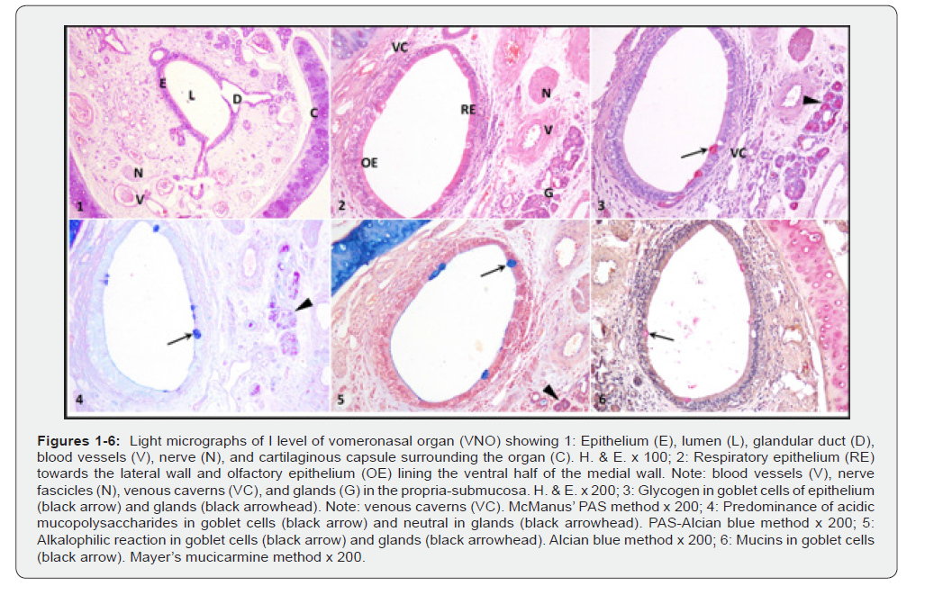
In all examined specimens, its rostral portion at the level of the third incisor was surrounded by a J-shaped hyaline cartilaginous capsule with its larger limb towards the medial side and smaller towards the lateral side (Figure 1), as previously reported in canines [17- 21] and camels [22]. In Egyptian goats and sheep, the cartilaginous capsule appeared as an inverted C-shaped structure in its rostral part [23, 24]. The VNO cartilage appeared slightly flattened medio-laterally and was almost completely enclosed, except for a narrow opening on the dorsal aspect in Angora goats [9]. In sheep, this organ was surrounded by an incomplete U-shaped cartilaginous capsule [25, 21]. The cartilage appeared C-shaped due to its incomplete structure in the dorso-lateral area in the pig [26]. However, it was pear-shaped, which remained open on the lateral aspect in buffalo [27]. The VNO tube was almost entirely encased in cartilage, except for an incomplete dorsolateral section in sheep [28]. In goats, it was surrounded by a fully formed, oval-shaped cartilage at the level of the fourth palatine rugae [29]; however, this ovoid cartilage was incomplete in Iranian goats [30]. The smaller lateral limb reached about halfway up the height of the tubular organ. The free tips of both limbs gradually reduced in thickness, and their distal ends were widely separated from each other. In canines, the dorso-lateral mucosal wall of the VNO lacked cartilaginous support [17, 20]. However, cartilage was not observed in the medial mucosal wall in dogs [18]. The cartilage was absent on the dorso-medial side in sheep [24]. The capsule was intact along the proximal portion of its length, with the lateral wall being thicker than the medial wall in Lori sheep [31]. In cattle, a lateral cleft was present in the rostral part, while another cleft appeared dorsally in the caudal portion [32].
The lumen of the VNO was elongated and oval in shape, with narrow dorsal and ventral ends as reported in mithun and zebu [34]. The VNO had a crescent-shaped in cross-section in Balady dogs [19]. A crescent-shaped lumen has also been described along most of its length in canines [17, 18] and Angora goats [9]. In sheep, several lumen configurations have been documented. An irregular crescent or C-shaped lumen caused by epithelial folding [25], while [28] observed a C-shaped lumen shifted towards the medial side of the cartilage. The lumen was characterized as comma-shaped, being narrow dorsally and wider ventrally [21]. In pigs, the vomeronasal duct showed regional variations, appearing narrow and elliptical rostrally and gradually widening caudally [26]. In goats, the VNO featured an irregular elliptical lumen with irregular epithelial folds extending into the cavity, characterized by a longer dorso-ventral axis and a shorter medio-lateral axis [29].
Structurally, the organ appeared as a tubular formation with distinct medial and lateral walls. The lateral wall was lined by pseudostratified columnar ciliated epithelium with only a few goblet cells (Figure 2) as also reported earlier in canines [17]. This same epithelium was described, extending up to the sixth transverse rugae in sheep [24]. In this study, the epithelium consisted of basal cells, non-ciliated supporting columnar cells, ciliated columnar cells, and a few goblet cells. However, in buffalo, alpaca, and sheep, the non-sensory epithelium consisted of ciliated, non-ciliated secretory, and basal cells [35, 36, 28, 11]. The lateral wall in dogs, buffaloes, and Korean native cattle was covered with respiratory-type pseudostratified columnar ciliated epithelium, comprising ciliated cells, non-ciliated columnar cells, and goblet cells [19, 27, 37, 38]. The rostral region was covered with non-sensory epithelium interspersed with goblet cells in goats [23, 29] and Corriedale sheep [39].
At the initial segment of the VNO, non-keratinized stratified squamous epithelium has been reported in camels, Alawasi Iraqi sheep, and pigs [22, 25, 26]. In pigs, this epithelium was reported to transition dorsally into respiratory epithelium at the level of the first transverse rugae, while both ventral walls retained non-keratinized stratified squamous epithelium [33]. Similar epithelial patterns were also documented in pigs, horses, and cows [40]. In bovines, the lining epithelium in the rostral part was predominantly non-neurosensory pseudostratified columnar epithelium, having few goblet cells, with occasional areas of stratified cuboidal epithelium [32]. The basal cells were spaced apart, having round to oval nuclei close to the basement membrane. These nuclei showed condensation of chromatin material, especially towards the outer nuclear membrane. Their darkly stained nucleoli were centrally placed. The cytoplasm of these cells was finely granular and eosinophilic in appearance. A small number of lymphocytes were observed between these cells, a finding that was consistent with reports in dogs [17]. In contrast, pigs exhibited more pronounced lymphoid infiltration by underlying lymphoid tissue, and in some areas, this infiltration was so extensive that epithelial and lymphoid cells were difficult to distinguish [33]. The nuclei of the non-ciliated columnar cells were oval to elongated with tapering ends and were vertically oriented. The nucleoplasm showed condensation of chromatin in smaller clumps, while the rest of the nucleoplasm had fine dusting of chromatin. The finely granular cytoplasm of these cells was eosinophilic in appearance.
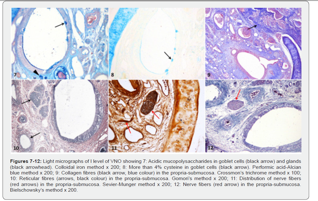
The nuclei of the ciliated columnar cells were round to oval and generally placed towards the basal part of the epithelium. These nuclei were horizontally placed with their chromatin also arranged in small, irregular clumps closer to the nuclear membrane. These nuclei were relatively lightly stained with nucleoli that were typically single and either centric or eccentric in position. The cytoplasm of these cells was generally more eosinophilic towards the supranuclear region. The free surface showed the presence of non-sensory cilia towards the luminal surface. The goblet cells exhibited nuclei at varying levels within the epithelium. Their round to oval nuclei, with distinct, darkly stained nucleoli, were centrally or eccentrically positioned. The cytoplasm was weakly eosinophilic and showed a vacuolated appearance. The goblet cells were PAS positive indicating the presence of glycogen by McManus’ PAS stain (Figure 3), as also reported earlier in goat [9, 23, 29], sheep [24, 39], and pig [33]. By using the PAS-Alcian blue method, acidic mucopolysaccharides were observed in the secretions of goblet cells (Figure 4), corroborating the results in sheep [24] and pig [33]. The goblet cells exhibited a strongly positive Alcianophilic reaction with Alcian blue staining method (Figure 5) in confirmation with the findings in sheep [24, 39] and pig [33]. These cells tested positive for mucins, as shown by Mayer’s mucicarmine method (Figure 6) as has been documented previously in pigs [33]. The goblet cells showed a positive reaction with colloidal iron method indicating the presence of acidic mucopolysaccharides (Figure 7), but a moderate positive reaction was observed in pigs [33]. Furthermore, using the performic acid-Alcian blue method, the goblet cells displayed a positive response, suggesting the presence of more than 4% cysteine in their secretions (Figure 8).
The propria-submucosa in the subepithelial portion contained loose, irregular connective tissue with various connective tissue cells, a small amount of collagen, very fine elastic, and isolated reticular fibres, along with delicate blood capillaries and venous caverns (Figures 3,9,10). The reticular fibres were prominent, constituting the basal lamina (Figure 11). The deeper part had comparatively dense connective tissue comprising large amounts of collagen fibers, glandular clusters of sero-mucous acini encased by fine reticular fibers, delicate blood capillaries, thin-walled blood vessels of various dimensions, venous cavern-like structures, and several nerve fascicles, which confirmed the earlier descriptions in dog [18] and goat [29]. Similar structures were noted in sheep, except that the propria-submucosa showed a dense form of connective tissue [24, 28]. However, in dogs, the lateral side had fewer blood vessels but more glands compared to the medial side [19, 21]. In Angora goats, mucous glands and blood capillaries were abundant, whereas nerve fibers were sparse [9]. Iraqi sheep exhibited several nerve plexuses, mucous glands, and multiple wide veins [25]. In buffalo, glands were more heavily concentrated along the lateral wall [27]. The dense propria-submucosa of sheep contained more glands, nerve fascicles, and blood vessels on the lateral side [21]. Three to seven large veins with thick walls were noticed in sheep [39], while in bovines, two to seven profiles of veins were identified [32].
Some exceptionally large nerve fascicles were also noted (Figure 12). At places, the glandular ducts lined by simple to stratified cuboidal epithelium opened towards the luminal surface of the epithelium, as also recorded previously in dogs [19]. However, the ducts were lined by simple cuboidal epithelium in Egyptian goats [23]. These ducts generally opened at the dorsalmost or ventral-most portion, where the medial and lateral walls of the organ joined as reported earlier in sheep [28] and canines [17, 20]. In buffalo, ducts terminating in the dorsal junctional region conveyed the glandular secretions into the organ’s lumen [35]. The glandular excretory ducts opened into both the lateral and medial surfaces of the vomeronasal epithelium in Corriedale sheep [39]. However, in bovines, the glands opened via ducts through the VNO epithelium from all sides [32]. The connective tissue became denser near the hyaline cartilage, with a significant increase in the concentration of collagen and elastic fibers. The glands showed a positive PAS reaction, suggesting glycogen in their secretions with a stronger affinity towards the luminal surface, while the glandular ducts lacked any evidence of secretory activity (Figure 3), which was in agreement with the studies in Korean native cattle [38], dog [19], buffalo [35], and sheep [24, 25, 28, 39]. The combined PAS-Alcian blue technique demonstrated the predominance of neutral mucopolysaccharides, particularly concentrated towards the luminal surface as previously observed in Egyptian goats [23] whereas, the cartilage displayed a strong reaction indicating acidic mucopolysaccharides (Figure 4). In contrast, the glandular units exhibited a mixed composition of both acidic and neutral mucopolysaccharides in sheep [24]. The glands showed weak to moderate Alcian blue positivity (Figure 5), corroborating reports in sheep [24, 39] and Korean cattle [38]. Conversely, no Alcian blue reactivity was observed in dogs [19]. The mucous acini exhibited a weak to moderate positive reaction revealing the presence of mucins in their secretions by Mayer’s mucicarmine stain. Using colloidal iron staining, the glands exhibited weak positivity for acidic mucopolysaccharides in our study (Figure 7), while glands showed mild positivity in pigs and buffaloes [33, 35]. The glandular tissue lacked proteinaceous components in its secretions.
The dorsal half of the medial wall was also covered with pseudostratified columnar ciliated epithelium with isolated goblet cells or the non-sensory epithelium (Figure 2). This epithelium was composed of basal cells, supporting ciliated columnar cells, and goblet cells, constituting about 3-4 rows of nuclei in the epithelium with similar cellular details as observed towards the lateral wall earlier. Epithelial height was reduced towards the dorsal portion of the organ at the point of union of the medial and lateral walls. Towards the ventral half of the medial wall, olfactory epithelium was observed, where three cell types could be recognized: basal, supporting, and receptor (sensory/olfactory) cells; however, the medial wall’s pseudostratified epithelium could be divided into three zones in canines: a deep layer having basal and receptor cells’ nuclei, a middle layer with sustentacular cell nuclei, and a superficial layer made up of the apical parts of receptor and sustentacular cells [17]. In buffalo, alpaca, and sheep, the VNO was surfaced medially by sensory epithelium [35, 36, 28, 11]. In dogs, buffaloes, and Korean native cattle, it consisted of sensory epithelium made up of supporting cells, neuroreceptor cells, and basal cells [19, 27, 37, 38]. The VNO was covered with non-sensory epithelium interspersed with goblet cells on medial wall in goats [23, 29]. Similar epithelium extended up to the sixth transverse rugae in sheep [24].
However, the organ exhibited respiratory epithelium throughout the length, except for a small ventro-medial region with olfactory epithelium in Angora goats [9]. In Corriedale sheep, the ventral two-third of the medial side was covered by sensory epithelium, whereas the dorsal one-third was covered by nonsensory epithelium [39]. The basal cells exhibited histological features similar to those found in the lateral wall. A few infiltrating lymphocytes were scattered among these cells. The supporting cells possessed comparatively smaller, oval to elongated nuclei with tapering ends. Their orientation was vertical, and their nucleoplasm showed condensation of chromatin material in small clumps, while the remaining nucleoplasm displayed a fine dusting pattern. These nuclei typically occupied the mid-region of the epithelium with a single and usually centric nucleolus. Some of the nuclei reached near the surface of the epithelium. The cytoplasm of these cells appeared finely granular and comparatively more eosinophilic than that of other cells. The sensory or receptor cells had large, round to oval nuclei, usually distributed near the basal region of the epithelium, and these were horizontally placed. Their chromatin was also assembled in smaller clumps irregularly, often concentrated near the nuclear membrane, resulting in lightly stained nuclei. Single, large, darkly stained nucleoli, either centric or eccentric, were generally observed in these cells. The cytoplasm exhibited accentuated eosinophilia in the supranuclear region of the epithelium. The free surface of the epithelium displayed sensory cilia of varying shapes and sizes arising from the sensory cells.
In our study, the epithelial height was generally greater on the medial wall as compared to the lateral wall. Comparable findings were reported in pigs [26], where the epithelium of the medial wall was slightly taller than that of the lateral wall. On the contrary, greater epithelial height was observed in the lateral wall than the medial wall in pigs [33].
The subepithelial portion of the propria-submucosa had loose, irregular connective tissue with various connective tissue cells and fibres, along with blood capillaries. These features were in accordance with observations in buffalo [27, 35], goat [29], and camel [22]. In pigs, the propria-submucosa was described as having dense connective tissue dominated by collagen and reticular fibers, with few elastic fibers and lymphoid tissue aggregates beneath the epithelium [26, 33]. Comparable structural features were reported in Alawasi Iraqi sheep, although lymphoid aggregations were absent in this species [25]. In canines, micro vessels were found to be more concentrated in the propriasubmucosa near the medial epithelium compared to the lateral side [17]. A highly vascular connective tissue with plenty of collagen fibers, glands, and nerve fascicles was observed in Egyptian goats [23]. The remaining propria-submucosa was denser, containing sero-mucous glands and their associated ducts, numerous fine capillaries, thin-walled blood vessels of varying sizes and shapes, venous caverns, and nerve fascicles. This region showed a notable increase in collagen fiber density, with fibers surrounding both blood vessels and glandular clusters. Similar observations were noted in dog [19, 21], Iranian goat [30], buffalo [35], Korean native cattle [38], alpaca [36], and pig [26]. However, in pigs, the mucous acini were dominant in these glands [33].
In Alawasi Iraqi sheep and buffalo, these glands were described as tubuloacinar and predominantly mucous [25, 27]. In contrast, buffalo glands were characterized as compound, branched, tubuloacinar, and serous [35], whereas; in camel they were mainly serous and tubuloalveolar, with some mucous secretory units [22]. In Egyptian goats, yak, zebu, and mithun, the glands were of serous type [23, 34]. Conversely, mucous-type glands have been reported in goats [29]. A complete absence of glands has been reported in sheep [28]. Sheep and Angora goats exhibited fewer blood vessels and mucous glands, yet a greater concentration of nerve fascicles than in the lateral propria-submucosa [9, 24]. In canines and bovines, many small, unmyelinated nerve fascicles were found in the deeper regions [17, 20, 32]. In contrast, sheep exhibited large bundles of unmyelinated nerve fibers [28]. In dogs, well-formed and extensive venous plexuses were observed [11, 17]. The perichondrium was dense and rich in collagen fibres. A large number of chondrocytes within lacunae were enveloped by extracellular matrix, and multiple isogenous groups were evident. Fine reticular fibres encased the glandular acini, nerve fascicles, and blood vessels. Nerve fascicles were more concentrated in the mid-ventral region of the propria-submucosa (Figures 1, 11).
In this study, the propria-submucosa was of similar thickness on both medial and lateral sides. However, pigs exhibited a relatively thinner lateral propria-submucosa, although the distribution of glands, associated ducts, blood vessels, and venous caverns was nearly the same [26, 33]. Contrary to this, sheep and goats showed a thicker lateral propria-submucosa; in sheep, it contained abundant mucous glands as well as both myelinated and unmyelinated nerve fibres [28], whereas; in goats it was dominated by mucous glands and numerous venous caverns [29]. Only small, unmyelinated nerve fascicles were observed in the lateral propria in bovines [32]. The propria-submucosa ventral to the point of union of medial and lateral walls epithelium also contained loose, irregular connective tissue rich in elastic fibres oriented in different directions. This region contained all usual components of the propria-submucosa except clusters of seromucous glandular units and large venous caverns. The propriasubmucosa towards the dorso-lateral region of the organ was comparatively denser and showed a greater distribution of sero-mucous glands, thin-walled blood vessels, venous caverns, and small to medium-sized blood vessels as has been described previously in dogs [19]. Similarly, the glandular acini were mainly located on the dorsal and lateral sides of the organ in canines [17, 21], buffalo [35], and Egyptian goat [23]. In the current study, the density of nerve fascicles decreased significantly towards the dorsal aspect, whereas elastic fibres were markedly increased. In contrast to this, nerve fascicles in the alpaca were predominantly localized to the dorso-medial region of the organ [36].
Level-II (Cranial to the First Premolar Tooth)
At the second level, the medial and lateral limbs of J-shaped cartilage surrounding the vomeronasal organ, gradually tapered towards their tips with a reduction in the distance between the two free ends, as also reported previously in dogs [19]. The dorsal free ends of the U-shaped cartilage in Iraqi sheep were positioned closer at this level [25]. In pigs, the C-shaped cartilage almost completely encircled the organ, leaving only a small dorsal opening [26]. The medial limb extended further dorsally at this level, while the lateral limb ended lower, creating a narrow gap between them in the pig [33]. The cartilaginous capsule in Egyptian goats appeared as a J-shaped structure in its middle section [23]. In goats, a small part of the oval-shaped cartilage was missing dorso-laterally at the level of eighth palatine rugae and mid-first premolar tooth [29].
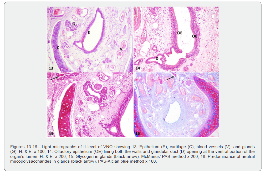
The vomeronasal organ itself was almond-shaped, with a pointed dorsal tip and a blunt, crescent-shaped ventral end, enclosing a wide lumen (Figure 13). However, in pigs, the vomeronasal duct displayed a narrow, vertically oriented lumen [26]. Both the medial and lateral walls were lined by sensory (olfactory) pseudostratified columnar epithelium consisting of basal cells, supporting cells, and sensory or olfactory cells, forming about 4-6 rows of nuclei in the epithelium. These cells displayed histological characteristics largely similar to those observed at the preceding level. The free surface of the epithelium presented sensory cilia towards the luminal surface (Figure 14). In pigs, this same epithelium covered both surfaces at the level of the third transverse rugae [33]. At that level, however, the medial wall had olfactory epithelium, while the lateral wall exhibited respiratory epithelium in the pig [26]. The middle section was lined medially by sensory olfactory epithelium and laterally by non-sensory epithelium in Egyptian goat [23], dog [18], buffalo [37], sheep [24, 25] and camel [22]. In goats, both the lateral and medial walls at the level of the eighth palatine rugae were surfaced by respiratory epithelium [29].
The propria-submucosa on the lateral side consisted of all typical components as observed at level I; however, the glands were more abundant than at the previous level. These glandular clusters were more numerous compared to those observed in the propria-submucosa of the medial wall. Glandular ducts typically opened into the dorsal and ventral extremes of the organ’s lumen (Figure 14). The glands exhibited similar histochemical reactions with different staining methods as recorded earlier in the previous level (Figure 15, 16). The medial propria-submucosa was also composed of highly vascularized connective tissue and all the structures noted at level I. Ventral to the point of junction of the medial and lateral wall epithelium, the propria-submucosa housed abundant elastic fibres, which were variably oriented and sectioned in different planes, along with all the constituents seen in the rest of the propria-submucosa. In pigs, the propriasubmucosa occupied only a small area ventrally relative to the epithelium at the level of the third transverse rugae [33]. In the dorso-lateral region, the propria-submucosa was similar to that observed at the previous level; however, several differently-sized nerve fascicles were also noticed. However, in pigs, the propriasubmucosa at the level of the third transverse rugae was extended upward to form an apex dorsally, containing numerous glandular ducts, venous caverns, and blood vessels [33].
Level-III (Cranial to the Second Premolar Tooth)
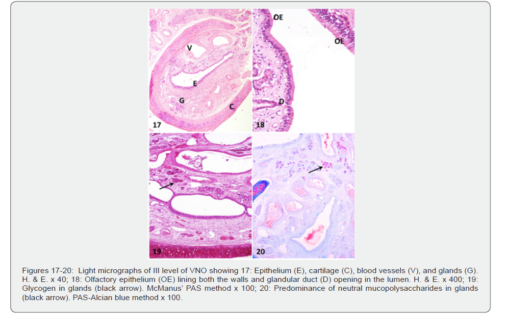
The hyaline cartilage surrounding the organ exhibited the usual J-shape, with the distance between the two free ends of the limbs of the cartilage being smaller compared to the previous two levels (Figure 17). The cartilaginous capsule in Iraqi sheep and Egyptian goat formed a complete ring enveloping the duct in its caudal part [23, 25]. In yaks, the VNO was surrounded entirely by thin, C-shaped cartilage that was dorsally incomplete, enclosing an oval lumen [34]. The organ was encased by oval or elliptical cartilage, except for the dorsal opening in pigs and goats [26, 29]. The thickness of the hyaline cartilage was greater in the medial limb than in the lateral limb. The cartilage gradually became narrow towards the free tips. The organ’s lumen was elongated and irregular in appearance, with a narrow dorsal end at this level. The vomeronasal organ exhibited pseudostratified columnar epithelium with sensory cilia on both its medial and lateral walls. It shared cellular architectural features similar to those described previously, although the epithelial height was reduced towards its dorsal portion (Figure 17, 18) which was in accordance with the observations reported in camel [22], Egyptian goat [23], goat [29], cattle [32], and mithun, yak, and zebu [34]. At the level of the fifth transverse rugae in pigs, olfactory-type pseudostratified columnar epithelium was observed on both surfaces [26, 33]. An olfactory epithelium was reported towards lateral and medial walls at the level of the first molar tooth in sheep [24]. In our study, the McManus’ PAS method showed no reaction in the epithelium, revealing the absence of glycogen (Figure 19).
The medial propria-submucosa contained all the structural components observed at the preceding levels. The lateral propriasubmucosa presented all the constituents found at the previous level, with minor variations such as an increased presence of seromucous glandular clusters. Notably, vascularity was markedly enhanced at this level, with large-diameter blood vessels being more prominent than at the previous two levels. Histochemically, the glands contained glycogen (Figure 19), predominantly neutral mucopolysaccharides (Figure 20), weakly sulfated mucopolysaccharides, hyaluronic acid, and sialomucins, weak to moderate amounts of mucins, and acidic mucopolysaccharides. The glandular tissue lacked affinity towards the performic acid- Alcian blue stain. The dorso-lateral propria-submucosa showed an increased presence of glands, blood vessels, nerve bundles, venous caverns, and small capillaries similar to those described in dogs [21]. In pigs, nerve bundles were relatively more abundant in this area [26, 33-40].
Conclusion
The vomeronasal organ demonstrated a well-organized and region-specific structural differentiation along its rostro-caudal extent, evident through progressive variations in epithelial height, lumen configuration, and glandular distribution from level I to III. The consistent presence of a J-shaped hyaline cartilaginous capsule provided robust structural support, and the gradual narrowing of its limbs caudally reflects increasing compactness of the organ. The predominance of sensory pseudostratified epithelium along the medial wall confirmed its principal chemosensory role. Histochemical analyses revealed different intensities of reactions for glycogen, acidic mucopolysaccharides, and mucins in goblet cells and sero-mucous glands, suggesting an active secretory role essential for maintaining luminal moisture and facilitating pheromone detection. The consistent positioning of glandular duct openings at dorsal and ventral junctions suggested regulated secretion into the lumen, contributing to optimal chemical transport and detection. Overall, the morphological and histochemical attributes collectively suggested that the vomeronasal organ is highly specialized for chemosensory perception, with its structural organization supporting both sensory transduction and the secretory mechanisms necessary for pheromonal communication. make food safety surveillance a proactive and successful process.
Acknowledgements
The first author gratefully acknowledges the financial support received in the form of Senior Research Fellowship from the Indian Council of Agricultural Research, New Delhi, India, and also extends appreciation to Lala Lajpat Rai University of Veterinary and Animal Sciences, Hisar, Haryana, for the assistance and facilities extended during this investigation.
References
- Lledo PM, Gheusi G, Vincent JD (2005) Information processing in the mammalian olfactory system. Physiological Reviews 85(1): 281-317.
- Taniguchi K, Mikami SI (1985) Fine structure of the epithelia of the vomeronasal organ of horse and cattle: a comparative study. Cell Tissue Research 240(1): 41-48.
- Torres MV, Ortiz-Leal I, Sanchez-Quinteiro P (2023) Pheromone sensing in mammals: a review of the vomeronasal system. Anatomia 2(4): 346-413.
- Dulac C, Axel R (1998) Expression of candidate pheromone receptor genes in vomeronasal neurons. Chemical Senses 23: 467-475.
- Jacobs VL, Sis RF, Chenoweth PJ, Klemm WR, Sherry CJ (1981) Structures of the bovine vomeronasal complex and its relationships to the palate: tongue manipulation. Acta Anatomica 110: 48-58.
- Troiter D, Doving KB, Eloit C (1996) The vomeronasal organ – a rediscovered sensory organ. Tidsskrift Norske Legeforening 116(1): 47-51.
- Salazar I, Lombardero M, Sanchez-Quinteiro P, Roel P, Cifuentes JM (1998) Origin and regional distribution of the arterial vessels of the vomeronasal organ in the sheep. Annals of Anatomy 180(2): 181-187.
- Abbasi M (2001) Vomeronasal organ in Lori sheep. Lorestan, Khorram Abad, Iran.
- Besoluk K, Eken E, Boydak M (2001) The vomeronasal organ in Angora goats (Capra hircus). Veterinary Arhives 71(1): 11-18.
- Abbasi M, Khosravinia H (2002) Vomeronasal organ in Lori sheep. Journal of the Faculty of Veterinary Medicine University of Tehran 58 (3): 279-282.
- Kostov DL (2007) Vomeronasal organ in domestic animals. Bulgarian Journal of Veterinary Medicine 10(1): 53-57.
- Buzek A, Serwanska-Leja K, Zaworska-Zakrzewska A, Kasprowicz-Potocka M (2022) The shape of the nasal cavity and adaptations to sniffing in the dog (Canis familiaris) compared to other domesticated mammals: a review article. Animals 12(4): 517.
- Pageat P, Gaultier E (2003) Current research in canine and feline pheromones. Veterinary Clinics of North America: Small Animal Practice 33(2): 187-211.
- Luna LG (1968) Manual of Histologic Staining Methods of Armed Forces Institute of Pathology. 3rd ed. McGraw-Hill Book Co., New York.
- Crossmon G (1937) A modification of Mallory’s connective tissue stain with a discussion of principles involved. Anatomical Record 69: 33-38.
- Pearse AGE (1968) Histochemistry: Theoretical and Applied. Churchill Livingstone, London.
- Adams DR, Wiekamp MD (1984) The canine vomeronasal organ. Journal of Anatomy 138(4): 771-787.
- Yilmaz B, Yildiz H, Akkoc CO, Arican I (2008) Vomeronasal organ in Labrador retriever dog (Canis familiaris). Bulletin of the Veterinary Institute in Pulawy 52: 185-188.
- Mahdy EAA, El Behery EI, Mohamed SKA (2019) Comparative morpho-histological analysis on the vomeronasal organ and the accessory olfactory bulb in Balady dogs (Canis familiaris) and New Zealand rabbits (Oryctolagus cuniculus). Journal of Advanced Veterinary and Animal Research 6(4): 506-515.
- Coli A, Stornelli MR, Giannessi E (2016) The dog vomeronasal organ: a review. Dog Behaviour 2(1): 24-31.
- Allouch GM, Alshanbari FA (2024) Comparative anatomy of the vomeronasal organ (VNO) in sheep (Ovis aries) and dogs (Canis familiaris) with simple reference to its histological structure and vasculature supply. International Journal of Morphology 42(2): 374-381.
- Karimi H, Hashem RMA, Ardalani G, Sadrkhanloo R, Hayatgheibi H (2014) Structure of vomeronasal organ (Jacobson organ) in male Camelus domesticus var. dromedarius persica. Anatomia Histologia Embryologia 43(6): 423-428.
- Moawad UK, Awaad AS, Abedellaah BA (2017) Morphological, histochemical and computed tomography on the vomeronasal organ (Jacobson’s organ) of Egyptian native breeds of goats (Capra hircus). Beni-Suef Univeristy Journal of Basic and Applied Sciences 6(2): 174-183.
- Ganganaik S, Jain RK, Kumar P, Gupta AN (2006) Histological studies on the vomeronasal organ of sheep (Ovis aries). Indian Journal of Animal Sciences 76(1): 21-23.
- Abass TA, Al-Mayahi MS, Al-Hussany BF (2012) Anatomical and histological investigate of vomeronasal organ (VNO) in Iraqi sheep Alawasi. Kufa Journal of Veterinary Medical Sciences 3(1): 98-112.
- Nikhila KPSN, Raja K, Lakshmi MS, Kumar RVS, Damini V (2024) Histomorphological studies on the vomeronasal organ in domestic pig (Sus scrofa domesticus). Journal of Veterinary and Animal Sciences 55(2): 432-439.
- Abbasi M (2007) The vomeronasal organ in buffalo. Italian Journal of Animal Sciences 6 (Suppl 2): 991-994.
- Kratzing JE (1971) The structure of the vomeronasal organ in the sheep. Journal of Anatomy 108(2): 247-260.
- Kumar Pawan, Kumar S, Singh Y (1992) Histology of the vomeronasal organ in goat. Indian Journal of Animal Sciences 62(11): 1052-1054.
- Karimi H, Ardalani G, Abbasi M, Larijani SD, Shi Mohammadi F (2007) The anatomy and histology of vomeronasal organ (VNO) of male Iranian helical horn goat (Capra persica). Journal of Animal and Veterinary Advances 6(11): 1291-1295.
- Khosravinia H, Abbasi M (2014) Sexual dimorphism in Lori sheep vomeronasal organ dimensions and their relationships with external body measurements. Animal Research International 11(2): 1939-1945.
- Adams DR (1986) The bovine vomeronasal organ. Archivum Histologicum Japonicum 49(2): 211-225.
- Parkash T, Kumar P, Singh G (2016) Gross anatomical and light microscopical studies on vomeronasal organ of young pigs (Sus scrofa). Veterinary Research International 4(2): 84-88.
- Kalita HC, Kalita PC (2004) Comparative histological studies on the vomeronasal organ in mithun (Bos frontalis), yak (Bos grunniens) and zebu (Bos indicus). Indian Journal of Animal Resarch 38(2): 102-106.
- Kassab A, El-Shafey A (2012) Light and scanning microscopic study on the vomeronasal organ of the buffalo (Bos bubalis). Global Veterinarian 8(5): 491-497.
- Hong S, Ahn M, Moon C, Ortiz-Leal I, Sanchez-Quinteiro P (2024) Histological evaluation of the alpaca (Vicugna pacos) vomeronasal organ. Journal of Veterinary and Medical Sciences 86(5): 458-462.
- Kumar S, Dhingra LD, Singh Y (1981) Anatomy of the vomeronasal organ of buffalo (Bubalus bubalis). Journal of Anatomical Society of India 30(2): 63-66.
- Jang S, Kim B, Kim JS, Moon C (2021) Histology and lectin histochemistry in the vomeronasal organ of Korean native cattle (Bos taurus coreanae). Journal of Animal Reproduction and Biotechnology 36(4): 270-284.
- Ibrahim D, Nakamuta N, Taniguchi K, Taniguchi K (2013) Lectin histochemical studies on the vomeronasal organ of the sheep. Journal of Veterinary and Medical Sciences 75(9): 1131-1137.
- Salazar I, Quinteiro PS, Cifuentes JM (1997) The soft-tissue components of the vomeronasal organ in pigs, cows, and horses. Anatomia Histologia Embryologia 26(3): 179-186.

























