Damage to Camel Meat by Onchocerca Fasciata Nodules in Jordan
FK Al-Ani1 and Z Amr2
1College of Applied & Health Sciences, A’Sharqiyah University, Oman
2Department of Biology, Jordan University of Science and Technology, Jordan
Submission: May 18, 2019; Published: June 14, 2019
*Corresponding author: FK Al-Ani, Biology Unit, College of Applied & Health Sciences, A’Sharqiyah University, Sultanate of Oman
How to cite this article: FK Al-Ani, Z Amr. Damage to Camel Meat by Onchocerca Fasciata Nodules in Jordan. Dairy and Vet Sci J. 2019; 12(3): 555840. DOI: 10.19080/JDVS.2019.12.555840
Abstract
Like other livestock camels are exposed to and affected by a range of Onchocerca species. Of particular regional importance to camels are the Onchocerca fasciata causing worm nests in the ligamentum nuchae and subcutaneous tissues of the body. Onchocerciasis also infects human but the caused species reported is Onchocerca fasciata volvulus that has been reported mainly in Africa, with additional foci in Latin America and the Middle East. Symptoms include severe itching, bumps under the skin, and blindness. The parasite worm is spread by the bites of a black fly of the Simulium type. To study the prevalence of the damage in camel meat by Onchocerca fasciata fasciata infection a total of 97 camels slaughtered at Ramtha slaughterhouse of Jordan were studied. After slaughtering the skin is removed and gross examination was performed on the subcutaneous tissues, muscles, ligamentum nuchae, blood vessels and internal organs. The presence of cutaneous nodules was carefully examined and then opened. Living parasites were removed and sent to the Department of Parasitology for diagnosis. The gross damage to the local tissue was described and photographed. Selectively tissue samples were collected, preserved in buffered 10% formalin solution and processed by standard histopathological techniques and stained with haematoxylin and eosin (H&E).
Results indicated that out of 97 camels examined 8camels were infected (8.24%) with Onchocerca fasciata. Most of older camels (over 4 years) had higher infection rate while younger one (1 to 2 years old) were not seen infected. Following slaughtering parasite nodules were found grossly visible on ligamentum nuchae as well as in the subcutaneous tissues of the abdominal cavity on the muscles of abdomen including the rectus abdominis muscle and the external oblique abdominis muscle. Also, nodules of the parasite were seen in the head region close to the eye in the cervicoscutularis muscle, under the ear in the zygomaticoscutularis muscle, and in the occipitofrontalis muscle. Because of the damage in the camel meat by parasite nodules, 6 carcasses were passes the inspection with local condemnation of the affected parts. Two carcasses were total condemned because of the extensive nodule involvements of the body.
Keywords: Onchocerciasis; Onchocerca fasciata; Camel meat; Jordan
Introduction
Cutaneous onchocerciasis is a filarial parasitic infection of animals, with world-wide distribution, caused by different species of Onchocerca fasciata. The adults of Onchocerca fasciata cervicalis are found in the ligamentous tissue adjacent to the nuchal attachment of the thoracic vertebral spinous processes and in and around the supraspinous bursa of horses [1]. In cattle, O. gutturosa locates in the ligamentum nuchae, and O. lienalis in the gastrosplenic ligament [2]. In camels three species of Onchocerca fasciata have been reported. These include O. fasciata, O. armillatta and O. gutturosa [3-5]. One investigator [6] confirmed the aortic onchocercosis due to O. armillata in 45 (41%) out of 109 Sudanese camels. Other authors who surveyed the prevalence of onchocercosis in camels found O. fasciata in 2.75% of camels in Egypt [7], 33.3% in camels in Saudi Arabia [8]. The purpose of our investigation was intended to record the epidemiological and pathological changes observed on camel meat infected with Onchocerca fasciata nodules.
Materials and Methods
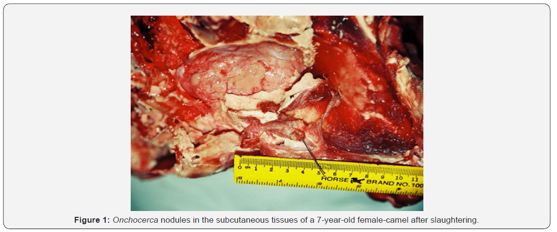
During a 1-year period, a total of 97camels slaughtered were examined for camel onchocerciasis. The camels were slaughtered at a local slaughterhouse at Ramtha province and the owners replaced the herds by buying camels of different ages from animal auctions located in different parts of Jordan. All camels are raised for slaughtering purposes. Routine clinical examinations of all camels were performed before slaughtering with special emphasis on the skin. Following slaughtering samples were collected one time a week in which 3 to 8 camels were slaughtered weekly over 12 months. After slaughtering the skin is removed and gross examination was performed on the subcutaneous tissues, muscles, ligamentum nuchae, blood vessels and internal organs. The presence of cutaneous nodules was carefully examined and then opened. Living parasites were removed and sent to the Department of Parasitology for diagnosis. The gross damage to the local tissue was described and photographed. Selectively tissue samples were collected, preserved in buffered 10% formalin solution and processed by standard histopathological techniques and stained with hematoxylin and eosin (H&E) (Figure 1).

Results
Out of 97 camels examined 8 camels were infested with Onchocerca fasciata (8.24%). In living animals no clinical signs of onchocerciasis were seen except two camels showed cutaneous swelling on the ventral abdominal wall. Whereas following slaughtering parasite nodules were found grossly visible on ligamentum nuchae as well as in the subcutaneous tissues of the abdominal cavity on the muscles of abdomen including the rectus abdominis muscle and the external oblique abdominis muscle. Also, nodules of the parasite were seen in the head region close to the eye in the cervicoscutularis muscle, under the ear in the zygomaticoscutularis muscle, and in the occipitofrontalis muscle. However, vision and hearing were not affected. No parasites lesions were seen in the blood vessels or internal organs. On palpation of the nodules, they were firm and of variable sizes. On incision, most of the nodules were filled by living parasites with little fluid. Some nodules were so hard and contained degenerated parasites with calcification. Both male and female parasites were present in the nodules. Most parasite still alive and move freely in the nodules. Parasite refereed to parasitology Department came up with the diagnosis of Onchocerca fasciata.

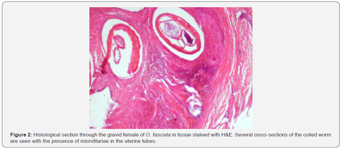
Histological changes were as desquamation of the epithelial cells with infiltration of different types of leukocytes such as the neutrophils, eosinophils and lymphocytes in the inflamed areas. Most of these worms are fertile females with microfilariae in the uterus. Inflammatory infiltration of varying composition is present around live and degenerated worms (Figure 2). In the early stages, lymphocytes, eosinophils and histocytes are prominent, but later, eosinophils decrease while plasma cells and giant cells increase in number. Heavy infiltrations by neutrophils occur in nodules containing degenerated or dead worms.
Discussion
Three of species of the nematode genus Onchocerca occur in the subcutaneous tissues of camel throughout the world [3]. O. fasciata is a nodule forming filaral nematodes in the ligamentum nuchae and other parts of the body, especially the subcutaneous tissues of the head and neck regions. In the present study 8.24% of the camels examined have parasite nodules on ligamentum nuchae as well as in the subcutaneous tissues of the abdominal cavity on the muscles of abdomen including the rectus abdominis muscle and the external oblique abdominis muscle. Also, nodules of the parasite were seen in the head region close to the eye in the cervicoscutularis muscle, under the ear in the zygomaticoscutularis muscle, and in the occipitofrontalis muscle. It is reported in camels in Saudi Arabia, Kuwait, Sudan, and Australia [9-11]. O. fasciata and O. gutturosa occur in the subcutaneous tissue of camels and cattle. O. armillata occurs in the aorta of camel as tortuous tunnels of parasitic tracks readily visible in the intimal surface of the vessels [6].
O. cervicalis occurs in the ligmantum nuchae and subcutaneous tissues of the neck, and in some cases, the flexor tendons and ligaments of the fetlock of horses. O. volvulus occurs in the sub of human [12]. The adult parasites reside in subcutaneous tissues, whereas microfilarias circulate in the blood. Life cycle is indirect and the intermediate host and vector may be one of a number of species of Culicoides. The parasite microfilariae are of 245 to 294μm long and are present in the skin and connective tissue spaces. Upon biting an infected camel, the Culicoides ingests microfilariae, which have a developmental cycle in the insect similar to that of other filarial larvae, transforming into infective forms that may enter a new host when the Culicoides again takes a blood meal [13]. After introduction into the new host, the developing worms wander through the subcutaneous tissues and then settle down, usually in groups of two or more; most worms finally become encapsulated. Onchocerca gutturosa occurs in the ligamentum nuchae of cattle and camels. Males are 2.9 to 3.0cm and females 6.0 cm long. O. gibsoni occurs in cattle, zebu and camels in Asia, Australia and South Africa [2]. The worms are usually found in nodules, which may occur especially on the brisket and the external surface of the hind limbs. The male is 30 to 53mm long while the female 140 to 190mm. Life cycle is indirect and the intermediate host is midge Culicoides pungens.
Various nonspecific skin changes have been observed in association with the filarial nematodes. It is not clear whether the parasites induced the dermatitis or the lesions are due to a hypersensitivity reaction to antigens released by dying microfilaria [14]. Camels affected by onchocerciasis usually develop no clinical signs and generally the parasite nodules are seen at slaughtering or postmortem examination. If the subcutaneous nodules are large, detection of these nodules by palpation is possible. Diagnosis is by identification of the microfilariae in skin snips. Biopsy of the subcutaneous nodules may be taken and histopathological sectioning of these nodules reveals the presence of worms enclosed in smooth fibrous capsules. Fibrinoid material has been associated with immune complex deposition. For this reason, Schmidt suggested that an immune reaction between the host and the Onchocerca species or its metabolic products results in the host inflammatory response adjacent to the parasites [15- 17]. Slightly higher than one-tenth of the camels in this study had evidence of adult Onchocerca fasciata infection. This is significantly lower than the 76.9% prevalence previously reported for camel onchocerciasis in Saudi Arabia.
Conclusion
This study has shown that camels in Jordan are at risk of being infected by onchocerciasis. This may cause economic loss to camel industry due to meat rejection of the infected carcasses. Therefore, occurrence of the infection can be reduced by treatment the camels with Ivermectin and controlling fly population as the vector of the parasite.
Acknowledgements
Financial assistance from Jordan Bidia Research and Development Center is highly appreciated.


