Morphometric and Histological Characterization o Goat Fetal Ovaries
Aruna Kumari G1*, Rooh Ul Amin2 and Sadasiva Rao K2
1Department of Veterinary Gynaecology & Obstetrics, College of Veterinary Science, India
2NTR College of Veterinary Science, India
Submission: June 22, 2017; Published: August 23, 2017
*Corresponding author: Aruna Kumari G, Department of Veterinary Gynaecology & Obstetrics, College of Veterinary Science, Korutla, PVNRTVU, Rajendranagar, Hyderabad-500 030, Telangana, India, Email: aruna.gangineni@gmail.com
How to cite this article: Aruna K G, Rooh Ul Amin, Sadasiva Rao K .Morphometric and Histological Characterization of Goat Fetal Ovaries. Dairy and Vet Sci J. 2017; 3(1): 555605. DOI: 10.19080/JDVS.2017.03.555605
Abstract
The objective of the present work was to study the morphological development and histo morphometry of goat fetal ovaries. Goat- fetal ovaries were collected from slaughter house and divided in to three groups based on age of the fetus as determined by crown rump length method. Gestation day (GD) 60-90 as group-I, GD 91-120 as group-II and GD 121-145 as group-III. Following recording the weight of ovaries, they were fixed, processed routinely and stained with Hematoxylene and Eosin. The fetal ovarian weights were 10.6±0.4mg, 30.6±1.53mg and 36±1.4mg respectively of group-I, group-II and group-III. Histologically in group I, ovaries had no distinct medulla and cortex. Oogonia were numerous and few primordial follicles are present. The average diameter of the follicle was 42.33±1.3μ and oocyte diameter was 10±1.5μ. In group-II, ovaries will have clear demarcation between cortex and medulla and also observed more primordial follicles, few preantral and very few antral follicles. In group-III along with primordial and preantral follicles, the antral follicles also present and the size of the antral follicle was 346.33±8.8μ, oocyte size is about 87.33±2.2μ. This 87.33±2.2μ size of the oocyte from antral follicle is similar with the results of our in vitro produced oocytes from cultured follicles of fetal ovaries.
Keywords: Goat fetal ovary; Development; Morphology
Introduction
The normal development and differentiation of the ovary determine the reproductive potential of the female. The pattern of ovarian differentiation is of two types depending on whether the germ cells of the ovary undergo immediate meiosis without previous steroid production in case of mouse, rat and hamster or delayed meiosis with steroid produced before meiosis starts in pig, sheep, dog and cow [1]. The gonadal development was initiated by the migration of primordial germ cells from the yolk sac into the gonadal ridge [2].
The morphogenesis of the fetal ovary may include colonization of primordial germ cells, interaction of primordial germ cells with somatic cells, formation of ovigenous cords and finally disappearance of ovigenous cords and establishment of definite number of primordial follicles. Although species differences in the timing of specific events are found, the general chronologies of developmental events that culminate in the formation of primordial follicles appear to be similar in all mammals. The ability to produce embryos from fetal oocytes offers the potential for shortening the generation interval of valuable animals and thus eliminates the necessity for adult animals in passing traits from one generation to the next. Besides shortening the interval between generations, this may avoid exposure of oocytes to undesirable environmental influences that may affect adult ovary. In addition this ability would have a great impact on assisted reproduction in endangered species and valuable animals in which death of the dam and or fetus is premature or sudden.
The basic knowledge on the physiological processes of the follicular development in fetal stages may allow for the development of strategies to increase reproductive efficiency in domestic ruminants. The development patterns of sheep, cattle and buffalo ovaries were studied [3-5]. There is paucity of information on ovarian development in goat fetuses. The ovarian developmental features of indigenous goats may be similar or vary from those of other ruminants. The objective of this work, therefore, is to study the morphological development and histomorphometry of goat fetal ovaries.
Materials and Methods
Forty five ovaries were collected from goat fetuses of different age groups. The age of the fetus in days was determined by crown-rump length [6] by using foetal age prediction equation X=2.1(Y+17) [7], where X is developmental age in days and Y is the crown rump length in centimeters. The ovaries were divided in to three different groups using specific age intervals as GD 60-90, 91-120, 121-145.
Fetuses were recovered at days 60 to 145 of gestation. Ovaries were fixed in Bouins solution and embedded in paraffin (n=45 ovaries) each from a separate fetus and a minimum of fifteen per age group and serially sectioned at a thickness of 5μm for histological evaluation, stained with haematoxylin and eosin [8]. The sections were studied under light microscope. Histologically, the follicles were classified according to the system of [9,10] in to the following: oogonia-germ cells devoid of granulosa cells with an intact nuclear membrane, primordial follicle: with one layer of flattened granulosa cells surrounding the oocyte, preantral follicles with more than two layers of cuboidal granulose cells antral follicles are those having more than five layers of cuboidal granulosa cells with antram formation. The diameters of the ovarian follicles and oocytes were measured using stage andocular microscope. The data were tested statistically by Analysis of variance and Duncans multiple range Test using SPSS windows version 16.00.
Results and Discussion
Weight of the ovary
In each group fifteen ovaries were studied. The ovaries were identified caudal to the kidneys in the fetuses. By GD 126-145 each of the ovaries was more caudo-laterally placed near the tip of the adjoining uterine horn. The two ovaries were resembled pin head, oval in shape with smooth surface and uniformly cream in color. The mean ovarian weights increased significantly (p<0.01) with the age of the fetuses.
In the GD 60-90 ovaries, the weight ofthe ovary was 10.6±.04a mg, where as in case of GD 91-120 the ovary was weighing about 30.6±1.5b mg and in GD 121-145 the weight of the ovary was 39.3±1.4c mg. The weights of the ovaries in group-I, II and group- III were significantly different from each other. With increase in age of the fetus the ovarian weight was also increased (Table 1).

Histological features
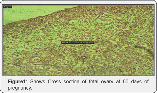
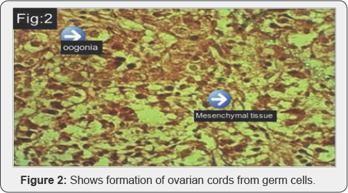
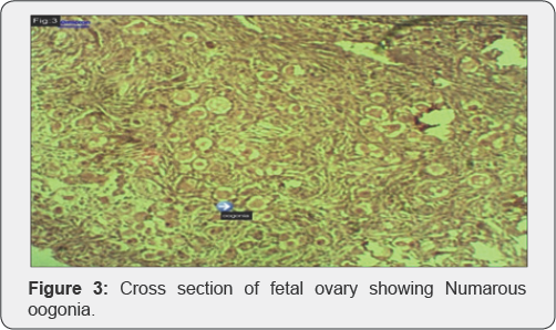

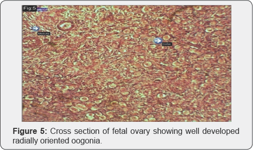
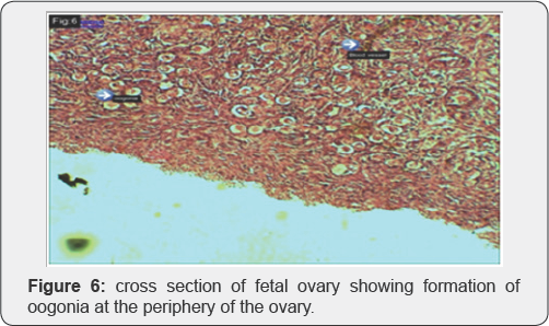
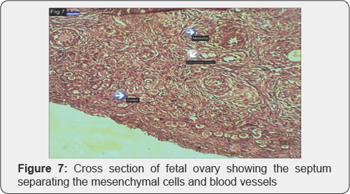
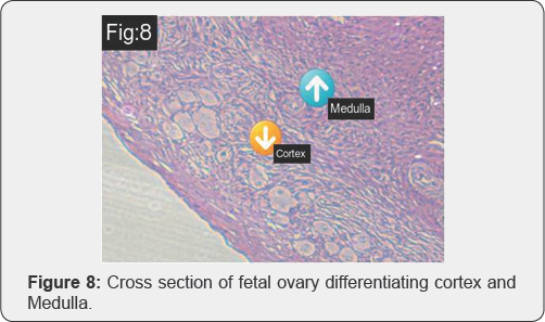
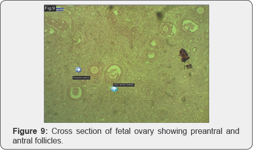
The development of the goat fetal ovary was studied from GD 60 to term GD 145. Histologically, in group-1, the ovary had no distinct cortex and medulla (Figure 1). The germ cells form ovarian cords (Figure 2) and the cords consisting of somatic or pregranulosa cells and germ cells or oogonia are of irregular shape and are separated from the surrounding loose mesenchymal tissue by the basal lamina (Figure 2). Oogonia were numerous among other stromal cells toward the periphery of the ovary (Figure 3 & 4). The cortex richly populated with germ cell cords and the medulla consists mainly of somatic cells. The cortex shows well developed radially oriented oogonia (Figure 5) consists of germ cells (oogonia) and somatic cells which are separated from each other by septa consisting of mesenchymal connective tissue and blood vessels (Figure 6) at this stage the germ cells attain their maximum number evidenced by the full development of oogonia. The oogonia towards the periphery of the developing goat ovary (Figure 7). By the end of the delayed period the ovarian cords start to break up in the central regions of the ovary. This process is related to the start of meiosis. Meiosis in developing goat ovary is initiated at 91-120 days of gestation. At this period ovary can be distinguished in to cortex and medulla (Figure 8). Our results are in accordance with Erickson [11]. The intermediate regions separating the cortex from the medulla are relatively richer with blood vessels (Figure 9). This specific distribution of blood vascularity must be of significance in causing differential organization of developing ovary. At this stage of gestation the inner part of ovarian cortex is dominated by primordial follicles. This indicates that in goat the primordial follicles are first formed in the inner portions of ovarian cortex at 90-120 days of gestation. There are also some growing (preantral) follicles and a few Graafian or antral follicles (Figure 10) up to 107±4.1μ and 153.6±3.8μ in diameter respectively. At gestation day 12-45, the diameter of growing or preantal follicle and graafian or antral follicle increases up to 175.3±5.2μ and 346.33±8.8μ in diameter. This suggest that initiation of follicle growth in the goat fetal ovary is initiated earlier during the fetal life of goat and also suggesting the possibility of an earlier development and differentiation or hypothalamic pituitary gonadal interrelationship or of an early ovarian responsiveness to gonadotropins in goat fetus. The presence of growing follicles during fetal life of goat in the present study suggests that gonadotropins begin to be secreted by the fetus pituitary as also suggested for buffalo [1]. The result of this study provides data on the morphological development of ovaries in goats. Oogonia were numerous among other stromal cells toward the periphery of the ovaries at GD60-90. Absence of granulosa cells around them indicates their primitive stage in these fetuses. The presence of many primordial follicles and few preantral an graafian follicles at GD 91 to 120 in the cortex and presence of more antral follicles at 121 to GD 145 suggests that folliculogenesis starts with in this period in the goats. This observation is supported by the reports of [5,10,11] in sheep .The observation of graafian follicles with evidence of antrum formation at GD91 to 121 and 121-145 GD ovaries suggest that the ovarian follicles undergo structural development with increase in the age of the fetuses. Similar observation like presence of vesicular follicles in cattle fetal ovaries aged 250 days post coitum were investigated on developing cattle ovaries [12]. Ovarian development forms a gradual process which is accompanied by the extensive development of somatic cells and blood capillaries at the dorsal margin of the gonad.

From the present study it was concluded that the GD60-90 ovaries comprised many oogonia and no distinct cortex and medulla. By 91-120 they comprised distinct cortex medulla and more primordial, few preantral and antral follicles. By GD121-145 the diameter of preantral, antral follicles and oocyte diameters also increases with fetal age. There is a linear regression between ovarian weight and fetal age in goat. This result provides baseline information on ovarian development in goats. This study appears to be the first report on development of goat ovary [13,14].
Acknowledgment
Department of Biotechnology (DBT), Government of India supported this work through a research grant to Dr. G. Aruna Kumari, Assistant Professor, ARGO, CVSc, R'Nagar, Hyderabad.
References
- Guraya SS (1997) Ovarian biology in Buffaloes and Cattle. In: Heywood R, Swayer, Perter S, Derek A, Heath, et al. (Eds.) Formation of Ovarian Follicles during Fetal Development in Sheep. (2002) ICAR Publications Biology of Reproduction, India, 66: 1134-1150.
- Osuagwuh AIA, Aire TA (1979) Studies on the developmental age of the Caprine fetuses. External measurements and appearance. Trop Vet 4: 39-51.
- McNatty KP, Smith P, Hudson N, Heath D, Tisdall DJ, et al. (1995) Development of the sheep ovary during fetal and early neonatal life and the effect of fecundity genes. J Repro Fertil 49: 123-135.
- Borwick SC, Rhind SM, McMillen SR, Racey PA (1997) Effect of under nutrition of ewes from the time of mating on fetal development in mid gestation. J Reprod Fertil Dev 9(7): 711-716.
- Sawyer HRP, Smith DA, Heath JL, Juengel J, Wakefield, et al. (2002) Formation of ovarian follicles during fetal development in sheep. J Bio Repro 66(4): 1143-1150.
- Zamboni lF, Bezard MP (1979) The role of the mesonephros in the development of the sheep fetal ovary. Anim Biochem Biophys 19: 1153-1178.
- Nwaogu IC, Ezeasor DN (2008) Studies on the development of omasum in West African Dwarf goats (Capra hircus). Vet Res Commun 32(7): 543-552.
- Drury RAB, Wallington EA, Cameron RS (1976) General staining procedures. In: Carlenton's Histological Technique. Oxford University Press, London, England, pp. 114-137.
- Oxender WD, Colenbrader B, Van de Wiel DFM, Wensing CJ (1979) Ovarian Development in fetal and prepubertal pigs. Biol Reprod 21: 715-721.
- Lundy T, Smith P, O Connell A, Hudson NL, McNatty KP (1999) Populations of granulose cells in the sheep ovary. J Reprod Fertil 115(2): 251-262.
- Erickson BH (1966) Development and radio-response of pre-natal bovine ovary. J Reprod Fert 10: 97-105.
- McNatty KP, Fidler AE, Juengil JL, Quirk LD, Smith PR, et al. (2000) Growth and paracrine factors regulating follicular formation and cellular function. Mol Cell Endocrinol 163 (1-2): 11-20.
- Henricson B, Rajakoshi E (1959) Studies on oocytogenesis in cattle. Cornell Vet 49: 494-503.
- Guraya SS (1998) Cellular and molecular biology of gonadal development and maturation in mammals: Fundamentals and biomedical implications. Narora publishing house, New Delhi, India.


