




 JCMAH Home



Evaluation of in Vitro Immunomodulatory Activity Dexamethasone and A Standardized Polyherbal Formula by Measurement of Inflammatory Markers on Dendritic Cells and Phagocytosis in Macrophage Cells
Shalu Chauhan1*, Pramod Shukla, Vivek Srivastava and Suresh Garg
Research and Development, Zeon life science LTD University, Paonta Sahib, India
Submission:March 9, 2024;Published:March 26, 2024
*Corresponding author:Shalu Chauhan, Zeon life science LTD University, Paonta Sahib, India, Email: adl3@zeon.co.in
How to cite this article: Shalu C, Pramod S, Vivek S, Suresh G. Evaluation of in Vitro Immunomodulatory Activity Dexamethasone and A Standardized Polyherbal Formula by Measurement of Inflammatory Markers on Dendritic Cells and Phagocytosis in Macrophage Cells. J Complement Med Alt Healthcare. 2024; 12(4): 555844. DOI: 10.19080/JCMAH.2024.12.555844
Abstract
Background: The polyherbal formula was evaluated for its in vitro Immunomodulatory activity against LPS induced stress in dendritic cells and evaluated for its in vitro phagocytic property of the test substance in macrophage cells.
Methodology: Initially polyherbal formula was checked for cytotoxicity and non-toxic concentrations were identified and were selected for efficacy studies on dendritic cells. Dendritic cells were intoxicated with LPS followed by treatment with the test substance. After 24 hrs., the cell supernatant bound immune markers (TNF-alpha, IL-1 beta and MIP-1-α) were estimated. And also checked cytotoxicity and non-toxic concentrations were identified and were selected for efficacy studies on macrophage cells. Macrophage (Raw 264.7) cells were treated with the test substance for 1 h then exposed to green E. Coli slurry. The amount of phagocytosis observed in macrophage cells was estimated by fluorescent microscopy and flow cytometry.
Result: Results indicate normalization of immune markers by the test substance indicating its immunomodulatory properties in vitro.
Conclusion: The test substance promotes phagocytosis in vitro.
Keywords: Immunomodulatory Activity; In Vitro Study; Polyherbal Formulation; Macrophage Cells.
Abbreviations: MCR: Microbiology; CB: Cell Biology; MB: Molecular Biology; BC: Biochemistry; 0C: Degree Centigrade; %: Percentage; GM: Gram; Hr.: Hour; DTL: Drug Testing; PC: Pre-Clinical; CL: Clinical; NCCS: National Centre for Cell; FBS: Fetal Bovine; PBS: Phosphate Buffer; EDTA: Ethylenediaminetetraacetic; MTT : 3-(4,5-Dimethylthiazol-2-yl)-2,5-Diphenyltetrazolium; TPVG: Trypsin Phosphate Versene Glucose; DMFM: Dulbecco’s Modified Eagle; DMSO: Dimethyl Sulfoxide; CTC50: Cytotoxicity Concentration; M: Milligram; ML: Milliliter; N: Nanometer; μl: Microliter; μg: Microgram; RT: Room Temperature
Introduction
The human body has a remarkably sophisticated immune system consisting of white blood cells and specialized immune molecules that protect the body against invading pathogens 9 [1]. The immune system is made up of innate and adaptive immune immunity. Innate immunity provides first protection against pathogens, and then it will stimulate adaptive immunity to enhance the protection. Innate immunity is the most rapidly acting immunity. It mostly depends on neutrophils, macrophages, dendritic cells, and monocytes, while T and B cells are involved in adaptive immunity [2,3]. Macrophages also modulate adaptive immunity by presenting antigen to CD4 T cells through major histocompatibility complex (MHC) class II antigen. CD4 T cells perform their functions by four subpopulations, which include Th-1, Th-2, Th-17, and CD4 T regulatory (Treg) cells [4]. The cells help B cells develop into plasma cells which can produce antibody and activate T cells to become activated cytotoxic T cells [5].
Several cytokines also play essential roles in immune response, which consist of pro-inflammatory cytokines such as tumor necrosis factor-alpha (TNF-α), interleukin 1 (IL-1), IL-6, IL-11, IL-8, and anti-inflammatory cytokines or cytokines inhibitor such as IL-4, IL-10, and IL-13. Cytokines as intercellular messenger molecules have several functions, and these include stimulating phagocyte migration and coordinating early responses of monocytes, macrophages, dendritic cells, and lymphocytes during inflammatory states [6]. The release of pro-inflammatory cytokines is regulated by nuclear factor-kappa B (NF-κB) and mitogen-activated protein kinase (MAPK) pathways [7]. Our immune system, a network of intricate stages and pathways in the body, recognizes foreign invaders like bacteria, viruses, and parasites and takes immediate action. Immunity is always affected by our daily lifestyle. Prevention is a practicable and sensible route to take, as treatment options are currently limited to antiviral, health and stability of the immune system of infected person.
The herbal products today symbolise safety in contrast to the synthetic drug compounds that are regarded as unsafe due to their severe adverse effects on human body. Many studies reported in literature about use of single traditional ingredient for their usage in prevention of therapeutic attributes. But it has been observed that using single ingredients, you may end up using high quantities, which may lead to undesirable product attributes as well as potential toxicity issues. So, research focused on combination of natural ingredients & polyherbal formulation is needed of the modern day. Keeping these things in mind Zeon have come with a polyherbal formula with combination of 3 natural ingredients extract for achieving synergistic effects using novel delivery technologies to enhance its efficacy in synergy, viral– immune response. A combination of drugs is synergistic when the combined effect is larger than the additive effect of each individual drug.
Currently, herbs are applied to the treatment of chronic and acute conditions and various ailments and problems such as infection, inflammation, and good for immune system. Polyherbal formula performs one or more activities like prophylaxis and treatment therapy without any side-effect. No toxic effect of the same formulation was reported. Poly-herbal formula uses a cautious but evidence-based strategy. Zeon developed Polyherbal formulation and performed it for preclinical studies. The acute oral study was conducted in animal model (rats) by using OECD 423 guidelines where rats were administered orally and observed for clinical signs, behavioural and gross pathological studies individually. The studied dose did not cause any mortality or signs of toxicity in the rats tested during the observation period. Beside this another study was conducted for Bacterial reverse mutation test using 5 different strains of Salmonella typhimurium: TA 98, TA100, TA102, TA1535 and TA1537. The product was tested at different concentrations by direct plate incorporation method and by preincubation method, where this poly-herbal formula, did not exhibit mutagenicity with bacterial reverse mutation at the tested concentrations under the test conditions.
Similarly, immunomodulating and anti-inflammatory properties of the poly-herbal formulation which exhibited protection against LPS induced TNF- α, IL-1- β and MIP-1-α in mouse dendritic cell line was also observed and this was compared with Dexamethasone which was a glucocorticoid medication. Under Florescent microscope, the process of phagocytosis is observed minimally in the treatment groups compared against the LPS-cell control. The compounds therefore have little effect on the phagocytosis in the cell lines. Polyherbal formulation was also evaluated for their immunomodulatory potencies using carbon clearance assay, Carbon clearance evaluates the effect of drug and phytoconstituents of the reticuloendothelial system (RES). In this assay, the enhanced phagocytic activity of macrophage is evaluated by the rate of elimination of exogenously administered antigen such as carbon particles. Poly-herbal formulation was enhancing the rate of carbon clearance (phagocytic activity) when compared to that of normal control and standard group (Dexamethasone).
Based on the findings of above studies it is evident that the poly-herbal formulation shows multiple pharmacological activities. Immunomodulatory, Antiviral, Anti-Inflammatory are the major therapeutic effects which have been performed by the Polyherbal Formulation. During the flu season or times of illness, people often seek help from supplements that are believed to boost immunity. Globally peoples have suffered from pandemic like Covid-19, which have alarmed people to focus more towards maintaining a good immune function. So, from the observations & findings of clinical studies conducted for this polyherbal formulation it can help in performing multiple functions in human body alongside maintaining a good health without any adverse effects on Human body. The proposed research work would be a small contribution from Zeon research team to stand with our country in the pandemic situation of COVID -19. This will be a specific socio-economic research project to cater the population of all the financial start in our country and can help 140 Crore Indians as well as global population to fight against this crucial situation by maintaining a good Health.
Method
Outline of the method
The non-toxic concentrations of the test substance were selected to evaluate its Immunomodulatory activity against LPS induced toxicity on dendritic cells. After dendritic cells were stimulated by LPS and treated with the test substance, the cell supernatants were used for estimation of the immune markers (TNF-alpha, IL-1-β and MIP-1-α) by ELISA kits. The non-toxic concentrations of the test substance were selected to evaluate its phagocytic properties of the test substance on RAW 264.7 cells. After Macrophage cells were treated with the test substance prior to stimulation with the green E. coli; the measurement of the amount of phagocytosis was measured by fluorescent microscopy and flow cytometry.
Preparation of test solution
10 mg of test substance weighed and separately dissolved in DMEM-HG supplemented with 2% inactivated FBS, volume was made up with media to obtain a stock solution of 1 mg/ml concentration and sterilized by filtration. Serial two-fold dilutions were prepared from the stock solution to perform cytotoxic studies and further efficacy studies.
Cell line and Culture medium:
Raw 264.7 (Mouse Macrophage) cell line was procured from National Centre for Cell Sciences (NCCS), Pune, India. Stock cells were cultured in DMEM supplemented with 10% inactivated Fetal Bovine Serum (FBS), penicillin (100 IU/ml), streptomycin (100 μg/ml) and amphotericin B (5 μg/ml) in a humidified atmosphere of 5% CO2 at 37°C until confluent. The cells were dissociated with TPVG solution (0.2% trypsin, 0.02% EDTA, 0.05% glucose in PBS). The stock cultures were grown in 25 cm2 culture flasks and all experiments were carried out in 96 well microtitre plates or 6 well plates (Tarsons India Pvt. Ltd., Kolkata, India).
Isolation of DC from mouse spleen:
The C57 mouse was anaesthised with an overdose of isoflurane (adjust flow rate of isoflurane to >5% and continue exposure at least 2 min after breathing stops) and dissected to separate the spleen under aseptic conditions. The spleen was separated and transferred to a fresh Petri dish, washed with RPMI medium and was cut into small (~0.2 cm 2) pieces using a scalpel. The pieces were incubated for 30 min at 37°C with 10 ml of collagenase solution. The partially digested suspension of the splenic tissue was poured onto a 70 μm cell strainer. Using a 5 ml syringe the contents are compressed. The mesh was washed with 10 ml of fresh medium and the filter was discarded. The suspension was centrifuged at 300 x g for 10 min at 4°C to pellet the cells. The supernatant was aspirated, and the cells were resuspended in 2 ml of RBC lysis buffer and incubated at RT for 10 min. The suspension was centrifuged at 300 x g for 5 min at 4°C.
To examine the capacity of the spleen cells to proliferate in response to GM-CSF, 2 x 10⁶ cells were placed in each well of a 24-well plate in 1 ml of complete medium, supplemented with 10% FBS and with 4 ng/ml mouse recombinant GM-CSF (Sigma, #G0282). Until day 4, the cells were gently washed after every 48 h and the media was replenished with GM-CSF. After 4 days, the non-adherent cells were harvested and were collected by day 10. The dendritic cells were isolated by negative selection method using FACS.
Dendritic cells were cultured in DMEM-HG/RPMI media supplemented with 10% inactivated Fetal Bovine Serum (FBS), penicillin (100 IU/ml), streptomycin (100μg/ml) and amphotericin B (5μg/ml) in a humidified atmosphere of 5% CO2 at 37°C until confluent. The stock cultures were grown in 25 cm2 culture flasks and experiments were carried out in 96 microtiter plates for cytotoxicity.


Cytotoxicity Studies:
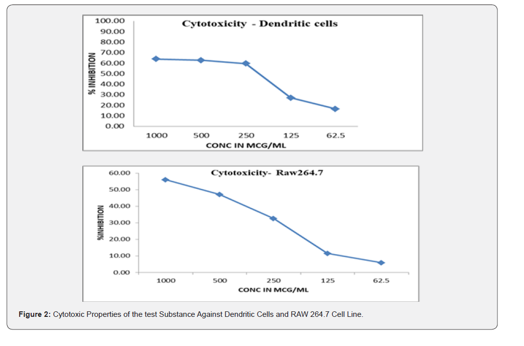
The cell viability is assessed by MTT reduction assay in semi confluent monolayer cultures. The drug solutions were added to cells and incubated at 37°C in 5% CO2 atmosphere. After 72 h of incubation the drug solutions in the wells were carefully removed and 100μl of MTT in PBS was added to each well. The plate was gently shaken and incubated for 3 h at 37°C in 5% CO2 atmosphere. The supernatant was removed as mentioned above and 100 μl of DMSO was added and the plate was gently shaken to solubilize the formed formazan. The absorbance was measured using a microplate reader at a wavelength of 540 nm. The plates were protected from light throughout the procedure. The percentage growth inhibition was calculated using the standard formula and concentration of test substance, needed to inhibit the growth of the cell by 50% i.e., CTC50 values were generated from the doseresponse curves. The inhibition was expressed as the percentage relative to the cell control.
Immunomodulatory Studies
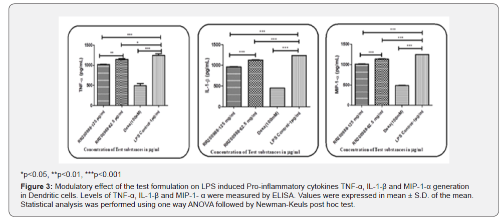
The dendritic cells, 2 x 10⁶ cells were seeded onto a 6 well plate and incubated for 24 h. After 24hrs, cell supernatants were replaced with non-toxic doses of test substances and LPS (1 μg/ml). Cell control and standard control (Dexamethasone) were maintained. After 24 h of incubation the supernatant was collected and centrifuged at 500g for 10 min and stored at -80°C until further use. Before estimation of the immune markers, the supernatants were brought to room temperatures and processed for immune markers TNF-α, IL-1-β and MIP-1-α by ELISA kit method according to the manufacturer’s instructions.
Phagocytosis Assay Studies
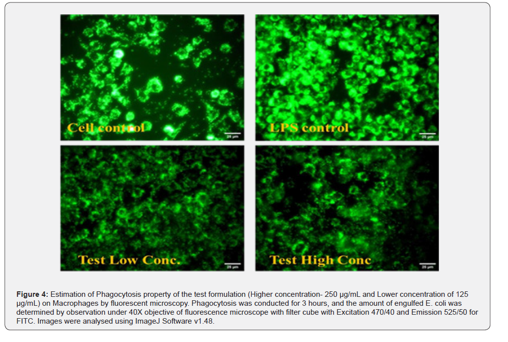
The Phagocytosis assay was processed according to the manufacturers’ instructions for analysis with fluorescent microscopy and flow cytometry. Briefly, 100μl the macrophage cells (1 x 10⁶cells/ml) were seed onto a 96 well plate and incubated for 24 h. After 24hrs, cell supernatants were replaced with non-toxic doses of test substances and incubated for 1 h at 37°C. After 1 h of incubation, 5 μl of E.coli slurry was added to each well and immediately the plate was incubated for 3 h at 37°C. After incubation, 50μl of quenching solution was added to each well and incubated for 2 min at room temperature. The wells were washed properly with phagocytosis assay buffer and observed under fluorescence microscope. For analysis with flow cytometry, 500 μl the macrophage cells (1 x 10⁶ cells/ml) were seed onto a 24 well plate and incubated for 24 h. After 24hrs, cell supernatants were replaced with 400μl of non-toxic doses of test substances and incubated for 1 h at 37°C. After 1 h of incubation, 20μl of E.coli slurry was added to each well and immediately the plate was incubated for 3 h at 37°C. After incubation, 200μl of quenching solution was added to each well and incubated for 2 min at room temperature. The wells were washed with phagocytosis assay buffer and were fixed with Methanol: Acetone (1:1). The fixed cells were observed under florescent microscope (Microscope - XD-RFL Fluorescence Microscope, SDPTOP, China) for the visible observation of Phagocytosis.
Discussion
The test substance was initially examined for their in vitro cytotoxicity studies against dendritic cells by MTT assay by exposing the cells to different concentrations of the test substance; furthermore, the non-toxic concentrations were identified and selected for further efficacy studies. The test substance exhibited immunomodulatory activity by inhibiting TNF-α, IL-1-β and MIP- 1- α in LPS treated dendritic cells. The examined in the current study offered protection against LPS induced TNF-α and exhibited protection by 26.53 ± 3.8 and 11.22 ± 1.0 percent at test doses 125 μg/ml and 62.5 μg/ml, respectively over the LPS control. Moreover, the test substance offered protection against LPS induced IL-1- β and exhibited protection by 21.94 ± 0.9 and 8.53 ± 0.6 percent at test doses 125 μg/ml and 62.5 μg/ml, respectively over the LPS control. Similarly, the test substance offered protection against LPS induced MIP-1-α and exhibited protection by 18.09 ± 0.9 and 7.75 ± 2.7 percent at test doses 125 μg/ml and 62.5 μg/ml, respectively over the LPS control. Against the three markers viz., TNF-α, IL-1-β and MIP-1-α the standard drug Dexamethasone exhibited significant inhibitory activity ranging between 85.16 ± 4.9, 63.67 ± 0.2 and 60.39± 6.5 over control. When the foreign pathogen infects higher organisms, phagocytosis serves as a first line defense mechanism.
For determining the effect of the test substance on phagocytosis, the test substance was initially examined for their in vitro cytotoxicity studies against macrophage cells by MTT assay by exposing the cells to different concentrations of the test substance; furthermore, the non-toxic concentrations (Higher concentration- 250 μg/mL and Lower concentration of 125 μg/ mL) were identified and selected for further phagocytosis studies. Bio Vision’s EZCellTM Phagocytosis Assay Kit (#K963) employs heat-killed, fluorescently pre- labeled E. coli particles as a tool for rapid and accurate detection and quantification of in vitro phagocytosis by fluorescent microscope. Phagocytosis is a specific form of endocytosis initiated by recognition and binding of foreign particles by cell surface receptors, followed by their engulfment, and formation of phagosomes. The observation of cells under fluorescent microscope (Figure 2) shows that the test substance has a minimal increase in the phagocytosis effect on macrophages and the effect on phagocytosis is not concentration dependent compared to the cell control.
Conclusion
The present study evaluated the immunomodulating and anti-inflammatory properties of the test Substance which exhibited protection against LPS induced TNF-α, IL-1-β and MIP- 1-α in mouse dendritic cell line. Collectively, the compounds are exhibiting a percentage protection from the inflammatory cytokines (TNF-α, IL-1-β and MIP-1-α) between 18-26% at the highest concentration, which is 125μg/ml. Compared to the standard chosen this protection is significantly less; however, it is to be noted that the compounds exhibit low to moderate protection against inflammation. Under Florescent microscope, the process of phagocytosis is observed minimally in the treatment groups compared over against the LPS-cell control. The compounds therefore have little effect on the phagocytosis in the cell lines. It’s mentioned that a patent for the tested substance has already been granted.
References
1. Tan B, Vanitha J (2004) Immunomodulatory and Antimicrobial Effects
of Some Traditional Chinese Medicinal Herbs: A Review. Current
Medicinal Chemistry 11(11):1423-1430.
2. Beutler B (2004) Innate Immunity: An Overview. Molecular
Immunology 40(12):845–859.
3. Saroj P, Verma M, Jha KK, Pal M (2012) An Overview on
Immunomodulation. Journal of Advanced Scientific Research 3(1):7-
12.
4. Chapel H, Haeney M, Misbah S, Snowden N (2006) Clinical Immunology.
5th Edn. Massachusetts: Blackwell Publishing.
5. Luckheeram RV, Zhou R, Verma AD, Xia B (2012) CD4+T Cells:
Differentiation and Functions. Clin Dev Immunol.
6. Shaikh P Z (2011) Cytokines & Their Physiologic and Pharmacologic
Functions in Inflammation: A Review. Int J Pharm Life Sci 2: 1247-1263.
7. Beyaert R, Beaugerie L, Van Assche G, Broche Renauld LJC, Renauld JC,
et al (2013) Cancer Risk in Immune-Mediated Inflammatory Diseases
(IMID). Molecular Cancer 12(1): 98.

























