Cupping Hijama Therapy Skin Marks: What Should We Know About Them?
Naseem Akhtar Qureshi*, Abdullah Mohammed Al-Bedah and Tamer Shaban Abushanab
National Center for Complementary and Alternative Medicine, Ministry of Health, Saudi Arabia
Submission: February 10, 2017; Published: August 18, 2017
*Correspondence author: Naseem Akhtar Qureshi, Consultant Psychiatrist and Public Health Specialist, National Center for Complementary and Alternative Medicine, Ministry of Health, Riyadh, Saudi Arabia, Tel: +966564542710; Email: qureshinaseem@live.com
How to cite this article: Naseem A Q, Abdullah M A B, Tamer S A . Cupping Hijama Therapy Skin Marks: What Should We Know About Them?. J Complement Med Alt Healthcare. 2017; 3(3): 555612. DOI: 10.19080/JCMAH.2017.03.555612
Abstract
Skin changes after cupping is one of the main characteristics of cupping therapy (CT). It usually disappears within three weeks. The aim of this article is to highlight the different explanations of these marks, and the pertinent studies done in this field. The article also discusses using these marks as a diagnostic method by Oriental medicine practitioners, and the factors affecting these skin changes after cupping therapy. The studies in this field will substantially help researchers in understanding cupping therapy (Hijama) and standardize its practice around the world.
Introduction
Cupping therapy (Hijama) is one of the oldest methods of healing. Its origin is controversial; however, from historical perspective ancient Egyptians are known to practice it for the first time. Subsequently, Chinese practitioners have practiced cupping therapy for thousands of years, and consequently cupping therapy became an important component of Traditional Chinese Medicine. CT involves using different types of cups and applying them on skin either by vacuum pump or by fire to create negative pressure inside the cups. The created pressure within the cup pulls the skin up. CT also may involve scarification of the skin to suck the blood out. There are many types of cupping therapies including wet cupping, dry cupping, flash cupping, sliding cupping and others [1]. CT has a relative good safety profile but scar formation, infection, abscess, blister and burns are its reported adverse effects especially when cupping is done without using aseptic methods by unqualified and untrained cupping therapists [2]. Dry cupping is a method of applying cups for 5, 10 or more minutes [3] without scarification, and the most common sites on which the cups have been applied are the back, chest, abdomen and other body areas [4]. Dry cupping can leave temporary bruised-like painful marks on the skin and can complicate course of eczema [4]. Cupping therapy needs to be avoided in some patients with eczema.
Cupping Marks and Explanations
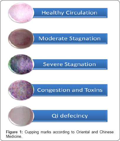
Cupping can leave skin marks, one of the commonest side effects of CT. Skin mark is usually round red to purple in color.There are many researches done in this field to analyze or explain these marks. Cupping marks can be explained by more than one theory. According to some researchers, these marks develop as a direct result of the suction pressure that lead to strong stress created within the skin [5]. Furthermore, Tham and colleagues suggested that the high stress in the skin layer is believed to be the primary cause of ecchymosis, a discoloration of the skin caused by the bleeding from the ruptured small blood vessels. Bleeding may accumulate just inside the cup rim, and in the middle because of high negative pressure in these areas, which lead to deeper skin marks with varying degrees of colors associated with bruising [5]. In Oriental medicine, cupping may be used for making various diagnoses through skin color (Figure 1). Purplish red cupping mark means severe damp heat. Red cupping mark signifies severe heat. Bluish purple cupping mark indicates severe cold dampness. Cupping mark with dark color means exuberance of the pathogenic qi, a life force. Cupping mark with light color implies mild pathogenic qi. No cupping mark means the absence of pathogenic qi. [6]. Kim & Lee [7] suggested that CT has been utilized in the diagnosis and for observing the skin hemoglobin, blister and oppressive pain responses. Among these, the hemoglobin response has been widely used to identify accumulated blood. According to this study, the pigmentation response, which is divided into four visual steps, assesses the state of internal organs indirectly Because cupping as a diagnostic method is still subjective, Kim and Lee suggested a new numerical analysis as an alternative to the visual diagnostic method, and used optical techniques in order to detect the changes in skin color incurred from cupping [7]. Kim and Lee analyzed the deep skin colored part and the light skin colored part after cupping, and found that the deep- skin colored part has higher levels of white blood cells, polymer pronuclear neutrophils leukocytes, red blood cells, hemoglobin, mean corpuscular hemoglobin, and hematocrit compared with the light skin colored part which was reported to have higher numbers of lymphocytes, monocytes, mean corpuscular volume, and platelets [7].

Many factors can affect the skin color after CT; however, time and pressure were evaluated as contributory factors. The stimulation of 10 minutes and the pressure of -0.04 mega pascalmay produce marked ecchymosison the cupping site, and the area gets darker and darker corresponding to the increase in the stimulation intensity. Thus, the effect of pressure on cupping mark color is characterized by a linear relationship. Conversely, the effect of time factor on cupping mark color is rather more complicated, and shows 10 minutes>30 minutes>20 minutes, which is possibly related with the cupping sites [8]. Histological smears were obtained from the cupping site and examined; the histological changes following cutting and bloodletting were mild edema, vacuolization and longitudinal fissure as a result of cutting in the epidermis. In the dermis, histological changes were dermal edema and bleeding in its upper and lower parts, and no cellular infiltration was noted in the dermis [9]. Healing of the skin and the return of cupping marks to the normal skin color usually happens to be within 3 weeks [9]. Laser therapy if used (energy density: 8J/cm2) may have a significant recovery of the skin erythema and skin pigmentation after cupping [10].
In summary, cupping can produce skin changes and marks, which were used by oriental medicine practitioners for the diagnosis of various ailments. Cupping skin marks were produced by the pressure inside the cup and the edema and bleeding in various skin layers. Many factors can determine the cupping marks such as pressure, and time. Marks usually disappear within 3 weeks of cupping, and laser therapy with special intensity is supposed to shorten this period. More studies are needed in this field to help researchers adequately understand cupping therapy marks, find answers for many questions about the relation between skin changes and factors like cupping site, cup type, suction, time, and patient related factors, and to evaluate the accuracy of using skin colors as an important parameter in oriental medicine diagnosis. Overall, this will help in standardization of cupping therapy practice around the world.
Conflicts of Interest:
No conflicts of interest reported in this work.
References
- Al-Bedah, AM, Shaban T, Alqaed MA, Qureshi NA, Suhaibani A, et al. (2016) Classification of Cupping Therapy: A tool for modernization and standardization. Journal Complementary Alternative Medical Research 1(1): 1-10.
- Al-Bedah AM, Shaban T, Suhaibani A, Gazzaffi I, Khalil M, et al. (2016) Safety of cupping therapy in studies conducted in twenty one century: A review of literature. British Journal Medicine Medical Research 15(8): 1-12.
- Yoo SS, Tausk F (2004) Cupping: east meets west. International Journal Dermatology 43(9): 664-665.
- Hon KL, Luk DC, Leong KF, Leung AK (2013) Cupping therapy may be harmful for eczema: a PubMed search. Case reports pediatrics 2013: 605829.
- Tham LM, Lee HP, Lu C (2006) Cupping: from a biomechanical perspective. Journal biomechanics 39(12): 2183-2193.
- Zhong L (2009) Joint Reinforcing-Reducing Effect of Acupuncture, Moxibustion and Cupping Therapies. Journal Traditional Chinese Medicine 29(2): 101-103.
- Kim S-B, Lee Y-H (2014) Numerical analysis of the change in skin color due to ecchymosis and petechiae generated by cupping: a pilot study. Journal Acupuncture Meridian Studies 7(6): 306-317.
- Zhao XX, Tong BY, Wang XX, Sun GL (2009) Effect of time and pressure factors on the cupping mark color. Zhongguo Zhen Jiu (Chinese Acupuncture & Moxibustion) 29(5): 385-388.
- Al-Rubaye, KQA (2012) The clinical and histological skin changes after the cupping therapy (Al-Hijamah). Journal Turkish Academy Dermatology 6(1): 1261a1.
- Kim S-B (2016) Laser acupuncture improves skin color deformation by cupping: A pilot study. Acupuncture & Electro-Therapeutics Research 41(3-4): 155-169.


