




 JAICM Home



Abstract
Aortic aneurysms are pathological dilations of the aorta that can lead to life-threatening complications if not properly managed. The risk factors for aneurysms formation are genetic, hypertension, and environmental factors. Genetic conditions such as Marfan syndrome, Ehlers-Danlos syndrome, and Loeys-Dietz syndrome are well-known risk factors for aortic aneurysm formation due to mutations in genes encoding structural proteins like fibrillin-1 and collagen. In contrast, amyloidosis, a rare but significant cause of organ dysfunction and aneurysms formation, is more challenging to diagnose, often requiring tissue biopsy and advanced imaging techniques. Amyloidosis can lead to degeneration of the thoracic and peripheral arteries, facilitating aneurysm formation, with increased risk for dissection. This case report and overview details a young male who underwent ascending aorta and aortic arch replacement for acute type A dissection and subsequently, he developed an aortic root and huge left subclavian artery aneurysms, requiring redo-surgery to treat both pathologies, including aortic valve replacement with mechanical valve prosthesis. During follow-up the patient presented with neurological symptoms and headaches. Following MRI, reveling micro cerebral hemorrhage, eventually aggravated by the anticoagulation therapy, and blood tests, amyloidosis was diagnosed. The prognosis in amyloidosis varies significantly depending on the type, extent of organ involvement, and the timeliness of diagnosis and treatment. The case highlights the importance of considering amyloidosis in patients with aortic aneurysms, particularly, in those without traditional connective tissue disorder features. It underscores the need for early diagnosis and intervention in these cases to prevent further complications.
Keywords: Peripheral Arteries; Aortic Aneurysms; Degenerative Thoracic; Marfan Syndrome; Cerebral Hemorrhage
Abbreviations: FBN1: Fibrillin-1; TTR: Transthyretin; AL: Amyloidosis; ATTR: Amyloidosis; LSA: Left Subclavian Artery; CT: Computed Tomography; CPB: Cardiopulmonary Bypass; CAA: Cerebral Amyloid Angiopathy; AD: Alzheimer's disease
Introduction
Aortic aneurysms are pathological dilations of the aorta, which can lead to life-threatening complications if left untreated. The formation and progression of aortic aneurysms are influenced by a complex interplay of genetic, hemodynamic, and environmental factors [1-3]. These factors can either increase the risk of aneurysm formation or exacerbate the condition once it has developed. Several genetic conditions and predispose individuals to aortic aneurysm formation. Marfan syndrome, Ehlers-Danlos syndrome, and Loeys-Dietz syndrome are examples of inherited connective tissue disorders that increase the risk of aortic aneurysms due to mutations in genes encoding structural proteins like fibrillin-1 (FBN1) and collagen [4-6]. Unlike the Marfan syndrome and the other genetic mutations, early diagnosis of patients affected by amyloidosis is more difficult [7,8]. Indeed, individuals with Marfan syndrome, in addition to a family history, often exhibit typical features such as tall stature, long limbs, arachnodactyly, joint hypermobility, ectopia lentis, and myopia.
The diagnosis of Marfan syndrome is primarily based on clinical criteria, as defined by the Ghent criteria, which include the evaluation of skeletal, cardiovascular, ocular, and family history. Genetic testing for mutations in the FBN1 gene can confirm the diagnosis. Imaging studies such as echocardiography and CT scan are used to assess aortic involvement and monitor for dilation or dissection [9]. In contrast, the diagnosis of amyloidosis is more complex, often requiring multiple steps. Tissue biopsy is the gold standard for confirming the presence of amyloid deposits, and it is typically performed on affected organs, such as the kidney and heart. Additionally, serum and urine protein electrophoresis can identify monoclonal light chains in AL amyloidosis. For ATTR amyloidosis, (caused by a protein called transthyretin or TTR), genetic testing and nuclear imaging techniques such as 99mTc-pyrophosphate scintigraphy are used to detect the transthyretin amyloid deposits.
Based on the risk factors, aorta diameter cut off for elective treatment is recommended, indeed, the actual guidelines to treat the dilated aorta differ between subjects with Marfan Syndrome and those with no gene mutation [10]. Early diagnostic of amyloidosis especially in young patients with aorta dilatations who are not included in the known connective tissue disorders might suggest early intervention with the same guidelines as for the known connective tissue disorders, thus, suggesting earlier replacement [11]. Indeed, amyloidosis exacerbate inflammation and oxidative stress, which may play a crucial role in linking amyloid aggregation to the deterioration of aortic wall integrity. Herein, we present the case of a young patient in whom a late diagnosis of amyloidosis was made. This patient initially underwent ascending aorta replacement for acute type A dissection and years later, required redo surgery due to an aortic root and huge left subclavian artery aneurysms. We also discuss connective tissue disorders, with a focus on amyloidosis to bring the attention and consideration when especially young individuals present with aorta aneurysm and not falling into the known connective tissue disorders.
Case Report
A 40-year-old male patient underwent ascending aorta and aortic arch replacement for acute type A aortic dissection. Ten years post-surgery, he presented to our institution with a diagnosis of aortic root aneurysm, severe aortic regurgitation, and a huge left subclavian artery (LSA) originating from the descending aorta at the level of the fourth thoracic vertebra (Figure 1,2). Additionally, computed tomography (CT) imaging revealed left hemi diaphragm paresis Figure 1. The patient subsequently underwent aortic valve and root replacement, Bentall-De Bono procedure. The surgical procedure also involved ligation of the origin of the LSA and an extra-anatomic bypass from the ascending aorta prosthesis to the LSA in an end-to-side fashion (Figure 3). Ligation of the LSA at its origin was performed via a midline sternotomy while the patient was placed on cardiopulmonary bypass.
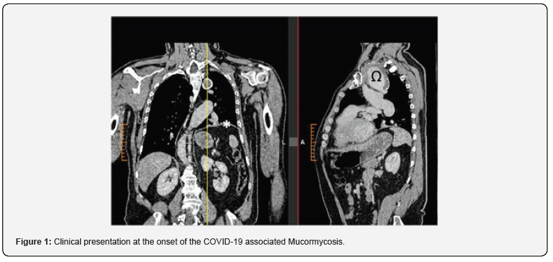
Briefly, under general anesthesia and invasive arterial blood pressure monitoring, prior to the re-sternotomy, the right common femoral artery is surgically exposed and for precautions reasons, percutaneously, a guide wire is inserted into the contralateral femoral vein to enable to promote CPB via peripheral vessels if needed. Left subclavear incision is also performed and the LSA is surgically exposed. During the re-sternotomy, the hemi-sterna are lifted vertically using Backhaus clamps which are placed temporarily on either side of the sternum, and systemic blood pressure is controlled pharmaceutically. Adhesions are dissected, allowing access to the heart and vessels.
After heparin administration, the patient is then connected to the CPB with the arterial return via the right femoral artery and venous drainage via two stage cannula which is placed in the right atrium. While cooling the body temperature to 32°C, the LSA was isolated proximally and legated. The Dacron graft from the first surgery was clamped and Cardiac arrest is achieved by blood cardioplegia, delivered to the aortic root. The “old” Dacron prosthesis is incised and the aortic root is replaced with composite graft, S. Jud Medical 25/28 mm (St. Jude Medical, Minnesota USA) according to Bental-De Bono technique. After cardiac de-airing, X-clamp is removed and while the body is re-warmed, an 8 mm ePTFE vascular graft (FlowLine Bipore, JOTEC GmbH, Hechingen, Germany) is anastomosed to the ascending Dacron graft, tunneled in the second left intercostals space and anastomosed end-to-end to the distal LSA.
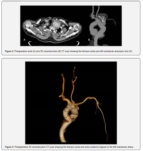
The patient is then wined from the ECC and at end of surgery moved to the ICU. Postoperative course was uneventful and the patient was discharged in 8th postoperative day. Several months later the patient complained of headaches. Following neurologic consultations, and MRI where micro cerebral bleedings were detected, the patient underwent blood controls and amyloidosis was diagnosed. To balance risks benefits, considering the mechanical valve in aortic position, anticoagulation regime was reduced and INR values are maintained between1.5-2. The patient is currently stable with a reduced anticoagulation regimen and ongoing monitoring
Discussion
Connective tissue disorders (Marfan syndrome and others) and amyloidosis are two distinct pathological conditions that affect the connective tissues and can result in significant systemic consequences. While they share some similarities in terms of their impact on various organ systems, they differ in their underlying pathophysiology, clinical features, and management. Meanwhile the Marfan syndrome is an autosomal dominant connective tissue disorder caused by mutations in the FBN1 gene, which encodes the fibrillin-1 protein. Fibrillin-1 is a critical component of the extracellular matrix and is essential for the proper formation of elastic fibers in connective tissues. Mutations in the FBN1 gene lead to structural abnormalities in fibrillin-1, causing impaired elasticity and tensile strength in various tissues and organs.
The defect in fibrillin-1 results in an impaired matrix integrity, particularly in connective tissues rich in elastic fibers, such as the aorta, skin, lungs, and eyes. The hallmark of Marfan syndrome is the weakening of the connective tissue scaffolding, leading to abnormal elongation and stretching of the affected organs. Amyloidosis, in contrast, is a heterogeneous group of diseases caused by the abnormal accumulation of amyloid fibrils in tissues and organs. Amyloidosis, first described by Rudolf Virchow in 1854 when he first identified amyloid deposits in tissues under a microscope diagnosis has transformed from a vague, observational phenomenon, to a precise, molecular, and genetic understanding. Amyloid fibrils are formed from misfolded proteins that aggregate into insoluble fibers, which then deposit extracellularly.
The most common types of amyloidosis are AL (light chain) amyloidosis and ATTR (transthyretin) amyloidosis, though other types, such as AA amyloidosis, exist. In AL amyloidosis, amyloid fibrils are composed of light chains produced by clonal plasma cells in the bone marrow, while in ATTR amyloidosis, they are formed from misfolded transthyretin proteins. The accumulation of these fibrils disrupts normal tissue architecture, leading to organ dysfunction. Amyloid deposits can infiltrate various organs, including the heart, kidneys, liver, and vessels wall. The clinical manifestations of amyloidosis depend on the organs affected by amyloid deposits. In particular, with regards to the cardiovascular system, amyloid deposits in the heart might lead to restrictive cardiomyopathy, characterized by impaired ventricular filling, diastolic heart failure, and arrhythmias.
In the present case, multiple procedures were required during the same surgery to treat the aortic valve and both aortic roots, and LSA aneurysms, where extra anatomic bypass to the distal LSA is mandatory. In particular, different surgical approaches have been considered for the exclusion of the LSA, including left thoracotomy, which would simplify visualization and potentially reduce the cardiopulmonary bypass (ECC) time limiting its potential negative impact on kidney and lung function [12]. However, considering the preoperative finding of left hemi diaphragm elevation and the potential risk of postoperative respiratory dysfunction, the choice was made to avoid additional thoracic wall trauma. This approach thought to minimized the risk of aggravate respiratory complications. An endovascular approach was also considered, given the favorable anatomy of the aorta, which offered an adequate proximal landing zone, especially since the LSA origin was located distal to the aortic arch and the left common carotid artery. This would have allowed for endovascular exclusion, but it was ultimately not pursued due to aortic wall pathology and the risk stent induced dissection or of late dilatation and endoprosthesis migration [13].
Indeed, although amyloidosis was not diagnosed at the time of surgery, the patient's early onset of acute type A aortic dissection, the presence of a large LSA aneurysm, and the aortic root aneurysm raised the suspicion of an underlying some connective tissue disorders. This possibility was taken into account when planning the surgical management. It appears that the combination of amyloidosis and the anticoagulation therapy required for the mechanical aortic valve prosthesis contributed to the development of micro cerebral bleeding diagnosed by MRI. Intracranial hemorrhage can arise from various underlying causes, each with different rates of disease progression and recurrence. Lobar intracerebral hemorrhage and spontaneous convexity subarachnoid hemorrhage are often associated with cerebral amyloid angiopathy (CAA).
Cerebral Amyloid Angiopathy is a progressive neurological disorder characterized by amyloid deposits in the blood vessel walls. In recent years, this form of amyloid angiopathy has gained significant attention and research focus. CAA involves the abnormal accumulation of amyloid beta (Aβ) protein in the cerebral blood vessels, which damages the vessels and disrupts normal blood flow. It is a leading cause of cerebral hemorrhage and cognitive decline, especially in older adults, and is closely linked to Alzheimer's disease (AD) [14]. Cerebral bleeding in individuals with CAA may become more frequent with advancing age. Gireud-Goss et al. reported that pathological signs of CAA are present in approximately 50% of individuals over the age of 70 [15]. While CAA is often asymptomatic, it can manifest with symptoms such as subacute cognitive decline, seizures, or headaches, as observed in the present case.
For patients with mechanical aortic valve, guideline recommended an international normalized ratio (INR) of 2.5 to 3.5, to balance bleeding/thrombosis risks. In order to reduce the risks of bleeding in patients with mechanical valves prostheses, requiring anticoagulation therapy, Eikelboom et al, in the Randomized, Phase II Study to evaluate the safety and pharmacokinetics of oral Dabigatran in patients after heart valve replacement, evaluated the use of Dabigatran in patients with mechanical heart valves. In this study the use of Dabigatran was associated with increased rates of thromboembolic complications, and no benefits [16]. We have considered also the use of direct anticoagulation therapy, however, case reports on its use in patients with mechanical heart prostheses are not favorable, reporting instances of thrombosis and obstruction of the mechanical aortic prosthesis associated with direct anticoagulation [17,18]. Recent publications suggests that low-dose oral anticoagulation may be effective in certain patient populations, offering a balance between thromboembolic prevention and reducing the risks of bleeding [19].
Consequently, based on the reported data, the patient’s international normalized ratio (INR) was maintained within a lower range, generally INR < 2. Given that the INR is maintained within the lower end of the therapeutic range, the patient has experienced no further complications and reports no symptoms of headaches. The decision to use low-dose anticoagulation should be individualized based on patient characteristics, including the type of mechanical valve, comorbid conditions, and previous history of thromboembolism. While some studies suggest that low-dose anticoagulation does not significantly increase the risk of thromboembolic events compared to standard therapy, ongoing research is needed to establish more definitive guidelines for its use in mechanical valve patients
Conclusion
Although Marfan syndrome and amyloidosis are both connective tissue disorders with systemic implications, they differ markedly in their etiology, clinical presentation, and treatment strategies. Marfan syndrome is primarily characterized by defects in fibrillin-1 and connective tissue structure, leading to cardiovascular, skeletal, and ocular abnormalities. In contrast, amyloidosis results from the deposition of misfolded proteins, leading to organ dysfunction through amyloid fibril accumulation. Amyloid aggregation, characterized by an increase in fibrils, disrupts the normal elasticity of the aorta by altering its microstructure. This disruption and less amortization of the forces applied on the aortic wall during the cardiac cycle, contributes to aneurysm formation and risk of dissection. Early diagnosis and treatment, especially in young population, will reduce the risk of dissection and related complications. Low-dose oral anticoagulation offers a promising alternative to standard high-dose regimens in selected patients with mechanical heart valve prostheses, potentially improving the safety and quality of life for these individuals. However, careful monitoring and patient-specific considerations remain essential in guiding anticoagulation therapy.
Declarations
Authors’ contributions Conceptualized the manuscript: JZ, PZ AI Contributed to data collection and literature review: EL, SK Contributed to the writing and editing of the manuscript: JZ MG.
References
- Zhongjie Wang, Nick Flores, Matthew Lum, Andrew D Wisneski, Yue Xuan, et al. (2021) Wall stress analyses in patients with 5 cm versus<5cm ascending thoracic aortic aneurysm. J Thorac Cardiovasc Surg 162: 1452-1459.
- Joon Bum Kim, Matthew Spotnitz, Mark E Lindsay, Thomas E MacGillivray, Isselbacher EM, et al. (2016) Sundt Risk of aortic dissection in the moderately dilated ascending aorta. J Am Coll Cardiol 68: 1209-1219.
- Lauren E Levy, Megan Zak, Jason P Glotzbach (2024) Current understanding of the genetics of thoracic aortic disease. Vessel Plus 8:4.
- Zhen Zhou, Alana C Cecchi, Siddharth K Prakash, DiannaM Milewicz (2022) Risk Factors for Thoracic Aortic Dissection Genes (Basel) 13(10): 1814.
- Gleason TG (2005) Heritable disorders predisposing to aortic dissection. Seminars in Thoracic and Cardiovascular Surgery 17(3): 274-281.
- Elendu C, Amaechi DCM, Elendu TCB, Ibhiedu JOM, Torubiri AOM, et al. (2023) Comprehensive review of aortic aneurysms, dissections, and cardiovascular complications in connective tissue disorders. Medicine 102: e36499.
- Larsson A, Malmström S, Westermark P (2011) Signs of cross-seeding: aortic medin amyloid as a trigger for protein AA deposition. Amyloid 18: 229-234.
- Jennifer H Pinney, Colette J Smith, Jessi B Taube, Helen J Lachmann, Christopher P Venner, et al. (2013) Systemic Amyloidosis in England: an epidemiological study. Br J Haematol 161(4): 525-532.
- Loeys BL, Dietz HC, Braverman AC, Bert L Callewaert, Julie De Backer, et al. (2010) The revised Ghent nosology for the Marfan syndrome. J Med Genet 47: 476-485.
- Garcia-Pavia P, Rapezzi C, Adler Y, Michael Arad, Cristina Basso, et al. (2021) Diagnosis and treatment of cardiac amyloidosis. A position statement of the European Society of Cardiology Working Group on Myocardial and Pericardial Diseases. Eur J Heart Fail 23(4): 512-526.
- EM Isselbacher, O Preventza, Black J Hamilton, John G Augoustides, Adam W Beck, et al. (2022) ACC/AHA Guideline for the Diagnosis and Management of Aortic Disease: A Report of the American Heart Association/American College of Cardiology Joint Committee on Clinical Practice Guidelines. Circulation 146(24): e334-e482.
- Zeitani J, Buccisano F, Nardella S, M Flaminio, P Prati, et al. (2013) Mini-extracorporeal circulation minimizes coagulation abnormalities and ameliorates pulmonary outcome in coronary artery bypass grafting surgery. Perfusion 28(4): 298-305.
- Schoenhoff FS, Schmidli J (2020) TEVAR in Patients with Marfan Syndrome: From Bailout to Strategy. Eur J Vasc Endovasc Surg 59: 586.
- Cozza M, Amadori L, Boccardi V (2023) Exploring cerebral amyloid angiopathy: insights into pathogenesis, diagnosis, and treatment. J. Neurological Sci 454: 120866.
- Monica Gireud-Goss, Alexis F Mack, Louise D, McCullough, Akihiko Urayam (2021) Cerebral Amyloid Angiopathy and Blood-Brain Barrier Dysfunction Neuroscientist. December 27(6): 668-684.
- John W Eikelboom, Stuart J Connolly, Martina Brueckmann, Christopher B Granger, Arie P Kappetein, et al. (2013) Dabigatran versus Warfarin in Patients with Mechanical Heart Valves Engl J Med 369: 1206-1214.
- Carvalho Silva DM, Braga A, de Jesus I, Neves J (2019) Mechanical prosthetic heart valve thrombosis in a patient receiving rivaroxaban. Cardiology 143:116-20.
- Ryoji Kinoshita, Taiju Watanabe, Ryumon Matsumoto, Kazunobu Hirooka (2024) Mechanical Aortic Valve Thrombosis During Anticoagulation Using Oral Rivaroxaban: A Case Report. Cureus 16(7): e65007.
- Heinrich Koertke, Armin Zittermann, Gero Tenderich, Otto Wagner, Mahmoud El-Arousy, et al. (2007) Low-dose oral anticoagulation in patients with mechanical heart valve prostheses: final report from the early self-management anticoagulation trial II. Eur Heart J 28(20): 2479-2484.

























