Abstract
Our facial expressions are actually like a mirror of our brain and thoughts. They are formed as a result of the synchronized work of facial muscles with stimuli coming from various points of the brain. It is thought that the facial and trigeminal nerves play a major role in the transmission. Studies have shown that the amygdala, basal ganglia and hypothalamus also regulate the transmissions to the cortex. While patients are being monitored for various cranial pathologies in the neurosurgery intensive care unit, their facial expressions may be different during the day. You can assume that this is a manifestation of cerebral disease. We want to introduce you to this tremendous information with examples and literature.
Keywords:Facial Expressions; Amygdala; Hypothalamus; Trigeminal-Facial Nerve Pathways
Abbreviations: FACS: Facial Action Coding System; AU: Action Unit; sEMG: Facial Surface Electromyography; CT: Computerized Tomography
Introduction
Facial expressions are crucial to social interactions [1]. Unfortunately, how they are managed to direct the coordinated muscle movements required to produce facial expressions has not been fully elucidated [2,3]. However, facial expressions are thought to consist of electrical activity carried by the cerebral cortex and sensorimotor pathways. It is necessary to generate new hypotheses about why facial expressions are atypical in various clinical populations. The sensorimotor mechanisms supporting facial expressions consist of neural pathways that regulate the motor control of facial sensation at the cortical, subcortical, and brainstem levels. Motor control of the muscles of facial expression involves the facial nerve (cranial nerve VII), while mechanosensory and proprioceptive feedback from these muscles involves the trigeminal nerve (cranial nerve V). The division of motor control and sensation between two separate systems differs from other skeletal muscles, where motor and sensory fibers are typically part of the same nerve [4].
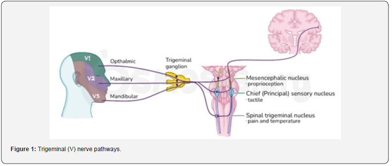
The Facial Action Coding System (FACS) for categorizing human facial expressions was described by Ekman and Friesen in 1978 [5]. The contraction of specific facial muscles is defined as an action unit (AU), and different facial expressions involve different combinations of these action units. Facial surface electromyography (sEMG) is another widely used method for characterizing facial expressions [6,7]. Facial expressions can be studied by directly recording muscle activity (as in sEMG) or by inferring muscle activity from the displacement of superficial facial features (as in manual or automated FACS). Sensorimotor control is generally defined as the integration of sensory input with motor commands and the generation of coordinated motor actions [8]. Facial expressions often require synchronous and symmetrical contraction of multiple muscle groups across the entire face (Table 1). Cerebral representations of faces are distinct; their mappings cover more cortical space than the mappings of other body parts. Facial somatosensory transmission is mediated via the trigeminal circuit, where electrical activity from different facial regions is topographically and serially coupled to the brainstem, thalamus, and neocortex [9] (Figure 1).
There is considerable evidence for peripheral connectivity between the facial sensory and motor systems. Cadaveric and surgical dissection of the human face has revealed synaptic connections between the facial nerve and the three major trigeminal nerve branches [10]. In particular, trigeminal afferents show uniquely robust communication with the facial nerve branches to the upper facial muscles, including the frontalis, orbicularis oculi, and zygomaticus major. Physiological studies have shown trigeminofacial functional connectivity, as stimulation of trigeminal sensory afferents leads to inhibition of motor activity in the lower facial muscles. Trigeminal-facial connections likely play an important role in, and may contribute to, proprioception and integrated facial sensory-motor control. The connectivity between the trigeminal sensory and facial motor systems is perhaps most evident at the brainstem level, via trigeminofacial reflexes. For example, the corneal reflex or “blink” reflex is elicited by the closing or “blinking” of both eyelids as a result of reflexive contraction of the orbicularis oculi muscles as a result of tactile stimulation of the corneal surface [11,12].


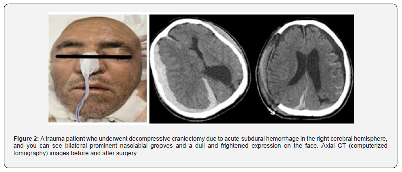
The hypothalamus plays an important role in the formation and regulation of emotional responses. The role of the hypothalamus in facial expression is related to its participation in both limbic and autonomic systems and its direct connection to the facial nerve [13]. The hypothalamus is known to be active during both laughter and crying, that is, behaviors that involve facial expressions, complex vocalizations, and tear secretion. The amygdala serves as a link between the sensory and limbic systems [14]. This is crucial for facial expression because the emotional salience of a sensory stimulus is critical for determining the type of expression it will trigger. The amygdala receives direct inputs from the primary sensory cortex and insula and sends direct outputs to various motor brain structures, including the basal ganglia, cerebellum, and middle cingulate cortex. It also plays a role in monosynaptic circuits connecting to brainstem trigeminal neurons. The amygdala also sends indirect projections to both sensory and motor cortical regions via the thalamus [15]. The insula is considered the primary interoceptive cortex because it receives body state-related input from the brainstem and spinal cord [16]. Intact insular projections to the amygdala are observed in both humans and animals and may represent a mechanism by which interoceptive information is integrated into facial motor control pathways (Figure 2).

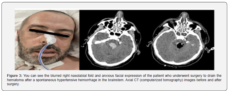
To date, numerous studies have been conducted to reveal the afferent and efferent connections of facial expressions and facial expressions with the cerebral cortex, intrinsic structures and cranial nerves. In particular, it has been shown that the cerebral cortex, amygdala-limbic system, basal ganglia and trigeminalfacial cranial pairs are in synchronous harmony with the facial muscles, especially in the formation of facial expressions. In his article in 2024, Bress explained all these anatomical structures in detail and provided readers with a nice experience about their relationship with facial expression [10]. Again, Erzurumlu et al. provided valuable information to readers in his article titled “Mapping the face in the somatosensory brainstem” in 2010 [9]. In our article, we would like to share with you the facial expressions of patients with various cerebral pathologies that we follow in the neurosurgery intensive care unit, by comparing them with the findings on brain computed tomography. These are presented to you, the readers, as evidentiary products of the facial-emotional communication pathways mentioned above (Figure 3).

Conclusion
In summary, facial expression is a complex behavior that involves a high degree of coordination among multiple neural systems across the cortex, subcortex, and brainstem. Despite the importance of facial expressions for social function, little is known about how they are produced and regulated as a unique sensorimotor behavior. Such a gap in knowledge is a major obstacle to studying atypical facial expressions in diverse clinical populations. Facial expression has become the basis of communication between humans, especially today with the advancement of technology. The current knowledge of how muscle contraction is directed to produce sensory prototypical facial expressions needs to be clarified. This work will require careful consideration of complex network interactions at multiple levels of the nervous system, including the cortex, subcortex, and brainstem.
References
- Barrett LF, Adolphs R, Marsella S, Martinez AM, Pollak SD (2019) Emotional expressions reconsidered: challenges to inferring emotion from human facial movements. Psychol. Sci. Public Interest 20(1): 1-68.
- Cattaneo L, Pavesi G (2014) The facial motor system. Neurosci. Biobehav Rev 38: 135-159.
- Morecraft RJ (1998) Van Hoesen GW Convergence of limbic input to the cingulate motor cortex in the rhesus monkey. Brain Res. Bull 45(2): 209-232.
- Hinsey JC (1934) The innervation of skeletal muscle. Physiol. Rev 14: 514-585.
- Ekman P, Friesen W (1978) Facial Action Coding System. Palo Alto: Consulting Psychologist Press, USA.
- Künecke J, Hildebrandt A, Recio G, Sommer W, Wilhelm O (2014) Facial EMG responses to emotional expressions are related to emotion perception ability. Plos One 9(1): e84053.
- Huang CN, Chen CH, Chung HY (2005) The review of applications and measurements in facial electromyography. J. Med Biol. Eng 25(1): 15-20.
- Ingram JN, Wolpert DM (2011) Naturalistic Approaches to Sensorimotor Control. In: in Progress in Brain Research 191: 3-29.
- Erzurumlu RS, Murakami Y, Rijl FMi (2010) Mapping the face in the somatosensory brainstem. Nat Rev Neurosci 11(4): 252-263.
- Bress KS, Cascio CJ (2024) Sensorimotor regulation of facial expression - An untouched frontier. Neuroscience and Biobehavioral Reviews 162: 105684.
- Kimura J, Powers J, Van Allen M (1969) Reflex response of orbicularis oculi muscle to supraorbital nerve stimulation: study in normal subjects and in peripheral facial paresis. Arch. Neurol 21(2): 193-199.
- Willer JC, Boulut P, Bratzlavskyt M (1984) Electrophysiological evidence for crossed oligosynaptic trigemino-facial connections in normal man. J. Neurol. Neurosurg. Psychiatry 47: 87-90.
- Veazey RB, Amaral DG, Cowan WM (1982) The morphology and connections of the posterior hypothalamus in the cynomolgus monkey (Macaca fascicularis). II. Efferent connections. J. Comp. Neurol 207: 135-156.
- Turner B, Mishkin M, Knapp M (1980) Organization of the amygdalopetal projections from modality-specific cortical association areas in the monkey. J. Comp. Neurol 191(4): 515-543.
- McDonald A (1998) Cortical pathways to the mammalian amygdala. Prog. Neurobiol 55(3): 257-332.
- Craig A (2002) How do you feel? Interoception: the sense of the physiological condition of the body. Nat. Rev. Neurosci 3: 655-666.


