




 GJO Home



Abstract
The carotid body paraganglioma is a rare benign neoplasm arising from the chemoreceptor cells of the carotid bulb. It is mostly asymptomatic in early presentation, but becomes symptomatic and difficult to manage when the tumor is large. We presented a 7 cm-sized shambling III, which was removed without complication.
Keywords: Giant Carotid Body; Shamblin; Chemoreceptor cells; Carotid bulb
Abbreviations: CBTs: Carotid Body Tumors; ICA: Internal Carotid Artery; CCA: Common Carotid Artery; CTA: CT Angiography; DSA: Digital Subtraction Angiography; ECA: External Carotid Artery
Introduction
Carotid body tumors (CBTs), also called paragangliomas or chemodectomas, are rare neuroendocrine neoplasms that are near the carotid bifurcation within glomus cells derived from the embryonic neural crest [1]. Incidence of CBTs is less than 1-2 in 100,000 [2], more common in females than males (1.9:1). The Majority of carotid body tumors are asymptomatic and usually noticed first as incidental findings on physical examination of the neck or during radiological studies. However, large tumors can produce symptoms secondary to the pressure effect and local invasion of the surrounding tissue, such as pain, difficulty with breathing, dysphagia, and autonomic dysfunction [3, 4]. We presented here a 6.5 cm size CBT Shamblin III, the clinical picture, the radiological findings, the surgical procedure performed and the outcomes.
Case Report
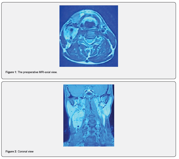
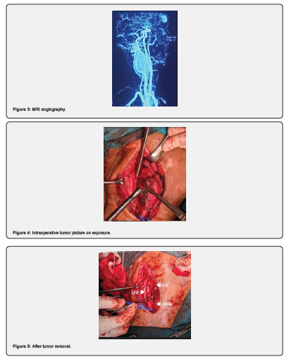
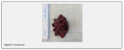
A 29-year-old patient presented to us with a history of right-sided neck swelling for 5 years, which had increased significantly in size in the last 3 to 4 months. The patient underwent radiological as well as biochemical investigations, including MRI angiography. The preoperative MRI-axial view (Figure 1), coronal view (Figure 2) and preoperative MRI angiography (Figure 3). Preoperatively, the patient underwent balloon occlusion test, and the patient tolerated the balloon occlusion test without deficits. The patient underwent surgical excision with a vascular surgeon on backup in case grafting was required. First, the common carotid artery was secured, and then the upper end of the internal carotid artery was also secured above the tumor. Next, the hypoglossal nerve was dissected and freed from the upper end of the tumor. Next, we began the tumor dissection from the posterior part of the tumor and proceeded medially. The tumor could be removed without the need to sacrifice the internal carotid artery. The external carotid artery was sacrificed in the process of the tumor removal. Intraoperative tumor picture on exposure (Figure 4), after tumor removal (Figure 5) and the specimen (Figure 6).
Discussion
Carotid body tumor with complete encasement of the internal carotid artery (ICA) & common carotid artery (CCA) is a complex, requiring a multidisciplinary team of head and neck surgeons, interventional radiologists and vascular surgeons for management. Surgical removal is the primary treatment option, as the tumor carries malignant potential and should be excised before it grows larger and becomes more challenging to remove. Complete surgical excision is the treatment of choice [5]. The location of CBT has made it one of the challenging neck tumors to manage in head and neck surgery. The size of the tumor is important for treatment. Shamblin I can be dissected from the vessels without injury, while Shamblin III, which are closely associated with the carotid vessels, are more difficult to resect and are associated with a higher rate of neurovascular complications [6-9]. Advanced imaging techniques, such as CT angiography (CTA) and Digital Subtraction Angiography (DSA), are essential for mapping vessel involvement. Pre-operative embolization to minimize intraoperative bleeding is advocated by some surgeons, while others do not advocate this due to the concerns about potential complications [10-12]. Pre-operative embolization is often performed to reduce intraoperative blood loss, as was done in our case. Surgery is the primary treatment, but it is complex due to vessel encasement and requires careful sub adventitial dissection (peeling off the artery) or, if necessary, sacrifice of the external carotid artery (ECA). In rare cases, carotid reconstruction is often required, using grafts to bypass or reconstruct the internal carotid artery or common carotid artery after tumor removal [13,14]. Any carotid body tumor more than 5cm has a higher perioperative mortality (1-3%) and morbidity, including stroke or nerve injury. With experienced teams, successful resection without complication is achievable. Though mostly benign, malignant tumors account for about 10% [15]; thus, the need to ensure complete resection during surgery, as done in our case.
Conclusion
Carotid body tumor Shamblin III size 6.5 cm resection is a relatively rare procedure and a challenge, but with proper preoperative evaluation and surgical planning, a safe resection can be done.
References
- Dixon JL, Atkins MD, Bohannon WT, Buckley CJ, Lairmore TC (2016) Surgical management of carotid body tumors: a 15-year single institution experience employing an interdisciplinary approach. Baylor University Medical Center Proceedings 29(1): 16-20.
- Hoang VT, Trinh CT, Lai TAK, Doan DT, Tran TTT (2019) Carotid body tumor: a case report and literature review. Journal of Radiology Case Reports 13(8): 19-30.
- Finocchiaro M, Benvegna S, Finocchiaro N, Alfano M, Benvegna G (2017) Chemodectoma: case report. Chirurgia 30(2): 73-77.
- Mokin M, Dumont TM, Kass-Hout T, Levy EI (2013) Carotid and vertebral artery disease. Primary Care: Clinics in Office Practice 40(1): 135-151.
- Davis FM, Obi A, Osborne N (2018) Carotid Body Tumors. Extracranial Carotid and Vertebral Artery Disease. Springer Cham pp. 253-260.
- Shamblin WR, ReMine WH, Sheps SG, Harrison EG (1971) Carotid body tumor (chemodectoma). The American Journal of Surgery 122(6): 732-739.
- Luna-Ortiz K, Rascon-Ortiz M, Villavicencio-Valencia V, Herrera-Gomez A, Herrera-Gomez A (2006) Does Shamblin’s classification predict post operative morbidity in carotid body tumour? A proposal to modify Shamblin’s classification. European Archives of Oto-Rhino-Laryngology 263(2): 171-175.
- Liu J, Mu H, Zhang W (2021) Diagnosis and treatment of carotid body tumors. Am J Transl Res 13(12): 14121-14132.
- Baysal BE, Myers EN (2002) Etiopathogenesis and clinical presentation of carotid body tumors. Microsc Res Tech 59(3): 256-261.
- Felbaum DR, Syed HR, McCullough MF, Armonda RA, Liu H (2016) Embolization of Carotid Body Tumors: Revisiting Direct Puncture Technique, Preliminary Experience and Literature Review. Cureus 8(2).
- Gonzalez-Urquijo M, Castro-Varela A, Barrios-Ruiz A, Hinojosa-Gonzalez DE, Garza Salas AK (2022) Current trends in carotid body tumors: Comprehensive review. Head Neck 44(10): 2316-2332.
- Arya S, Rao V, Juvekar S, Dcruz A (2008) Carotid body tumors: objective criteria to predict the Shamblin group on MR imaging. Am J Neuroradiol 29: 1349-1354.
- Mohammad Alqaim, Ajit S Puri, Alec E Vaezi, Andres Schanzer (2019) Carotid body tumor resection utilizing a covered stent graft to enable resection of the tumor en bloc with the internal carotid artery 135(4): 481-484.
- Muduroglu A, Yuksel A (2017) Carotid body tumors: a report of three cases and current literature review. Vascular Diseases and Therapy 2(5): 1-3.
- Maxwell JG, Jones SW, Wilson E, Kotwall CA, Hall T, Hamann S (2004) Carotid body tumor excisions: adverse outcomes of adding carotid endarterectomy. Journal of the American College of Surgeons 198(1): 36-41.

























