




 GJO Home



Abstract
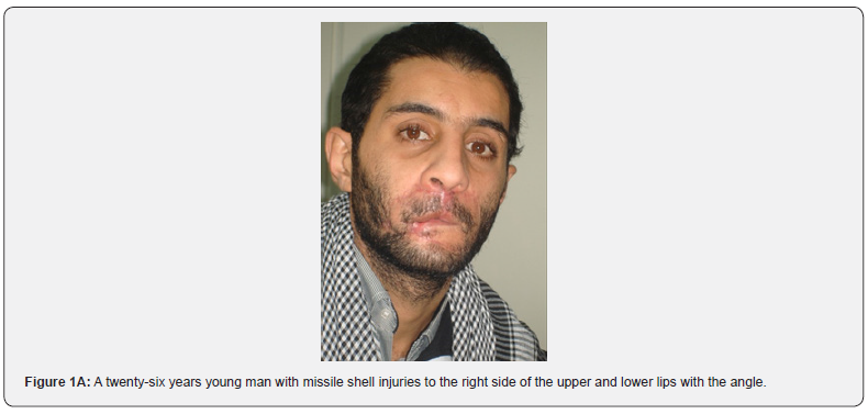
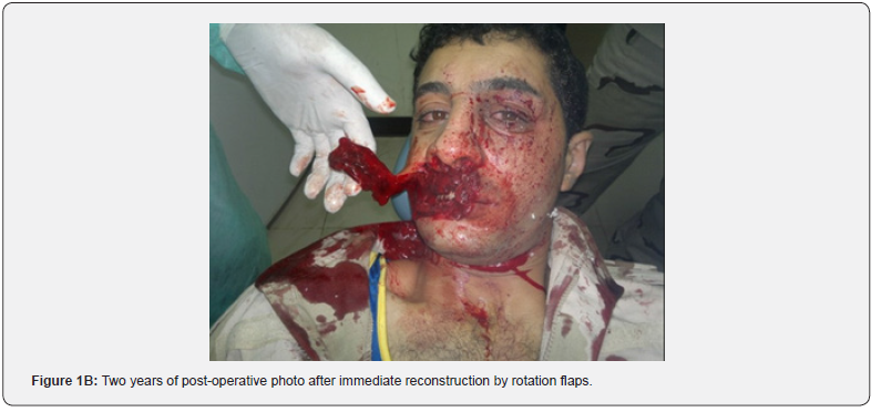
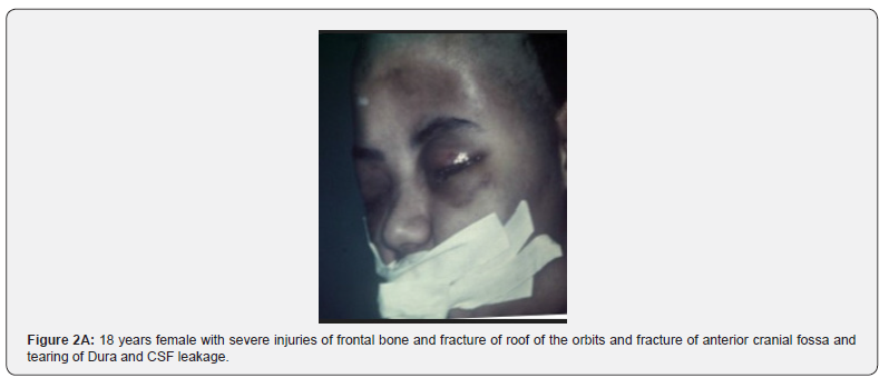
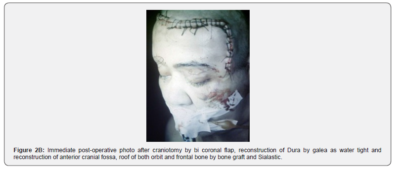
Role of mesenchymal stem cells were used for reconstruction of the Facial Skeleton. Stem cells were found in embryonic stages, formation and healing of bone grafting, distraction for elongation of short limbs in children in cases of poliomyelitis. Stem cells were played an important role in cancer therapy and shown promising in unimodality effect that could revolutionized cancer therapy. Stem cells in maxilla-facial surgery played and focuses on repairing hard and soft tissue. There was significant loss of trauma, missile war injuries, pathological lesion effecting the mandible and developmental deformities. Oral cancer was been played an important role in development or therapeutic effect and regenerative therapy. Series of cases were treated and reported like shell injuries to the face, traumatic traffic accident with severe damages to both roofs of the orbits with fracture anterior cranial fossa were treated by bone grafts. Intra uterine trauma to female child borne with ankylosis of the TMJ and First Arch dysplasia or hemifacial microsomia, were treated by Kummoona Chondr-Osseous graft for restoration of function and growth of the mandible and midface.
Keywords:Stem Cells; Regeneration; Reconstruction; Bone Graft; Distraction; Chondro-Osseous Graft; Cancer Therapy
Introduction
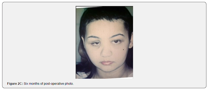
The aim of this research and studies to evaluate the role of mesenchymal stem cells, played transformative role in reconstruction of the facial skeleton and managements of oral cancer as therapeutic agent by promoting bone and soft tissue regeneration. Stem cells defined an organism that can develop into many types of cells during early life and growth and serve as repair system and play a great role in regenerative medicine. The facial skeleton consisting from fifty small bones articulate as a pyramid. These small cells forming the charisma of the person and the face itself carry the features of bountifulness, dullness and smart and stupidness. The face also carries the signs of disease like, paleness due to anemias, puffy face due to kidney disease and heart failure and redness might carry blood disease and loss of wight with paleness of the face might the patient feature serious disease like cancer. The facial skeleton might be subjected to RTA or to missile war injuries creating sever deformities of the face might associated with multiple injuries and loss of conscious due to head injuries [1] (Figure 1 (A),(B) & Figure 2(A-C)).
Malformation of congenital disease like First Arch Syndrome or traumatic injuries. to temporomandibular joint (TMJ) causing ankylosis of the joint with deformities and loss of function in both adult and children. Also, cancer of orofacial region constitutes about 4% of total cancer. Bone grafting and Distraction should not be forgot in. (Figure 1 (A),(B) & Figure 2(A-C)) All these cases required mesenchymal stem cells during development, healing process in bone graft and destruction required reconstruction of soft tissues and bones. The mesenchymal stem cells were used in regenerative medicine focusing in repairing both soft tissue and hard tissue were required varieties of flaps and bone grafting harvested from iliac crest might resulting morbidity. These types of surgery was a great challenge to the surgeons and may face psychologically suffering of the patients. This status required tissue engineering for reconstruction in different sites of Craniomaxillo- facial regions. Stem cells play now days an important role TMJ surgery, bone grafting, chondro-osseous graft for TMJ growth and function and elongation of limbs children were suffering from poliomyelitis [2].
Materials, Result and Methods
Series of cases were involved in these managements of cases
including young adults and children of males and females. The
cases enrolled in this study were suffering from previous trauma of
road traffic accident like TMJ ankylosis, congenital malformation
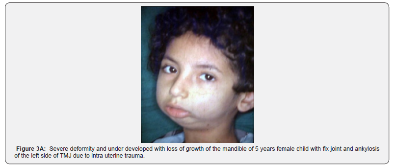
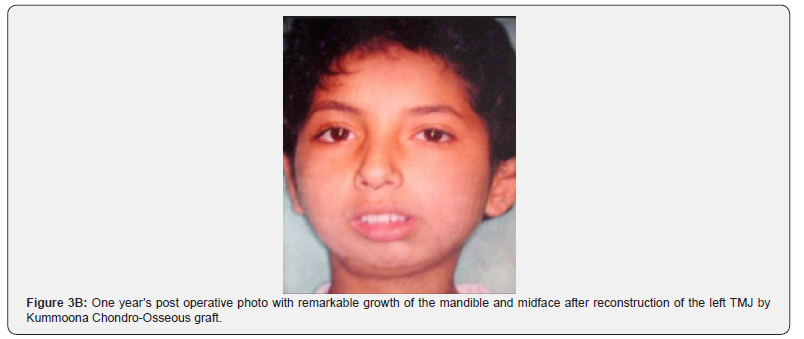
like first arch syndrome and hypoplasia of the condyle (Figure
3(A),(B)). Ankylosis is the most insult disease to human body due
to traumatic injuries to the TMJ both in children and young adults.
Experimental studied on young Rabbits of 3 months of age and
terminated in 3 months also and the aim were to [1];
a) Prove the condyle carry a growth center first was
reported by (John Hunter 1772-73). We did our experiment by
excision the head of condyle , three months after termination of
the Rabbits, we observed sever deformity of the jaw Rabbit in the
effected side.
b) After resection of the Rabbits condyle, we did reconstruct
the TMJ by chondro-osseous graft to restore the normal biological
function of growth, remodeling and repair [3-5].
c) Specimens of the resected chondro-osseous graft that
replaced the resected condyle and TMJ, the specimens were
immersed in buffer formalin of 10% with decalcified material
for two weeks, slide prepared and stained by Hematoxylin-Eosin
(H&E), to study the cytological changes of the chondro-osseous
graft that occurred after transferred from iliac crest as wight
bearing to new functional activity of masticatory process of the
new TMJ created and advocated by Kummoona Chondro-Osseous
Graft [6-13].
d) The aim of our experiment in Rabbits to study the
viability of bone graft. Also, the value of Distraction osteogenesis
for managements of leg with poliomyelitis o children and advance
jaw deformity.
Histopathology
We found during examination of the chondro-osseous graft by microscope with staining by (H&E) [4]; distinct layers were notice. The first layer consisting from thick fibrocartilage layer, it was thick because the hard food, the second layer consisting from several layers of round mesenchymal stem cells (growth layer), the third layer consisting from chondrocyte with series of hypertrophic layer and the fourth layer consist from osteoid tissue and bone marrow in between.
Histopathology of bone graft and distraction osteogenesis
Cytological and histopathological studies was quiet interesting started by formation of aggregation of plat lets as clot with liberation of platelets growth factor (PGF), and healthy granulation tissue, fine small vessels invading the graft and bone to grafted by creeping mechanism, mesenchymal stem cells were derived from bone marrow of the stump, periosteum and overlying muscles, with formation of large number of fibroblasts and tiny small vessels. Osteoid tissue and chondrocyte also seen. The cellular changes of distraction technique and bone grafting by inducing mesenchymal stem cells are the same in both bone grafting and distraction except distraction induced by osteotomy with stretching growth potential of bone based on Illizarov theory and bone grafting is based on decortication of both graft and stump of bone [4,5].
Discussion
This research with our clinical studies of role of mesenchymal stem cells in growth and reconstruction. Stem cells proved to be a revolution in engineering of reconstructive surgery of the facial skeleton. Tissue engineering was joined with medicine 1987 and it was the key point in tissue engineering by work of few doctors from Bosten and MIT and the wide benefit from tissue engineering are reduction of donor site and morbidity [13]. Also decrease in the procedure sensitivity of repaid and the capacity intimately adapt in vivo tissue environment into recapitulate normal craniofacial development. Stem cell plays a role in its effect in cancer therapy. It’s does show promising an uno modulatory effect that could revolutionize cancer therapy and treatment modality [3]. Stem cells play an important role in healing of bone grafting and distraction of bone. Stem cell also played in growth of jaws and the TMJ that was replaced by chondro-osseous graft for managements and fore replaced damaged joint in ankylosis. The chondro-osseous also been used for managements and reconstruction of the hypoplasia of the condyle and First Arch Syndrome.
The recent work in Oral cancer was able to modulate immune responses by reducing inflammation and enhancing the immune system against cancer cell and further to that the researcher been able to enhance the immune system against cancer cell and promoting tissue repair and might reduce the cancer recurrence [3]. It was found that stem cell had the ability to suppress the activities of immune cell that been able to promote cancer cell that attack cancer cell. Recently the work in engineering medicine that stem cell can attack cancer by making anti-tumor agent and targeting the cancer cell. Stem cells derived from mesenchymal tissues of bone marrow; we notice and found there are strong relation between the bone graft and distraction osteogenesis. Stem cells were derived from bone marrow, periosteum and overlying muscles. Growth factor released from platelets (PGF). Stem cells play an important role in reconstructed Kummoona Chondro-Osseous graft in growth, repair and remodeling of the TMJ.
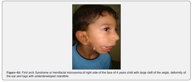
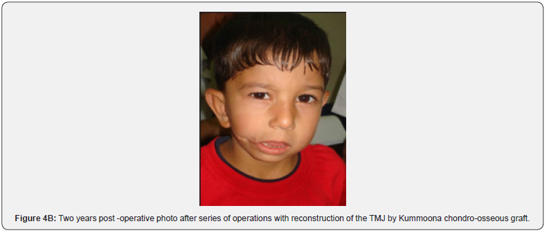
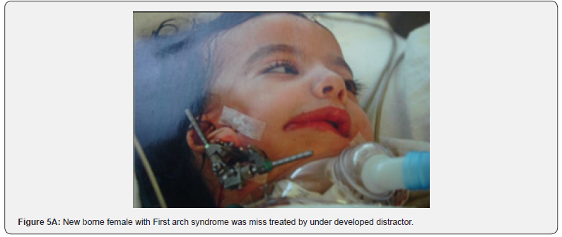
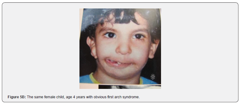
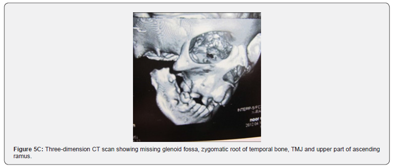
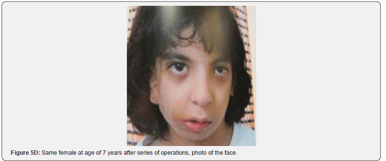
Furthermore, observation on stem cells were an important factor in bone grafting and distraction of bone for managements of short limb due to poliomyelitis based on theory created by Illizarov of Russia. The TMJ reconstruction was a great challenge to all craniomaxillofacial surgeons for the last 6 decades. The biological reconstruction of the TMJ damaged disease such as damaged TMJ in ankylosed joint or missing or under developed one. In cassis of First Arch dysplasia syndrome due to early occlusion of Stape dial artery, the main nutrient vessel to the First and Second branchial arches in the embryonic development (Figure 4(A),(B) & Figure 5(A-D)). Ankylosis of the TMJ is a serious crippling disease with stiff joint effect children due to traumatic injury to the TMJ in road traffic accident or fall from Hight with intra capsular fracture with fragmentation of the disc of the condyle. Miss treatment led to ankylosis.
The child complained from un ability to open or close the mouth with difficulty in chewing food, swallowing or speech. The consequences of this disease are damage to growth center in the condyle and effect on growth of the mandible and midface. Clinically the disease featured in three forms of First Arch dysplasia Syndrome (hemifacial microsomia) milled, moderate and sever form of the disease. The milled form appeared with under developed mandible and presence of tags in the preauricular area as remanent of Meckel’s cartilage. The moderate type featured as cleft in the angle of the mouth, underdeveloped masseter muscle, missing condyle, ear deformity and deficiency in growth of the mandible and midface. The severe form of the disease is very wide cleft of the angle of the mouth with partial or complete loss of the ear, missing of temporomandibular joint (TMJ), absence of glenoid fossa and zygomatic root of temporal bone, with missing upper part of ascending ramus with underdeveloped mandible and midface. Kummoona Chondro-Osseous graft with presence of mesenchymal stem cells from iliac crest was used for reconstruction of the TMJ in children for restoring growth of the mandible and midface after correction of angle of the mouth by commissuroplasty and cartilage graft for building the glenoid fossa and bone graft for reconstruction of the zygomatic root of temporal bone. This series of operation should be carried out before the child go to school.
References
- Kummoona R (2018) Reconstruction of lower jaw by iliac bone graft, experimental study on Rabbit and role of mesenchymal stem cells. J Stem Cell Reg Biol 4(1): 20-24.
- Illizarov GA (1988) The principle of Illizarov method. Bulten of the hospital of joint disease, Orthopedic Institute 48: 1-11.
- Perrone M (2024) Stem cell application in treating oral cancers: A comprehensive Review Stem Cell Reg 5(2): 65.
- Kummoona R (2025) Role of Stem Cells in Immediate Reconstruction by Bone Graft after Radical Resection of Malignant Tumors of the Jaw, Clinical and Experimental Studies. Genisis J Surg Med 4(2): 40.
- Kummoona R (2018) Bone Grafting, Recent Advances with Special References to Cranio-Maxillofacial Surgery. Book, ed, Raja Kummoona, Intech Open, British publication, London.
- Kummoona R (2020) The role of mesenchymal stem cells in Kummoona chondro-Osseous graft for reconstruction. J Stem Cell Rese 1: 1.
- Kummoona R (2017) Kummoona chondro-osseous graft for reconstruction of the temporomandibular joint in children. Arch Clin Expo Ortho 1: 001-003.
- Kummoona R (2020) The Role of mesenchymal stem cells in Kummoona chondro-osseous graft for reconstruction of the Temporomandibular joint. J Stem Cell Res 1(1): 1-3.
- Kummoona R (2017) Kummoona chondro-osseous graft for reconstruction of the Temporomandibular Joint in Children. Arch Clin Expo Orthop 1: 001-003.
- Kummoona R (2026) Role of Stem Cells of the Chondro-Osseous Graft in Growth of the Condyle of the Temporomandibular Joint. Arch of Case Report.
- Kummoona R (2026) Recent advances in the evaluation and managements of Craniofacial missile injuries. PubMed, National Institute of Health, (gov).
- Kummoona R (2021) The use of Kummoona Chondro-Osseous Graft for Surgical Reconstruction of the Temporo-mandibular joint of First Arch Syndrome. EC Orthopedic 12: 110-117.
- Kummoona R (2026) Role of stem cells of the Chondro-Osseous graft in the growth of the condyle of the Temporomandibular Joint. Arch Clinic case Rep J 1(1): 1002.

























