




 GJO Home



Abstract
Background: Platelet rich plasma (PRP) is an innovative material that is being used in many surgical specialties.
Objective: A literature review of the use of platelet rich plasma (PRP) in ontological surgery.
Data sources: search of the PubMed database was conducted using the terms “platelet rich plasma” and/ or “platelet rich plasma” in otology,
“platelet rich plasma” in ENT, “platelet rich plasma” ear diseases and ear surgery.
Methods: A literature review on the running uses of PRP in otological surgery and ear diseases was performed.
Results: There are few numbers of literatures on the use of PRP in otology compared with other specialties: only 22 studies were included in
our review.
Conclusion: Based on the few numbers of studies, we cannot conclude about the value of PRP in ontological surgery and diseases in spite of
that the available literature suggests that PRP bear our hopes for future use in a number of clinical applications.
Keywords:Platelet-Rich Plasma; Otology; Ear diseases; Ear surgery
Abbreviations:PRP: Platelet rich plasma; PDGF: Platelets derived growth factor; TGF-B: transforming growth factor beta; FGM: fat graft myringoplasty; HA: Hyaluronic acid; PMW: posterior meatal wall; CWD: canal wall down
Introduction
Platelets rich plasma (PRP) is an autologus material that is being used in many surgical specialties. It has numerous advantages include safety, low cost, short preparation time and the increased concentration of platelets in the healing area. PRP was first used in 1987 in open heart surgery to promote tissue regeneration and wound healing. Now PRP has varies applications in, plastic surgery, orthopedic and neurosurgery, also in dentistry and maxillofacial surgery [1]. PRP contains high platelets concentration and a number of growth factors. In particular, Platelets derived growth factor (PDGF) and transforming growth factor beta (TGF-B) facilitate rapid wound healing, heamostasis and decrease scarring [2]. PRP is a small volume of plasma having a platelet concentration above base line level. When PRP mixed with calcium, this will lead to degranulation of alpha granules and release of growth factors, forming a gel structure [3-5]. Platelets rich plasma is obtained by extracting 10 cc of peripheral blood which is centrifuged and separate plasma from blood “soft spin”, then PRP separated from platelet poor plasma after the hard spin centrifugation [1]. Although PRP is an established treatment that provides good results for different clinical conditions, but its use in ENT is not yet common. The first use of PRP in ENT was in 2005 in endoscopic sinus surgery [6]. In 2009 the first trial in otology surgery was done by Erkilet et al, it is experimental study reported the use of PRP to tympanic membrane healing in 44 rats after bilateral TM perforation [7]. This review describes the available evidence on the use of PRP in otology surgery.
Material and Methods
A literature search of the PubMed database was conducted using the terms “platelet rich plasma” and/ or “platelet rich plasma” in otology , “platelet rich plasma” in Otological surgery, “platelet rich plasma” and ear diseases. Data extraction comprised a narrative summary of each article.
Results
In 2011, Maria et al. [8] reported a prospective study of three patients with inactive central tympanic membrane perforation. For all patients platelet rich plasma plug was applied around the perforation followed by overlay type I tympanoplasty. The authors reported complete closure of all three perforation and advised this procedure to be proven in a large sample. In 2013, a prospective study conducted by G. Sankaranarayanan et al. [9] 50 patients with dry perforation included in the study. 25 patients underwent underlay tympanoplasty using PRP and a second group of 25 control patients who underwent tympanoplasty without PRP. After one month, closure rates were 72% in the first group versus 40% in the control group. After 2 months the success rate was 92% in the first group and 72% in the control group. After 3 months the success was 96% in the first group and 80% in the control group. The authors concluded that using PRP during tympanoplasty accelerate tympanic membrane closure, prevent graft displacement and improves the success rate of myringoplasty.
El- Anwar et al. [10] studied 64 patients underwent myringoplasty by underlay graft using conchal cartilage. All of them had large dry central tympanic membrane perforations. . Patients were classified into two groups: group (A) included 32 patients who had undergone myringoplasty with use of autologous PRP and control group (B) included 32 patients who had undergone myringoplasty without use of autologous PRP. Both groups were statistically matched with regard to age and sex. At 6 months postoperatively, success rate (graft taking) in group (A) (100%), was significantly higher than in group (B) (81.25%) ( P = 0.02). Success in terms of hearing gain (≥10 dB) was achieved in 21 patients (65.6%) in group (A) and 11 patients (34.4%) in group (B) with statistically non-significant difference ( P = 0.079). Infection rate in group (B) (12.5%) was found to be significantly higher than in group (A) ( P < 0.0001). They concluded that the use of topical autologous PRP during myringolpasty is safe, effective and promotes healing of TM perforation with no postoperative complications.
An Ayala et al. [11] studied the efficacy of topical phenytoin therapy versus PRP for tympanic membrane perforation repair. Ten patients were included and all of them fulfilled the selection criteria: Age over five years-old, indiscriminate sex with a diagnosis of perforated eardrum, eardrum perforation between 30 and 60% and presence of tympanic remnant; and no previous surgical treatment. Exclusion criteria were those patients with marginal eardrum perforations, patients who were not on antiplatelet drugs or anticoagulants, patients with known autoimmune, active neoplastic, atopictic conditions and those currently under immunosuppressive treatments. Patients were randomized into two groups according to treatment: (A) phenytoin (0.2 mg) or (B) platelet-rich plasma (0.2---0.5 cc). Results: Ten patients with unilateral tympanic perforation were recorded whose average age was 26.9 ± 14.9 years; the sex distribution was 1.5:1 male predominance. In group A, the average closing time was 3.8 ± 0.836 weeks; meanwhile, in group B the closure time was 5.2 ± 0.836 weeks. They concluded that PRP and phenytoin therapy are effective as topical treatment options for tympanic membrane closure.
El-Anwar et al. [12] in their prospective study to evaluate the effectiveness of PRP hourglass graft in small tympanic membrane perforations as an office based procedure. The study included 25 patients, aged 19 to 45 years who each underwent repair of one drum. Under local anesthesia, the margin of the perforation was freshened, PRP was placed in an hourglass configuration with equal portions laying medial and lateral to the perforation. Successful closure of perforation was achieved in 21 of the 25 ears (84%). No complications were detected. The authors concluded that the office based PRP mryingoplasty is an effective and safe minimally invasive procedure that is suitable for repairing small tympanic membrane perforation.
Saeedi et al. [13] conducted a double blind randomized clinical trial of 24 patients, 12 males and 12 females with mean age 43.33+/- 12.34 with tympanic membrane (TM) perforation were randomly divided to two groups; intervention group with mean age 43.33+/- 12.34 years underwent tympanoplasty with PRP enriched gel foams and control group with mean age 41.33+/- 10.02 underwent operation with conventional gel foam alone. Postoperative follow up was recorded after 4 and 12 months. The results showed complete healing of tympanic membrane in 8 (66.67%) patients in the operated group and 3 (25%) patients in control group four months postoperatively ( p = 0.031, OR = 5.98) . They concluded that mixing the conventional gelfoam with PRP in repair of TM perforation increases the healing rate with decrease the incidence of morbidity and complications.
Hosam et al. [14] studied the effectiveness of topical use of autologous platelet rich fibrin (PRF) in butterfly cartilage myringoplasty . Fifty patients with dry central perforation were classified into two groups: Case group included 25 patients who had undergone endoscopic inlay butterfly myringoplasty with the use of autologous PRF, and control group consisted of 25 patients who had undergone the same operation but without the use of autologous PRF. The study was performed during the period between 2013 and 2016. The follow-up period ranged from 1 to 14 months. All patients were assessed clinically and audio logically after the follow up period. The success rate was 96% (24 cases) in case group , whereas the graft take rate in control group was 76% (19 cases) with a statistically significant difference between the two groups. No postoperative complications were reported. They concluded that Inlay butterfly cartilage myringoplasty is a simple technique for repairing small-to medium-sized tympanic membrane perforation. The success rate of this technique has improved with topical application of PRF. The autologous PRF enhances healing of the graft and protects it from infection.
Fawzy et al. [15] In their prospective study, evaluated the effect of combining the fat graft with PRP in myringoplasty procedures. The study included forty patients, presenting with dry tympanic membrane perforation secondary to tubo-tympanic CSOM. 20 patients underwent fat graft Myringoplasty with PRP application (group A), while others served as control and received the fat graft only, without PRP (group B). Results In group A, eighteen cases (90%) significantly showed an intact tympanic membrane, while Eleven cases (55%) were successful in group B. P=0.044. They concluded that topical autologous PRP application during fat plug Myringoplasty is highly successful with minimal postoperative complications.
Foad et al. [16] In their retrospective study, investigated the effect of adding platelet rich plasma (PRP) or Hyaluronic acid (HA) to fat graft myringoplasty (FGM) for medium sized central tympanic membrane (TM) perforation. 69 patients with medium sized central TM perforations classified into three groups. In 21 patients, PRP was used with the FGM (group A) ; and in 23 patients, HA was used with the FGM (group B) ; while in 25 patients, pure FGM was performed (group C). Successful TM perforation repair was achieved in 18 ears (85.7) in group A and in 20 ears (87%) in group B and in 15 ears (60%) in .group C. They concluded that FGM with adding PRP or HA is successful TM perforation repair.
Yadav et al. [17] investigated the efficacy of PRP during underlay myringoplasty in 40 patients. Twenty patients underwent routine underlay myringoplasty with autologous PRP was applied in between temporalis fascia graft and tympanic membrane remnant. A control group with 20 patients underwent the same operation without PRP. After 3 months follow up raft uptake was 95 % in group I and 85% in group 2. The author concluded that, PRP with its availability, low cost, autologous nature, easy preparation and good graft uptake rate justifies its use in myringoplasty Another study of myringoplasty with a fat graft from the ear lobule and PRP was conducted by Ahmed et al. [18] The authors investigated 42 patients with small to medium sized perforation who underwent mryingoplasty with a single piece of ear lobule fat graft. Under local anesthesia, the margin of perforation was freshened, then the graft nearly double the size of perforation, was applied through the perforation , then the PRP was applied over the fat graft. Closure of the perforation was achieved in 36 ears (85.7). 100% success rate was obtained in small perforation and 79.3 in medium sized perforation. There was significant postoperative improvement in the mean air bone gap ( p=0.0016). The authors concluded that mryingoplasty with fat graft mixed with PRP is effective, safe, cost saving and minimally invasive procedures. It is suitable to repair small and medium sized TM perforations.
Elbary et al. [19] assessed the results of posterior meatal wall (PMW) reconstruction in 20 patients after canal wall down CWD mastoidectomy for cholesteatoma using titanium mesh and the mixture of PRP with bone pate. During the follow up of 12 to 36 months, the post-operative appearance of the external auditory canal showed smooth contour without pouches, irregularities or stenosis. No recurrence of cholesteatoma. Ruta et al. [20] introduced a prospective study to evaluate the results of myringoplasty with autologus PRP in 20 patients with dry perforation. Patients were undergoing myringoplasty with temporalis facia graft by underlay technique. PRP was applied into external auditory canal over the surface of the graft and remnant of tympanic membrane. Graft uptake was successful in all 20 patients with different sized perforations. Nithin et al. [21] on their randomized controlled prospective study evaluate efficacy and safety of platelet rich fibrin on success rate of myringoplasty. 10 ml of blood collected from every patient. The blood sample was centrifuged at 2,700 rpm for 12 minutes. This will lead to formation of a platelet rich fibrin in the middle of the tube between the plasma at the top and the RBC layer at the bottom. Then the middle layer was separated under aseptic precautions (4). Eighty-six patients were divided into two groups. Forty three patients underwent myringoplasty aided with platelet rich fibrin, and the other 43 patients in the control group underwent the same procedure but without the platelet rich fibrin. The patients were followed up for 3 months postoperatively. Postoperative infection was 4.7% in the studied group versus a rate of 19% in the control group. The success rate of graft uptake was found to be 97.7% in the study group versus 81% in control group. The author concluded that platelet rich fibrin is safe and effective. The postoperative results are better in the studied group than the control group. The postoperative infection rate was also lower in the same group.
Mandour et al. [22] compared the clinical and audiological outcome among fifty patients with medium sized tympanic membrane perforation (25% - 50% of its surface area) went throw myringoplasty with fat graft aided with PRP (group 1) versus cartilage perichondrium graft (group 2). The patients were classified into two groups with 25 patients in each group. After 3 months follow up, the rate of successful closure were 88% in group (1) and 92% in group (2). The mean overall improvement in pure tone average was 18.8dB for group 1 and 18.24 dB in group 2. The author concluded that PRP can be recommended as an alternative choice in management of medium sized central perforations of tympanic membrane. Graft uptake and audiological results were comparable to conventional cartilage perichondrium myringoplasty. Shiomi Y et al. [23] recorded a retrospective study of 118 patients who underwent trans canal myringoplasty under local anesthesia. An atelocollagen sponge injected with PRP was inserted into the perforation as a scaffold. Postoperative evaluation of the size of perforation, if decreased, the same operation was repeated. Perforation size was evaluated after the last surgery to assess the success rate of closure. In addition, the authors investigated the relationships of success rate with the cause, duration of perforation and patient age. The results of the study showed that the perforations was closed after initial or repeated surgery in 95.8% of cases with small sized perforations, 80% of cases with middle sized perforation and 85.7% of cases with large sized perforations. The number of reoperations required for closure significantly increased with increase in the size of perforation. The cause and duration of perforation were not prognostic for surgical outcome. Patient’s age was a significant predictor of the surgical outcome. Patients younger than 80 years had a significantly good success rate than older patients. Shrief et al. [24] reported the contribution of PRP mixed with cortical bone pate to mastoid reconstruction after canal wall down (CWD) mastoidectomy . The study included twenty one patients with chronic suppurative otitis media who need CWD mastoidectomy. Most of cases had unilateral diseases. Three patients had unilateral diseases. The authors only operated one ear at time. The patients had mastoid reconstruction with bone pate and PRP after CWD mastoidectomy. After one year of follow up, good healing of TM was seen in 18 patients and residual TM perforation were detected in only 3 patients. CT for all patients were done with no signs of recurrence was detected. The mastoid cavity was will aerated and smooth. The authors concluded that PRP and bone pate (autologous materials) after CWD mastoidectomy considered to be an effective method for mastoid reconstruction. Those materials give a smooth appearance to the mastoid cavity and improve healing with fewer complications. BPS Tyagi et al. [25] published a study about the effect of PRP in treatment of sensorineural hearing loss. Based on audiogram 200 patients were selected with pure sensorineural and mixed hearing loss over a period of 1 year. Intratympanic injection of 5 ml of PRP in the round window niche of the patients was done. Injections were repeated every 3 weeks with audiogram and compared with previous audiogram. Out of the 200 patients, 172 patients were having improvement in hearing after 5 times of repeated injection and 28 patients didn’t show any improvement in hearing. Out of 28 patients, 19 patients were above 70 years and with diabetes, and 9 were above 70 years old. The authors concluded that introduction of PRP in SNHL is a promising and a revolutionary line of treatment.
Taneja [26] Conducted a comparative study of tympanoplasty with and without PRP. A total of 82 patients having dry large central perforation underwent type I tympanoplasty by trans canal route under local anesthesia. Control group underwent surgery without PRP. The study group tympanoplasty was added with PRP soaked gel foam placed in middle ear, over the graft and in bony canal along with wetting the temporal fascia graft by PRP. Preoperative audiometry was done. Majority of the patients were of the age group of 20–30 years. Success rate of graft uptake was 85.3% in control group and 95.1% in PRP group. Hearing Improvements in control group was 46.3% and in PRP group was 78.0%. They concluded that addition of PRP in tympanoplasty increases the success rate in graft uptake and early healing with improvement in hearing.
Ersozlu et al. [27] evaluated the effect of autologous plateletrich plasma gel (PRPG) on fat graft myringoplasty (FGM) in tympanic membrane perforations caused by tubotympanic chronic otitis media. This retrospective study involved 63 patients. 32 patients underwent FGM with autologous PRP was performed with the transcanal approach as group A. group B, which included 31 patients who underwent FGM alone. Both groups were statistically matched regarding age and sex. The mean postoperative follow-up was 11.6 and 12.1 months for groups A and B, respectively. Four months postoperatively, the success rate of the graft in group A (100%) was significantly higher than that in group B (83.8%; P ¼ .03). The preoperative and postoperative median air– bone gaps of the groups were similar (P ¼ .653 and P ¼ .198, respectively). No worsening of the air–bone gap was noted postoperatively in either group. They concluded that autologous PRPG application during FGM allows for a higher success rate than FGM alone. Furthermore, the use of PRPG with FGM for large perforations increases the success rate.
Anwar et al. [28] analyzed the use of autologous PRP in myringoplasty. A total of 70 patients diagnosed with chronic suppurative otitis media tubotympanic type were divided into groups of 35 each. Patients in group one who underwent myringoplasty with PRP while in second group underwent myringoplasty without PRP. Graft status and pure tone audiometry were performed for all the patients postoperatively. Graft uptake in case group was 88.57% and graft uptake in the control group was 77.1%. Result was better in cases that underwent myringoplasty with PRP. Of 35 patients in case group, audiological improvement (>10 dB) was seen in 31 patients (88.57%), whereas in the control group of 35 patients, 27 (77.1%) had audiological benefits. They concluded that there is a great benefit of using PRP in myringoplasty. As the PRP can be easily prepared, PRP myringoplasty can be routinely performed.
Nida Riaz et al. [29] conducted prospective study with 50 participants in whom myringoplasty was done through post auricular approach using underlay technique. In 25 patients topical drops of PRF were used. Outcomes were compared after three months with the control group (n=25), who underwent myringoplasty without PRF. After three months follow-up, graft uptake was reported 78% and 52% in cases and controls, respectively (P=0.070). Mean hearing improvement was 18 dB and 6 dB in cases and controls, respectively (P=0.014). Postoperative infection occurred in 8% of the cases, and in 32% controls (P=0.037). Topical use of Platelet-Rich Fibrin during myringoplasty results in improved graft uptake. Hence, resulting in much improved hearing, with significant reduction in infection rates.
Discussion
This literature review included varies types of studies into PRP in ear surgeries. The studies in our review are small in number if compared with the number of studies into the use of PRP in other surgical specialties. Different control groups were used by each article with different evaluation methods so accurate conclusions cannot be obtained about the role of PRP in ear diseases. The conclusion of each study is summarized in Table 1. The studies of this review approved that PRP increases the healing process and tissue sealing by promoting growth factor aggregation that facilitate wound healing and decrease scaring (2) It promotes graft taking and prevent its displacement in cases of tympanoplasty (8). PRP increases the proliferation rate of human osteoblast like cells and stromal stem cells and so it promotes the regeneration of mineralized tissues and induces bone growth [30]. In treatment of chronic TM perforation, PRP is always combined with autografts to repair the TM [17,22,27]. It is mostly prepared and inserted into the external ear canal on the outer face of the TM remnant after an autograft is used to cover the perforation. According to the included studies, several types of autografts have been applied with PRP to explore their effectiveness, including temporalis fascia [22], conchal perichondrium [10] and fat grafts [16,22,27].
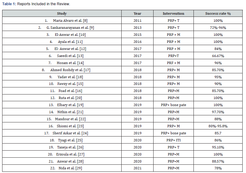
Abbreviations: PRP: Platelet rich plasma; M: Myringoplasty; T: Tympanoplasty; ITI: intratympanic injection; PRF: Platelet rich fibrin.
Some authors combined Gelfoam with PRP to repair perforations, and the closure rates in the PRP group (66.7%) and the control group (25%) were both lower than those in other studies using autografts [13]. Alhabib et al. [31] also inserted PRP directly through the perforation toward the middle ear rather than in combination with autografts to repair perforations. This study was aborted due to a low success rate of 18.2% [2/11]. The only two successful cases in the PRP group also had a tendency to retract medially because of a weak membrane. These results suggested that surgeons should consider the rigidity and adsorption of transplant materials when using PRP to repair TM perforation. Compared to other conventional surgical approaches (e.g., temporalis fascia tympanoplasty), FGM is performed as a minimally invasive approach without elevation of the tympan meatal flap and can be performed under local anesthesia. However, the success rate of FGM is associated with the perforation site and size [32]. For patients with large perforations, FGM alone may not be as satisfactory as other approaches due to adipose resorption before achieving complete closure [32]. There are many limitations of our study. One of this is the use of different types of grafts, different approaches for surgery and different periods of follow up. Although the studies of our review concluded that PRP is a promising material to be used in otology, more studies are needed to prove this material as a beneficial tool for the otology surgeon.
References
- Stavrakas M, Karkos PD, Markou K (2016) Platelets rich plasma in otolaryngology. The Journal of Laryngology and Otology 130: 1098-1102.
- Pomerants J, Dutton JM (2005) Platelet gel for endoscopic sinus surgery. Ann Otol Rhinol Laryngol 114: 699-704.
- Sclafani AP, Azzi J (2015) Platelet preparations for use in facial rejuvenation and wound healing: a critical review of current literature. Aesthetic Plast Surgery 39: 495-505.
- Boyapati L, Wang HL (2006) The role of platelet rich plasma in sinus augmentation: a critical review. Implant Dent 15: 160-170.
- Eppley BL, Pietrzak WS (2006) Platelet rich plasma review of biology and applications in plastic surgery. Plast Reconst Surg 118: 147-159.
- Pomerantz J, Dutton JM (2005) Platelet gel for endoscopic sinus surgery. Ann Otol Rhinol Laryngol 114: 669-704.
- Erkilet E, M Koyuncu, S Atmaca (2009) Platelet rich plasma improves healing of tympanic membrane perforations: experimental study. The Journal of Laryngology and Otology 123: 482-487.
- Maria LN, Ortiz N, Rodrigues L (2011) Pilot study on the efficiency of autologous plasma rich in platelet growth factors in otologic surgery (tympanoplasty type I). ISRN Surgery.
- Sancaranaraynan G, Prithivi Raj V, Kumar RV (2013) A study on efficacy of autologous platelet rich plasma in myringoplasty. Otolaryngology online journal 3: 2250-2259.
- El-Anwar MW, El Ahl MA, Zidan AA, Yacoup MA (2015) Topical use of autologous platelet rich plasma in myringoplasty. Auris Nasus Larynx 42: 365-368.
- Ayala-Montes SN, Alla JC, López-Valdés BD (2016) Effectiveness of topical phenytoin therapy versus platelet-rich plasma for tympanic perforations closure: Comparative study. Rev Med Hosp Gen Mex 79(4): 183-188.
- El-Anwar MW, El nashar I, Foad YA (2017) Platelet rich plasma myringoplasty. Ear Nose Throat J 96(8): 312-326.
- Saeedi M, Ajalloueian M, Zare E (2017) The effect of PRP enriched gelfoam on chronic tympanic membrane perforation. Int Tinnitus J 21(2): 108-111.
- Hosam M,Shaker M, Aboulwafa A (2017) Effect of use of platelet rich fibrin in repairing central tympanic membrane perforation using the endoscopic inlay butterfly cartilage myringoplasty technique. The Egyptian Journal of Otolaryngology 33: 557-563.
- Fawzy T, Hussein M, Eid S (2018) Sherif Guindy. Effect of adding platelet rich plasma to fat graft in myringoplasty. The Egyptian Journal of Otolaryngology 34: 224-228.
- Yasser Ahmed F , Mohammad A , Mohammad EA , Ezzat M (2018) Topical platelet rich plasma versus hyaluronic acid during fat graft myringoplasty. Am J Otolaryngol 39(6): 741-745.
- Roshdy A, Ahmed Y, Bedir M (2018) Myringoplasty of central tympanic membrane perforation with a fat graft from the ear lobule and platelet rich plasma. Z.U.M 24(2): 143-149.
- Yadav SPS, Malik JS, Malik P (2018) Studying the result of underlay myringoplasty using platelet rich plasma. J Laryngol Otol 132(11): 990-994.
- Elbary MEA, Nasr WF, Sorour SS (2018) Platelet rich plasma in reconstruction of posterior meatal wall after canal wall mastoidectomy. Int Arch Otolaryngology 22(2): 103-107.
- Ruta S, Yoganathan G, Vellavwdu U (2018) Myringoplasty with autologus platelet rich plasma. JMSCG 6(10): 1170-1173.
- Nithin P, AlexanderA, Abhishekh B (2018) Safety and efficacy of platelet rich fibrin on graft uptake in myringoplasty. Int Arch Otorhinolaryngol 23: 77-82.
- Mandour MF, Elsheikh MN, Khalil MF (2019) Platelet rich plasma fat graft versus cartilage perichondrium for repair of medium size tympanic membrane perforations. Otolaryngol Head Neck Surg 160(1): 116-121
- Shiomi Y, Shiomi Y (2019) Surgical outcomes of myringoplasty using platelet rich plasma and evaluation of the outcome associated factors Auris Nasus Larynx 19: 30116-30126.
- Sherif MA, Ibrahim M, Omar M (2019) Mastoid reconstruction with platelet rich plasma and bone pate after canal wall down mastoidectomy: A preliminary report. Ear nose and throat journal p: 1-5.
- Tyagi BPS, Rout M (2019) PRP: A revolutionary treatment of sensorineural hearing loss. Acta Scientific Otolaryngology (ASOL) 1(4): 2-5.
- Taneja MK (2020) Role of platelet rich plasma in tympanoplasty. Indian J Otolaryngol Head Neck Surg 72(2): 247-250.
- Tolga E, and Erdogan G (2020) A Comparison of the Autologous Platelet-Rich Plasma Gel Fat Graft Myringoplasty and the Fat Graft Myringoplasty forthe Closure of Different Sizes of Tympanic Membrane Perforations. Ear, Nose & Throat Journal 99(5): 331-336.
- Anwar FM, Shenoy VS, Kamas PM (2020) Study on use of platelet rich plasma in myringoplasty. Indian Journal of otology 26(2): 71-74.
- Nida R, Muhammad A, Muhammad SK (2021) Efficacy of Platelet Rich Fibrin in Myringoplasty. Pak J Med Sci 37(1): 212-216.
- Smith RG, Gassmann CJ, Campbell MS (2007) Platelet rich plasma: properties and clinical applications. J Lanc Gen Hosp 2: 273-277.
- Alhabib SF, Saliba I (2017) Hyaluronic acid fat graft myringoplasty versus autologous Platelet Rich Plasma. J Clin Med Res 9: 30-34.
- Lee DY, Kim YH (2018) Can fat-plug Myringoplasty Be a good alternative to formal myringoplasty. A systematic review and meta-analysis. Otol Neurotol 39: 403-409.

























