




 GJO Home



Abstract
Background and Objectives: Management of condylar fractures depends on well-defined indications and contraindications. While most
fractures are treated by closed reduction, open reduction is reserved for cases with absolute indications. Both open reduction and internal
fixation (ORIF) and closed reduction with intermaxillary fixation (IMF) have their own advantages and limitations. This study aimed to compare
the outcomes of ORIF with those of closed reduction using IMF in the management of moderately displaced condylar fractures.
Materials and Methods: The present study involved 20 patients (>18 years) with moderately displaced condylar fractures. Patients were
randomly allocated into surgical and nonsurgical groups and treated accordingly. Outcomes of conservative versus surgical management of
sub condylar and condylar neck fractures were evaluated based on seven parameters: maximal interincisal mouth opening, protrusive and
lateral excursive mandibular movements, occlusal status, mandibular deviation on mouth opening, pain (visual analog scale), and radiographic
assessment of ascending ramus height. Measurements were recorded preoperatively and postoperatively at various intervals up to 6 weeks.
Statistical Analysis: Data analysis was conducted using SPSS version 19. The Mann–Whitney U-test assessed differences between groups,
while the Wilcoxon signed-rank test evaluated paired observations within groups.
Results: Surgically treated patients demonstrated superior improvement in mouth opening, lateral excursion, and reduction of mandibular
deviation. They also reported earlier pain relief and better restoration of ramus height and facial symmetry. In contrast, patients managed
conservatively experienced prolonged pain, persistent deviation, and limited restoration of ramus height throughout the 6-week follow-up
period. The differences between the groups were statistically significant (P < 0.05).
Interpretation and Conclusion: Based on the findings of this study, surgical management is preferable to conservative treatment for
moderately displaced condylar fractures. ORIF provided improved functional outcomes, reduced symptoms, and better anatomical restoration.
The results underscore the need for further research with larger sample sizes to support definitive clinical recommendations.
Keywords:Condylar fracture; Deviation; Ramus height; Conservative Management; ORIF; Sub condylar fracture; Condylar neck fracture; Symmetry
Abbreviations:ORIF: Open Reduction and Internal Fixation; IMF: Intermaxillary Fixation; TMJ: Temporomandibular Joint; OPGs: Orthopantomography’s; CT: Computed Tomography; CBCT: Cone-Beam Computed Tomography
Introduction
Mandibular condylar fractures constitute approximately 20%–62% of all mandibular fractures [1]. The primary etiological factors include road traffic accidents (≈50%), falls (≈30%), and interpersonal violence (≈20%) [2]. The fundamental goal of treatment is to restore peritraumatic masticatory function, which requires re-establishing anatomical alignment, occlusion, and maxillofacial symmetry. Condylar fractures may be treated either conservatively (closed reduction with immobilization) or surgically (open reduction with internal fixation). Each modality has well-recognized indications, contraindications, and inherent advantages and disadvantages [3]. Historically, conservative management predominated, partly due to the perception of “satisfactory” outcomes and concerns regarding surgical risksparticularly facial nerve injury [4]. Conservative treatment preserves occlusion with relatively low morbidity and allows early mobilization, often leading to acceptable functional recovery.
However, long-term complications such as reduced mouth opening, malocclusion, and mandibular deviation have been frequently reported [4]. Additional disadvantages include airway compromise, oral hygiene difficulties, impaired speech, reduced nutritional intake, and disuse atrophy of masticatory muscles [3]. Recent advancements in surgical techniques and anatomical understanding have increased the adoption of ORIF. Open reduction enables precise repositioning of the condylar segment, restoration of ramus height, and re-establishment of stable occlusion with early functional rehabilitation [3]. Literature suggests that condylar fractures with >35°–45° displacement or >5 mm shortening of ramus height are strong candidates for surgical management [5–7]. Despite these developments, the optimal treatment approach remains debated, and clear consensus guidelines are lacking. Therefore, this study was undertaken to compare the outcomes of surgical and conservative management in moderately displaced sub condylar and condylar neck fractures.
Materials and Methods
The present prospective cohort study was conducted on 20 patients presenting with condylar fractures to the Outpatient Department over a period of 2½ years, beginning in May 2013. All patients were adults undergoing extraction of maxillary or mandibular teeth simultaneously as part of a split-mouth study design. The study protocol was reviewed and approved by the Institutional Ethics Committee in accordance with guidelines for research involving human participants.
Inclusion Criteria
Patients were included if they met the following criteria:
a) Age >18 years
b) Presence of condylar fractures, with or without
associated mandibular body or ramus fractures
c) Fracture line located in the condylar neck or sub
condylar region
d) Displacement of the condylar fragment between 10° and
45° in the frontal or sagittal plane; and/or
e) Shortening of the ascending ramus height ≥2 mm
Exclusion Criteria
Patients were excluded if they presented with any of the
following:
I. Condylar head fractures
II. Insufficient dentition to achieve or maintain normal
occlusion
III. Medical contraindication to undergoing surgery under
general anesthesia
IV. Presence of any associated midface fractures
V. History of temporomandibular joint (TMJ) dysfunction
Patient Preparation and Evaluation
All patients were informed in detail about the procedures involved and any potential complications associated with surgery or intermaxillary fixation (IMF). Written informed consent was obtained from every participant enrolled in both the surgical and conservative treatment groups. A comprehensive case history was recorded for each patient, including previous exposure to anesthetics and sedatives, prior surgeries, and any past hospital admissions. General physical examination and routine hematological investigations were performed for all participants, along with HIV and Hepatitis B surface antigen testing.
For patients assigned to the surgical group, additional preoperative assessments-including a chest radiograph and electrocardiogram-were performed. Further investigations were carried out as required based on the patient’s systemic condition. Preoperative clinical photographs and radiographs, including orthopantomography (OPGs) and computed tomography (CT) scans, were obtained for all subjects.
Surgical Procedure
Patients undergoing open reduction were treated under general anesthesia with endotracheal intubation following a standardized protocol.
Clinical and Radiographic Assessment
Both clinical and radiological parameters were evaluated
during follow-up appointments. The following parameters were
assessed:
a. Maximal interincisal mouth opening
b. Protrusive and lateral excursive mandibular movements
c. Occlusal discrepancies, determined by improper
intercuspation of the first molars
d. Pain
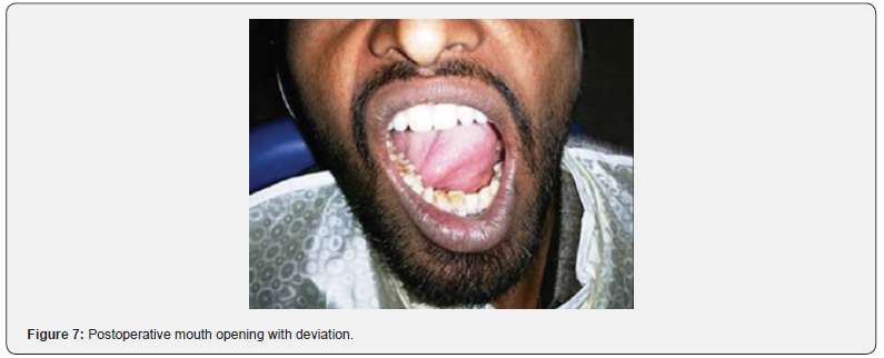
e. Mandibular deviation on mouth opening
f. Height of the ascending ramus
Maximal interincisal opening and protrusive movements were measured from the incisal edges of the upper and lower anterior teeth. Lateral excursions and deviation on opening were evaluated using a metallic scale, measured relative to the dental midline.
Follow-up Schedule
Surgical group
Parameters were recorded preoperatively and on postoperative day 3, and at weeks 1, 2, 4, and 6.
Conservative group
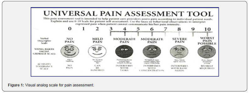
Measurements were taken preoperatively and at weeks 2, 4, and 6 postoperatively. Pain assessment for both groups was conducted using a visual analog scale (VAS) (Figure 1) preoperatively, and on postoperative day 1, day 3, and weeks 1, 2, 4, and 6.
Radiographic Measurements
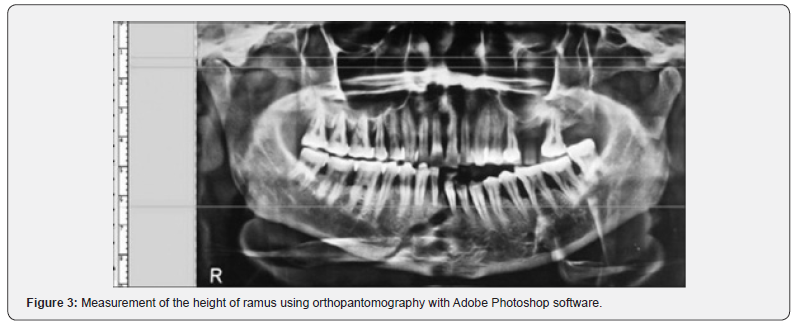
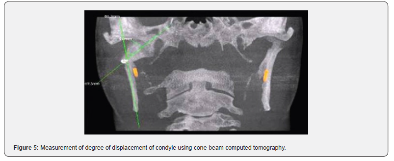
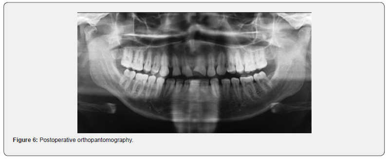
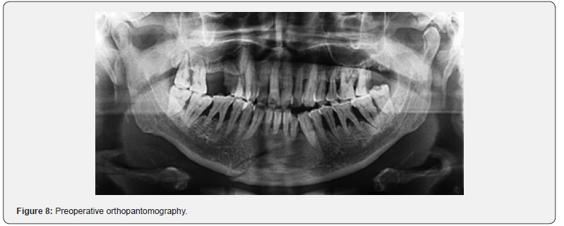
The height of the ascending ramus was measured on OPGs from the superior-most point of the condyle to the inferior-most point of the mandibular angle on the affected side. Measurements were performed using Adobe Photoshop software preoperatively, and on day 3 and week 6 postoperatively for both groups. Additionally, the degree of condylar displacement for all study participants was assessed using cone-beam computed tomography (CBCT) as part of the selection criteria.
Procedures
Closed Reduction
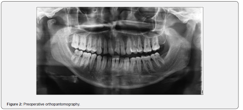
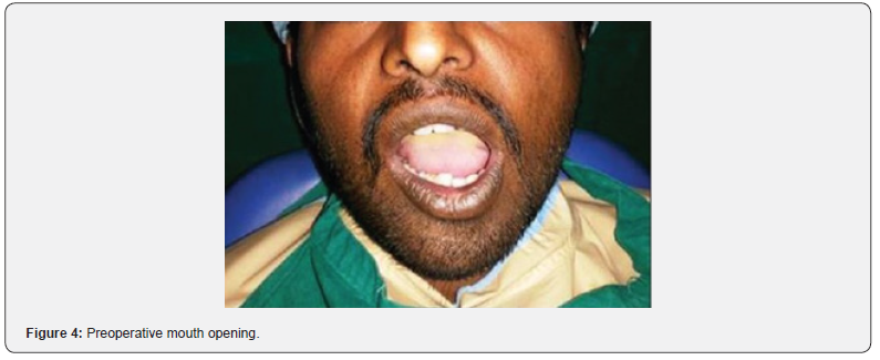
For all patients managed conservatively, arch bar splinting was applied to both the maxillary and mandibular dentition, followed by intermaxillary fixation (IMF) with the teeth placed in proper occlusion using guiding elastics. Patients were systematically evaluated during follow-up visits on postoperative day 1, day 3, and at weeks 1, 2, 4, and 6 (Figures 2–7).
Open Reduction
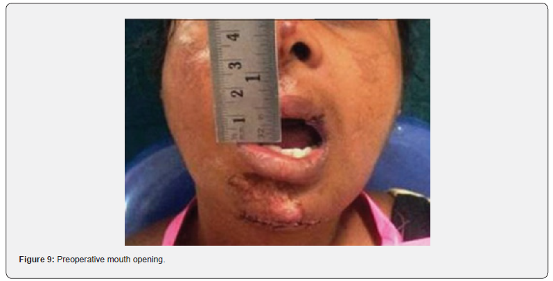
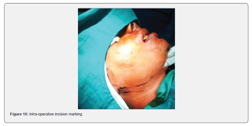
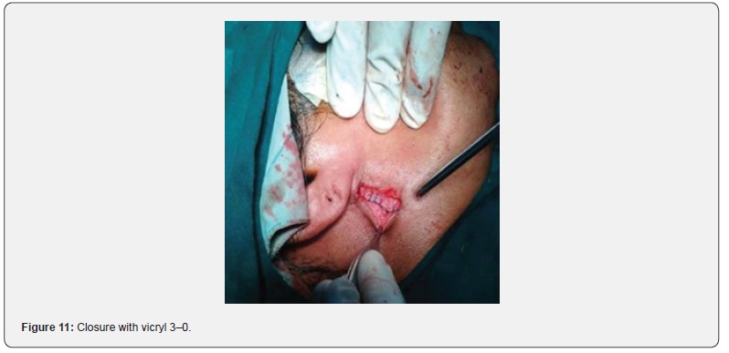
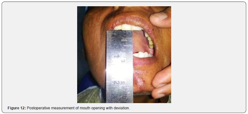
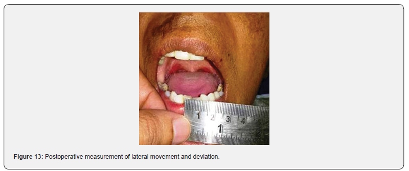
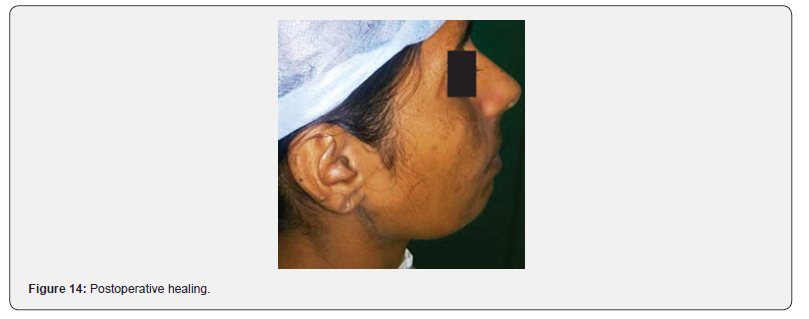
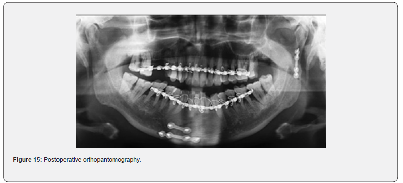
For all patients selected for surgical intervention, the retromandibular approach was utilized. Reduction of the condylar fracture was performed with the teeth maintained in proper occlusion, followed by fixation using appropriate titanium miniplates and screws. Postoperative follow-up was conducted at standardized intervals-day 1, day 3, and weeks 1, 2, 4, and 6-similar to the conservative group (Figures 8–15).
Statistical Analysis Used
Descriptive and analytical statistics were computed using the Statistical Package for the Social Sciences (SPSS) version 19 (SPSS Inc., Chicago, IL, USA). The Mann–Whitney U-test was employed to evaluate the significance of differences between the two treatment groups, while the Wilcoxon signed-rank test was used to assess the significance of differences between paired observations within each group.
Results
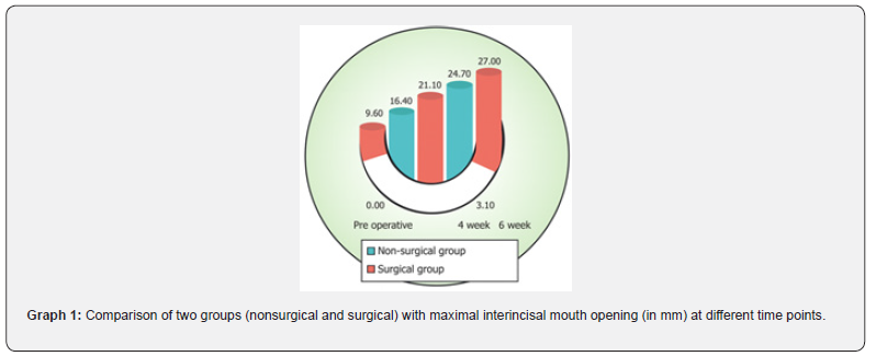
The present study included 20 patients with condylar fractures, with 10 patients managed conservatively and 10 managed surgically. Among these, 80% (n = 16) were male and 20% (n = 4) were female. The mean age of the study population was 33 years, with all participants being older than 18 years. Preoperatively, the maximal interincisal mouth opening measured 9.60 mm in the nonsurgical group and 6.10 mm in the surgical group. By the 2nd, 4th, and 6th postoperative weeks, the mean increases in maximal mouth opening were 6.80 mm, 9.90 mm, and 12.50 mm, respectively, in the nonsurgical group, compared with 15.00 mm, 18.60 mm, and 20.90 mm in the surgical group (Graph 1). Immediate anatomic restoration of ramus height, along with improved symmetry, was noted as an additional benefit in surgically managed patients. Protrusive and lateral excursive movements were also compared. For protrusive movements, the mean improvements at weeks 2, 4, and 6 were 0.40 mm, 2.60 mm, and 3.00 mm in the nonsurgical group and 1.60 mm, 2.20 mm, and 2.60 mm in the surgical group, respectively (Graph 2). For lateral excursive movements, the mean increases at weeks 2, 4, and 6 were 0.90 mm, 4.60 mm, and 5.30 mm in the nonsurgical group, while the surgical group demonstrated mean increases of 2.80 mm, 6.60 mm, and 8.90 mm, respectively (Graph 3).
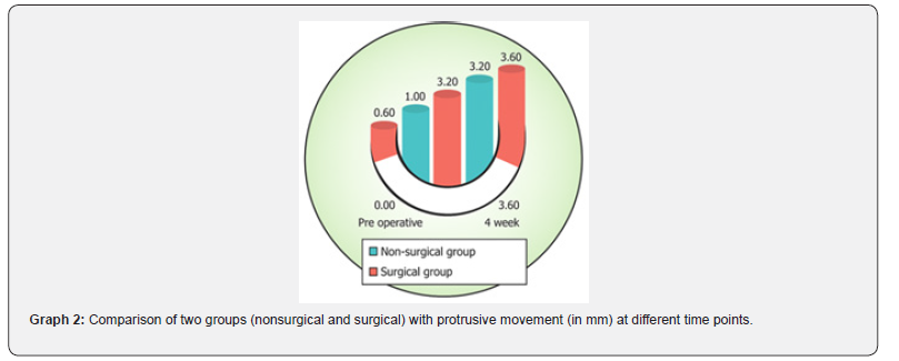
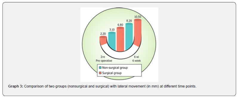
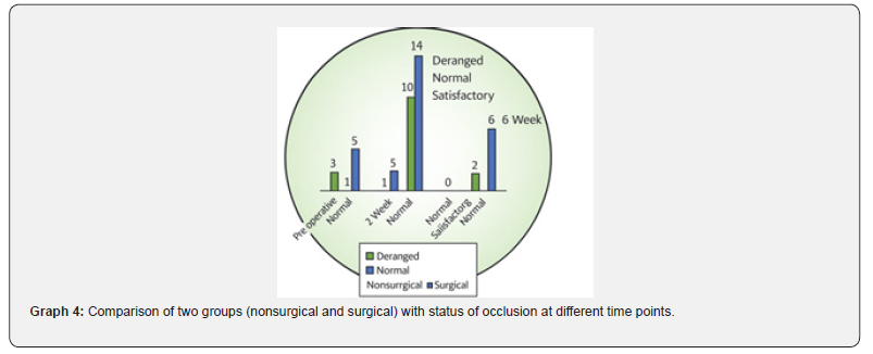
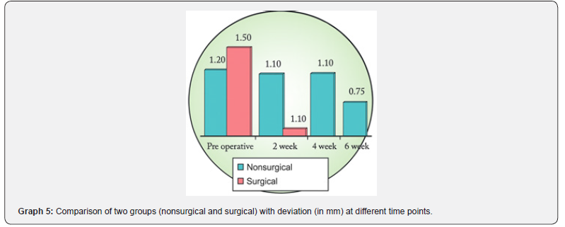
These findings indicate a clear functional advantage of surgical treatment, particularly in restoring lateral mandibular movements by 6 weeks. Assessment of protrusive and lateral movements was more challenging in the IMF group during weeks 2 and 4 due to fixation restrictions. In the nonsurgical group, six patients presented with deranged occlusion preoperatively. By the 2nd week, two patients achieved normal occlusion and three showed satisfactory improvement. By week 6, all patients attained normal occlusion. In the surgical group, seven patients had deranged occlusion by the 2nd postoperative week; however, all seven achieved normal occlusions, and by week 6, all 10 patients demonstrated normal occlusion (Graph 4), reflecting earlier occlusal restoration following surgical repositioning. Mandibular deviation during mouth opening was assessed preoperatively and at postoperative weeks 2, 4, and 6. Preoperatively, deviation measured 1.20 mm in the nonsurgical group and 1.50 mm in the surgical group. By week 2, deviation had reduced to 1.10 mm and 0.10 mm, respectively (Graph 5).
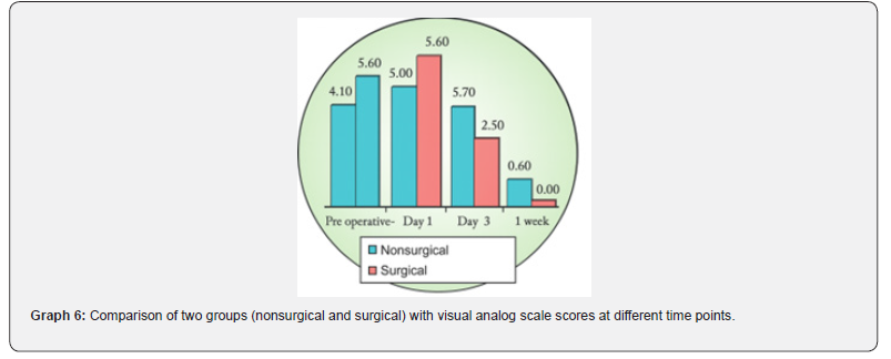
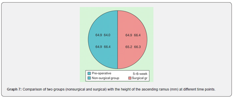
The difference between the groups was statistically significant (P = 0.0173). Pain perception based on the Visual Analog Scale (VAS) was recorded preoperatively, on postoperative day 1 and day 3, and at weeks 1, 2, 4, and 6. Preoperative mean VAS scores were 5.60 for the nonsurgical group and 4.10 for the surgical group. Pain scores were comparable between groups on day 1 and at weeks 2, 4, and 6, showing no significant difference. However, on postoperative day 3 and week 1, the nonsurgical group exhibited higher pain scores (5.70 and 3.60, respectively) compared with the surgical group (2.70 and 0.90, respectively), yielding statistically significant differences (P = 0.0082 and P = 0.0376) (Graph 6). The height of the ascending ramus was measured radiographically on the fractured side preoperatively and on postoperative day 3 and week 6 in both groups. By day 3, the nonsurgical group showed no restoration (mean = 0 mm), whereas the surgical group demonstrated a mean restoration of 2.40 mm (Graph 7), a statistically significant difference (P = 0.0002).
Discussion
Condylar fractures constitute one of the most frequently encountered mandibular injuries, accounting for nearly 20%– 62% of all mandibular fractures. The choice between surgical and nonsurgical management of these fractures continues to be a subject of debate. Historically, conservative management was preferred due to concerns about postoperative infection in the pre-antibiotic era, the presence of vital anatomical structures, and the lack of advanced osteosynthesis technology. However, with modern developments such as improved anesthesia, effective antibiotic coverage, and better surgical instruments and fixation systems, open reduction has gained wider acceptance. Achieving proper occlusion and restoring function remain the primary objectives in treating mandibular fractures. Currently, open approaches are often recommended for dislocated fractures. The advent of reliable osteosynthesis techniques-including miniplates (Pape et al., 1980), lag screws (Wackerbauer, 1962; Petzel, 1980; Eckelt & Gerber, 1981; Krenkel, 1992)-and enhancements in surgical access have made open reduction both safer and functionally advantageous by enabling early mobilization of traumatized tissues. Nevertheless, for moderately displaced fractures, conservative management is still considered a viable option.
Several authors, such as Baker (1998), have advocated surgical management due to better postoperative outcomes in mouth opening and mandibular movements. Improved anatomical knowledge and advancements in surgical instrumentation have contributed to making procedures in the condylar region more predictable. On the other hand, proponents of conservative treatment highlight its advantages, such as avoiding the complexities of TMJ access, eliminating the need for condylar repositioning, and reducing the risk of facial nerve injury. This study compared conservative and surgical management using seven clinical and radiographic parameters, including maximal interincisal opening, lateral and protrusive movements, occlusal status, deviation on opening, VAS pain scores, and radiographic ramus height, evaluated at various intervals pre- and postoperatively. Hyde et al. [6], in a prospective study, reported significantly greater mouth opening in the surgical group (42 mm) compared to the conservative group (32 mm), aligning with the findings of the present study. Eckelt et al. [8] similarly reported superior lateral excursive movements in surgically treated patients (up to 16 mm) compared to those who underwent closed treatment (up to 13 mm). These results were consistent with those observed in the present study. However, Carneiro et al. [9] found no significant difference in protrusive and lateral excursive movements between the two treatment modalities.
Regarding occlusion, Ellis et al. [10] concluded that closed treatment was associated with a higher rate of postoperative malocclusion compared with open reduction, despite greater initial displacement in surgically managed cases. Contrary to this, Haug & Assael [11] reported no significant difference in occlusal outcome between ORIF and closed reduction with maxillomandibular fixation (CRMMF). Pain analysis in various studies reveals further support for surgical management. Hyde et al. [6] demonstrated significantly lower VAS scores in surgically treated patients, a finding consistent with Haug & Assael [11] who also reported reduced pain levels in patients undergoing CRMMF. The present study correlates closely with these observations, again showing significantly lower pain levels in the surgical group.
Radiographically, Danda et al. [1] reported superior anatomical reduction in surgically treated patients compared to those managed conservatively. Similarly, Eckelt et al. [8] demonstrated that correct anatomical positioning was achieved more consistently through surgical intervention. These findings were in agreement with the present study, in which restoration of ramus height and anatomical symmetry was significantly better in the surgical group by the 6-week postoperative period. Similar outcomes were also reported by Ebenezer & Ramalingam [12]. Overall, the present study strongly favors surgical management over conservative methods for moderately displaced condylar fractures, offering valuable insights for maxillofacial surgeons in treatment planning. Despite its limitations-including a relatively small sample size and treatments performed by different surgeons-the study supports surgical intervention as the preferred modality. Future research with larger sample sizes and standardized surgical expertise would further strengthen the reliability of conclusions.
Conclusion
The present study demonstrated the clear superiority of surgical management over conservative treatment for moderately displaced condylar fractures. Among the seven evaluated parameters, five-maximal interincisal mouth opening, lateral excursions with minimal deviation, earlier pain relief, and restoration of ramus height with improved symmetry-showed statistically significant improvements in the surgical group during the 2–6-week follow-up period. Although conservative management remains a viable option, particularly because it avoids the complexity of surgical access and reduces the risk of facial nerve injury while still yielding acceptable outcomes, advancements in surgical instrumentation and techniques have made operative treatment more predictable and functionally advantageous. Considering the benefits of early rehabilitation, superior functional outcomes, greater patient comfort, and better anatomical reduction with symmetry, the findings of this study support surgery as the preferred treatment modality over conservative approaches for moderately displaced condylar fractures.
References
- Danda AK, Muthusekhar MR, Narayanan V, Baig MF, Siddareddi A (2010) Open versus closed treatment of unilateral sub condylar and condylar neck fractures: A prospective, randomized clinical study. J Oral Maxillofac Surg 68: 1238-1241.
- Sawazaki R, Lima Júnior SM, Asprino L, Moreira RW, De Moraes M (2010) Incidence and patterns of mandibular condyle fractures. J Oral Maxillofac Surg 68: 1252-1259.
- Rasheed A, Mumtaz M, Bhatti M (2010) Comparison of surgical with non-surgical treatment for fractured mandibular condyle: A study. Pak Oral Dent J 30: 295-298.
- Ellis E, Throckmorton GS (2005) Treatment of mandibular condylar process fractures: Biological considerations. J Oral Maxillofac Surg 63: 115-134.
- Santler G, Kärcher H, Ruda C, Köle E (1999) Fractures of the condylar process: Surgical versus nonsurgical treatment. J Oral Maxillofac Surg 57: 392-397.
- Hyde N, Manisali M, Aghabeigi B, Sneddon K, Newman L (2002) The role of open reduction and internal fixation in unilateral fractures of the mandibular condyle: A prospective study. Br J Oral Maxillofac Surg 40: 19-22.
- De Riu G, Gamba U, Anghinoni M, Sesenna E (2001) A comparison of open and closed treatment of condylar fractures: A change in philosophy. Int J Oral Maxillofac Surg 30: 384-389.
- Eckelt U, Schneider M, Erasmus F, Gerlach KL, Kuhlisch E, et al. (2006) Open versus closed treatment of fractures of the mandibular condylar process-a prospective randomized multi-centre study. J Craniomaxillofac Surg 34: 306-314.
- Carneiro S, Vasconcelos B, Caldas A, Jr, Leal J, Frazao M (2008) Treatment of condylar fractures: A retrospective cohort study. Med Oral Patol Oral Cir Bucal 13: E589-94.
- Ellis E, Simon P, Throckmorton GS (2000) Occlusal results after open or closed treatment of fractures of the mandibular condylar process. J Oral Maxillofac Surg 58: 260-268.
- Haug RH, Assael LA (2001) Outcomes of open versus closed treatment of mandibular sub condylar fractures. J Oral Maxillofac Surg 59: 370-375.
- Ebenezer V, Ramalingam B (2010) Treatment outcome of unilateral condylar fractures surgical versus conservative approach. Indian J Multidiscip Dent 1: 8-11.

























