




 GJO Home



Abstract
Objective: To evaluate hearing outcomes and functional success following tympanoplasty with and without platelet-rich plasma (PRP) in patients with chronic tympanic membrane perforation. Methods: A total of 30 patients were divided equally into two groups: one underwent tympanoplasty with PRP (n=15), and the other without PRP (control group, n=15). Preoperative and postoperative air-bone gap (ABG) values were compared to assess hearing improvement. Functional success was defined as an ABG gain >10 dB. Hearing results were also compared to findings from previously published studies. Results: Postoperative average ABG gains were higher in the PRP group compared to the control group. Functional success was achieved in 11 of 15 patients (73.3%) in the PRP group and 9 of 15 patients (60.0%) in the control group. The difference in success rates between the groups was not statistically significant (p = 0.699, Fisher’s exact test). These findings align with previous studies, which reported functional success rates of 65.6-90% in PRP-treated patients and 40.6-77.1% in controls. Conclusion: The use of PRP in tympanoplasty may enhance hearing improvement and increase the rate of functional success. However, the difference compared to conventional tympanoplasty did not reach statistical significance. Larger studies with standardized frequency reporting are needed to confirm these findings
Keywords:Tympanoplasty; Platelets; Plasma; Fascial; Otitis media; Posterior perforations
Abbreviations:PRP: Platelet-Rich Plasma; ABG: Air-Bone Gap; TM: Tympanic Membrane; CSOM: Chronic Suppurative Otitis Media; PTA: Pure-Tone Audiometry; BC: Bone Conduction; EMMs: Estimated Marginal Means; AC: Air Conduction; BC: Bone Conduction; ABG: Air-Bone Gap
Introduction
Tympanic membrane (TM) perforation is a common condition resulting from trauma or chronic suppurative otitis media (CSOM), often leading to hearing loss, recurrent infections, and reduced quality of life. When spontaneous healing fails, tympanoplasty is indicated to restore TM integrity and middle ear protection. Among perforations, anterior ones are particularly challenging due to limited blood supply and surgical access. In chronic otitis media (COM) cases-especially prevalent in developing countries-successful outcomes depend heavily on surgical technique and graft material selection [1,2]. Historically, the first documented tympanoplasty was performed by Berthold in 1878, who introduced a technique involving de-epithelialization and skin grafting of the TM remnant (Berthold, 1878). In modern otologic practice, myringoplasty refers specifically to TM repair without ossicular manipulation, while tympanoplasty may involve additional middle ear reconstruction if pathology is present [3,4].
The underlay technique, a widely accepted and relatively straightforward method, is ideal for posterior perforations. It involves positioning the graft medial to the TM annulus and ossicles. Despite its popularity, limitations such as restricted visualization, poor anterior margin coverage, and higher failure rates due to inadequate graft vascularity persist. In contrast, the overlay technique, which places the graft lateral to the annulus, provides superior access to anterior defects and yields higher closure rates (>90%), making it the preferred method for anterior and subtotal perforations or when underlay fails [5-7]. Surgical success also varies based on perforation size, surgeon experience, and the adjunctive use of biological agents. Emerging studies explore agents like hyaluronic acid, pentoxifylline, and fibroblast growth factors to enhance TM regeneration [8]. Common graft materials include temporalis fascia, cartilage, fat, and perichondrium, with temporalis fascia being the gold standard due to its ease of harvest and favorable integration [9]. Recently, Platelet-Rich Plasma (PRP) has garnered attention as a potential regenerative adjunct in tympanoplasty.
PRP, an autologous concentration of platelets suspended in plasma, was first applied in medicine in the 1980s and has since been widely used in orthopedic, dental, and cosmetic procedures. PRP provides a potent mix of growth factors-including PDGF, VEGF, and TGF-β-that stimulate fibroblasts, promote angiogenesis, and accelerate epithelial and stromal healing. It also exhibits adhesive, mitogenic, and anti-inflammatory properties that make it an ideal biological scaffold [10,11]. Despite its growing use in regenerative medicine, limited clinical studies have evaluated PRP’s role in tympanoplasty, particularly in direct comparison with conventional underlay techniques using fascia alone. Thus, the current study aims to evaluate the efficacy of PRP-augmented hourglass grafts compared to the standard underlay temporalis fascia technique in the repair of TM perforations. This comparison may clarify whether PRP offers a meaningful improvement in surgical outcomes and postoperative healing.
Patients and Methods
This study is designed as a prospective randomized controlled trial aiming to evaluate the impact of platelet-rich plasma (PRP) in tympanoplasty outcomes. The research was conducted at the Otorhinolaryngology Department of Helwan University Hospital (Badr Hospital) over a duration of nine months, from April 2024 to December 2024. Approval from the Research Ethics Committee of Helwan Faculty of Medicine was obtained. All participants provided informed written consent, including information on study purpose, design, duration, procedures, risks, and confidentiality. Official administrative permission was secured from Helwan University Hospitals. Participants were selected from patients attending the outpatient otorhinolaryngology clinic who are candidates for tympanoplasty. All patients were assessed through general and local endoscopic ear examinations, pure-tone audiometry (PTA), speech audiometry, and air-bone gap analysis. Inclusion criteria comprised patients aged under 12 or over 60 years, of both sexes, who are diagnosed with tympanic membrane perforation requiring tympanoplasty and are willing to undergo the operation after providing informed consent. Exclusion criteria included patients between 12 and 60 years of age, those with active middle ear infections, chronic systemic illnesses affecting wound healing such as ischemia, diabetes mellitus, or venous stasis disease, and patients deemed non-compliant. Randomization was carried out using opaque sealed envelopes containing sequential numbers, ensuring allocation concealment. Patients were randomly assigned to one of two groups at a 1:1 ratio. Group A (n = 15) have undergone underlay myringoplasty using temporalis fascia graft only, while Group B (n = 15) have undergone conventional fascial tympanoplasty aided with PRP. Sample size was determined using the formula [n = Z² × P(1-P) / d²], where Z = 1.96 (for 95% confidence), P = expected proportion based on graft success rates from a prior study (Taneja, 2020)- 85.3% in the control group versus 95.1% in the PRP group-and d = precision of 0.05.
To account for potential dropouts, six additional participants were added, resulting in a final sample size of 30 patients. The sampling technique applied will be probability sampling using simple random selection [12]. Study procedures began with preoperative assessments, including comprehensive history taking (name, age, sex, marital status, residence), general clinical examination (vital signs, signs of anemia, cyanosis, jaundice, or lymphadenopathy), and full ENT examination including otoscopic, oropharyngeal, nasal, and cervical evaluations. Laboratory tests included complete blood count, liver and kidney function tests, and coagulation profile. Radiological evaluation included a lateral soft tissue X-ray of the nasopharynx, and audiological investigations encompassed pure-tone audiometry, tympanometry, and speech audiometry. The surgical procedure for all patients was performed under general anesthesia. A post-auricular approach was used. After freshening the perforation edges, the tympanomeatal flap was elevated and the ossicular chain evaluated. An underlay fascia graft was placed, and the middle ear was packed with gelfoam. In the study group, PRP-impregnated gelfoam pieces were placed in the middle ear under the fascia graft, and additional PRP-soaked gelfoam pieces were placed lateral to the graft in the external canal. In the control group, dry gelfoam was used in both positions. In both groups, the tympanomeatal flap was repositioned, a fusidic acid-painted ear wick was inserted, and the wound was closed in two layers, followed by a pressure dressing on the first day. PRP was prepared by drawing 9mL of venous blood with 1mL of anticoagulant, followed by a first centrifugation at 1500rpm for 15 minutes. The resulting plasma supernatant was transferred and subjected to a second spin at 300rpm for 15 minutes. The platelet-rich pellet was resuspended in 1mL of plasma. Just before application, 0.1mL of calcium gluconate was added to activate growth factor release. PRP was used to moisten gelfoam and fascia graft pieces, which were then placed in appropriate positions during the operation. Postoperative care included alternate-day pack observation and suctioning, with ribbon pack removal on the 7th postoperative day.
Patients received 2 days of injectable antibiotics and a 10-day oral course, along with analgesics, antihistamines, and vitamin C and D supplementation. Postoperative assessments focused on tympanic membrane closure status at 1- and 3-months following surgery. Relationships between closure rate and variables such as perforation size, etiology, and presence of otorrhea were statistically analyzed using the chi-square test. Additionally, postoperative hearing outcomes were assessed via air-bone gap measurements on audiometry. Statistical analyses were conducted using R (version R-4.5.1) with the ezANOVA package for repeated-measures mixed ANOVA. The primary outcomes-air conduction (AC), bone conduction (BC), and air-bone gap (ABG)- were measured at three time points (preoperatively, 1 month, and 3 months postoperatively) and compared between two groups: PRP-assisted tympanoplasty and conventional tympanoplasty. For each outcome, a mixed-design repeated-measures ANOVA was performed with Group as the between-subjects factor and Time as the within-subjects factor. Mauchly’s test was used to assess the sphericity assumption. When sphericity was violated, Greenhouse-Geisser and Huynh-Feldt corrections were applied. Significance was set at p < 0.05. Effect sizes were reported using generalized eta squared (ges) to quantify the magnitude of observed effects. Continuous variables were expressed as means ± standard deviations, or median (interquartile range), while categorical variables were presented as frequencies and percentages. Between-group comparisons were conducted using appropriate tests based on data distribution and type. Specifically, the independent t-test was used for normally distributed continuous variables, while the Mann-Whitney U test was applied to non-normally distributed continuous variables. For categorical variables, either the Chi-square test or Fisher’s exact test was used, depending on the expected cell frequencies. A two-tailed p-value of less than 0.05 was considered statistically significant. When significant main or interaction effects were observed, post-hoc comparisons were conducted with Bonferroni-adjusted p-values to control for multiple testing.
Results
Baseline Characteristics
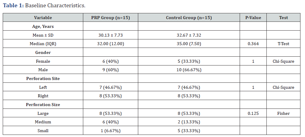
The baseline characteristics (Table 1) of the two groups were comparable. The mean age was 30.13 ± 7.73 years in the PRP group and 32.67 ± 7.32 years in the control group, with no statistically significant difference (p = 0.364). Gender distribution was similar between groups, with 40% females in the PRP group and 33.3% in the control group (p = 1.000). The site of tympanic membrane perforation was equally distributed, with 46.67% involving the left ear and 53.33% the right ear in both groups (p = 1.000). While not statistically significant (p = 0.125), there was a trend toward a higher proportion of small perforations in the control group (33.3%) compared to the PRP group (6.7%), and a higher proportion of medium perforations in the PRP group (40%) than in the control group (13.3%).
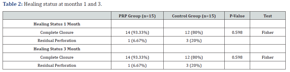
Healing Outcomes and Audiological Improvements: At both 1 and 3 months postoperatively, complete tympanic membrane closure was observed in 93.3% of the PRP group compared to 80% in the control group, with no statistically significant difference (p = 0.598).
Audiological Outcomes
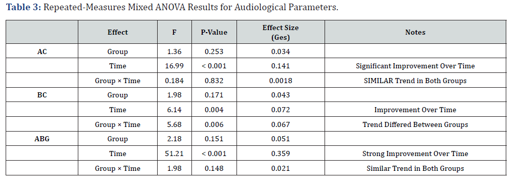
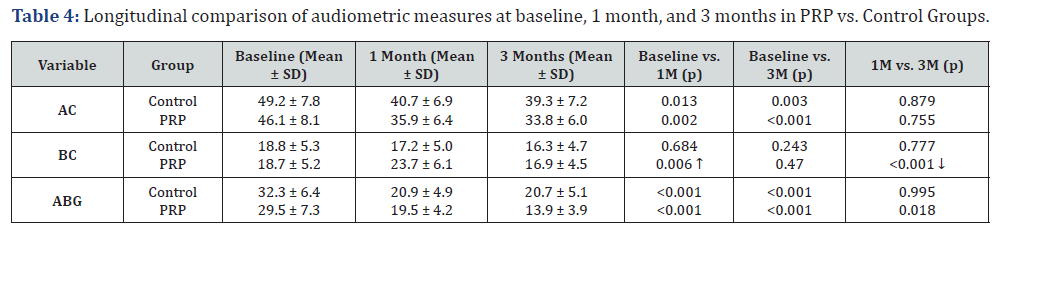
Audiological assessments demonstrated improvement over time in both groups. Repeated-measures ANOVA showed a significant main effect of Time for all variables-air conduction (AC), bone conduction (BC), and air-bone gap (ABG)-indicating significant improvement across the 3-month period (Table 2). For AC, there was a significant Time effect (p < 0.001), but no significant differences between groups or interaction effects, suggesting both groups improved similarly. For BC, both the Time effect (p = 0.004) and the Group × Time interaction (p = 0.006) was significant, indicating that improvement patterns differed between groups. For ABG, a highly significant Time effect was observed (p < 0.001), with no group or interaction effects. These findings suggest that both interventions led to significant auditory improvement over time, with the PRP group showing a more favorable trend in BC recovery. Estimated marginal means (EMMs) revealed a progressive decrease in air conduction (AC) scores over time in both groups. In the control group, the mean AC score declined from 49.2 dB at baseline to 40.7 dB at 1 month and 39.3 dB at 3 months. The PRP group demonstrated a more marked improvement, with mean AC scores decreasing from 46.1 dB at baseline to 35.9 dB at 1 month and 33.8 dB at 3 months (Table 3). Within-group pairwise comparisons using Tukeyadjusted p-values confirmed significant improvements from baseline to 1 month (p = 0.013) and to 3 months (p = 0.003) in the control group, whereas the change between 1 and 3 months was not significant (p = 0.879). Similarly, the PRP group showed significant improvements from baseline to 1 month (p = 0.002) and from baseline to 3 months (p < 0.001), with no significant change between 1 and 3 months (p = 0.755).
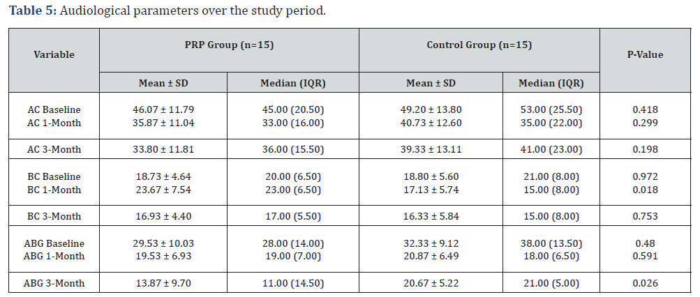
Bone conduction (BC) scores remained relatively stable in the control group, with no significant differences between time points (baseline: 18.8 dB; 1 month: 17.1 dB; 3 months: 16.3 dB; all p > 0.24). In contrast, the PRP group exhibited a transient increase in BC thresholds at 1 month (23.7 dB) compared to baseline (18.7 dB, p = 0.006), followed by a significant reduction by 3 months (16.9 dB, p < 0.001). However, the overall difference between baseline and 3 months was not statistically significant (p = 0.470). Air-bone gap (ABG) values also showed significant improvement over time. In the control group, the mean ABG dropped from 32.3 dB at baseline to 20.9 dB at 1 month and 20.7 dB at 3 months, with significant reductions observed from baseline to 1 month and to 3 months (both p < 0.0001), while the difference between 1 and 3 months was not significant (p = 0.995) (Table 4). The PRP group experienced a greater reduction in ABG, decreasing from 29.5 dB at baseline to 19.5 dB at 1 month and further to 13.9 dB at 3 months. All within-group comparisons in the PRP group were statistically significant: baseline vs. 1 month (p < 0.0001), baseline vs. 3 months (p < 0.0001), and 1 month vs. 3 months (p = 0.018) (Figure 1). To further clarify the group differences across time, post-hoc comparisons at individual time points were performed. Regarding audiological parameters, both groups showed progressive improvement over time. Although baseline air conduction (AC), bone conduction (BC), and air-bone gap (ABG) were comparable between groups, significant differences emerged at certain follow-up points (Figure 2). At 1 month, BC thresholds were significantly better in the control group (mean 17.13 ± 5.74 dB) compared to the PRP group (mean 23.67 ± 7.54 dB; p = 0.018). By 3 months, the PRP group demonstrated significantly greater ABG improvement (mean 13.87 ± 9.70 dB vs. 20.67 ± 5.22 dB in the control group; p = 0.026) (Table 5). Other changes, including AC thresholds at 1 and 3 months, showed a trend toward improvement in the PRP group but did not reach statistical significance ().
Functional success rate
Functional success, defined as an air-bone gap (ABG) gain greater than 10 dB, was achieved in 11 out of 15 patients (73.3%) in the PRP group and 9 out of 15 patients (60.0%) in the control group. While the PRP group showed a numerically higher success rate, the difference between groups was not statistically significant (p = 0.699, Fisher’s exact test).
Discussion
Platelet-rich plasma (PRP), one of the bioactive materials used during the last three decades, functions as a sealant and haemostatic agent during surgery. It has been shown to be effectively accelerating TM healing [13]. PRP has a regenerative power as it accelerates endothelial tissue regeneration and enhances tissue healing. In most of human trials it shoed promising benefits. However, the TM perforation closure rate differs among studies. Therefore, both healing and hearing improvements outcomes require further studies [14]. This study compared the outcomes of underlay fascial tympanoplasty assisted with PRP to conventional underlay fascial tympanoplasty. The two groups were comparable in terms of baseline characteristics, including age, gender, perforation site, and perforation size. While the PRP group had a slightly younger mean age and a higher proportion of medium-sized perforations, these differences were not statistically significant.
Closure of the Tympanic Membrane
The results of the current study showed that the tympanic membrane healing rates were high in both groups, with complete closure observed in 93.3% of patients in the PRP group and 80% in the control group at both 1 and 3 months postoperatively. Although the difference did not reach statistical significance, the higher closure rate in the PRP group suggests a potentially beneficial role of PRP in enhancing healing, consistent with PRP’s known regenerative properties. However, the small sample size may have limited the power to detect a statistically significant difference. In agreement with our findings, Huang et al. [13] conducted a review of published articles on the subject, reporting 93.4% of complete closure cases in patients who received PRP after surgery compared to 78.6% in patients who had surgery alone, with a low incidence of complications [14].
Graft rejection was described in one case where a surgical repair failed. In the same study, PRP bactericidal capabilities were postulated since four cases in the control group had postoperative infections but none of those with PRP application [15]. Navarrete Alvaro et al. [16] reported using PRP as an adjuvant in tympanoplasty, resulting in complete closure of tympanic membrane perforations [16]. Sankaranarayanan et al. discovered that a PRP clot applied during tympanoplasty decreased transplant displacement [17]. In 32-patient research, PRP formulations were found to help in healing of acute tympanic membrane perforations [18]. Elbary et al. [19] found that employing titanium mesh and PRP combined with bone material to reconstruct the posterior meatal wall after canal wall-down mastoidectomy for middle ear cholesteatoma was effective [19]. PRP, continuous hyperbaric oxygen, and polydeoxyribonucleotide were employed in a unilateral total ear amputation. It has been established that PRP can successfully salvage practically the whole auricle [20]. Vozel et al. [21] confirmed in a randomized controlled clinical trial that autologous PRP is an effective treatment modality for chronic postoperative temporal bone cavity inflammation in patients whose disease could not be treated surgically to maintain serviceable hearing loss and a reasonable disease-related quality of life [21].
Audiological Improvements
The results of our current study demonstrated that both groups showed significant improvements in hearing over time, as reflected by reductions in AC thresholds and air-bone gaps ABG. Repeated-measures ANOVA confirmed a strong effect of time on AC, BC, and ABG in both groups, indicating that tympanoplastyregardless of technique-effectively restored auditory function. Importantly, the PRP group demonstrated a more favorable trend, particularly in ABG improvement, which was significantly better at 3 months compared to the control group (13.9 dB vs. 20.7 dB; p = 0.026). This suggests enhanced ossicular chain function and better conductive hearing outcomes with PRP. Interestingly, BC thresholds showed a transient increase in the PRP group at 1 month, possibly due to temporary middle ear inflammation or measurement variability. However, BC values normalized by 3 months, and no long-term adverse impact was observed. The control group, on the other hand, showed stable BC values throughout the follow-up. Post-hoc comparisons also supported these trends. While AC improvements were not statistically different between groups, the PRP group consistently showed lower mean thresholds at each time point. This indicates a more pronounced, though not statistically significant, benefit of PRP in auditory recovery. In our study, postoperative hearing improvement was observed in both the PRP and control groups.
Ges: Generalized Eta Squared.
AC: Air Conduction; BC: Bone Conduction; ABG: Air Bone Gap.
The average hearing gain (preoperative ABG - postoperative month-3 ABG) in the PRP group was 15.6 dB, compared to 11.6 dB in the control group. These findings are consistent with those reported in earlier studies where PRP-treated patients showed average gains ranging from 10.3 to 18.62 dB, while control groups showed gains between 7.23 and 15.64 dB [22]. For instance, Yadav et al. reported a gain of 18.62 dB in the PRP group versus 13.15 dB in the control group. Similarly, El-Anwar et al. [15] found a mean gain of 10.5 dB in the PRP group versus 7.43 dB in controls. In terms of functional success, defined as ABG gain >10 dB, our study achieved a success rate of 73.3% in the PRP group and 60% in the control group, aligning with prior reports where the improvement rates ranged from 65.6% to 90% in PRP-treated patients and 40.6% to 77.1% in controls [22-24]. For example, Anwar et al. [22] reported success rates of 88.6% in the PRP group and 77.1% in the control group. Although our findings demonstrated significant improvement between pre- and postoperative ABG values within each group, no statistically significant difference was noted between the PRP and control groups in terms of hearing gain, which is also in agreement with previous systematic reviews [e.g., Ersozlu et al. [25], Fouad et al. [23]. This may reflect variability in frequency selection, surgical techniques, or small sample sizes that limit statistical power. Taken together, our results reinforce the trend that PRP, when combined with autologous graft materials, contributes to clinically meaningful hearing improvements, even if the statistical differences between treatment groups remain inconsistent across studies.
Interpretation and Clinical Implications
The findings suggest that PRP may provide modest but clinically relevant advantages in tympanic membrane healing and functional hearing outcomes. Its autologous nature, ease of preparation, and regenerative potential make it an appealing adjunct in tympanoplasty. However, due to the limited sample size and lack of statistically significant differences in most outcomes, these results should be interpreted cautiously. Future studies with larger cohorts and longer follow-up periods are needed to validate these findings and explore the mechanistic basis of PRP’s effect on middle ear healing. Additionally, evaluating cost-effectiveness and patient-reported outcomes will be essential for assessing PRP’s role in routine otologic practice.
References
- Faramarzi M, Kazemi T, Shishegar M, Zargerani O, Faramarzi A, et al. (2021) Does intraoperative ciprofloxacin-soaked gelfoam have adverse effects on graft success rate? A randomized, double-blind controlled trial. Laryngoscope Investigative Otolaryngology 6(5): 1182-1187.
- Visvanathan V, Vallamkondu V, Bhimrao Sk (2018) Achieving a Successful Closure of an Anterior Tympanic Membrane Perforation: Evidence-Based Systematic Review. Otolaryngology Head and Neck Surgery 158(6): 1011-1015.
- Pontillo V, Cavallaro G, Barbara F, Mastrodonato M, Murri A, et al. (2023) Recurrent tympanic perforation after myringoplasty: a narrative literature review and personal experience. Acta Otorhinolaryngol Ital 43(Suppl 1): S41-S47.
- Abdelaal TM, Ibrahim AA (2021) Endoscopic tympanoplasty using Tutoplast versus conventional microscopic tympanoplasty. The Egyptian Journal of Otolaryngology 37(1): 88.
- Shishegar M, Faramarzi M, Biniaz D, Rabiei N, Babaei A, et al. (2023) Comparison of the underlay and over-underlay tympanoplasty: A randomized, double-blind controlled trial. Laryngoscope Investigative Otolaryngology 8(2): 518-524.
- Barake R, El Natout T, Bassim M, El Natout Ma (2019) Loop underlay tympanoplasty for anterior, subtotal and total tympanic membrane perforations: a retrospective review. J Otolaryngol Head Neck Surg 48(1): 12.
- Arora Rd, Thakur N, Kamble P, Jati M, Nagarkar Nm, et al. (2022) Circumferential subannular tympanoplasty: surgical and hearing outcome in 224 years with subtotal perforation. Acta Otolaryngol 142(3-4): 254-258.
- Lou Z, Lou Z, Jiang Y, Chen Z (2021) FGF2 and EGF for the Regeneration of Tympanic Membrane: A Systematic Review. Stem Cells International 2021(1): 2366291.
- Alzahrani M, Saliba I (2015) Hyaluronic acid fat graft myringoplasty vs fat patch fat graft myringoplasty. European Archives of Otorhinolaryngol 272(8): 1873-1877.
- Yadav Sps, Malik Js, Malik P, Sehgal Pk, Gulia Js, et al. (2018) Studying the result of underlay myringoplasty using platelet-rich plasma. The Journal of Laryngology and Otology 132(11): 990-994.
- Verma R, Kumar S, Garg P, Verma Yk (2025) Platelet-rich plasma: a comparative and economical therapy for wound healing and tissue regeneration. Cell Tissue Bank 24(2): 285-306.
- Taneja MK (2020) Role of Platelet Rich Plasma in Tympanoplasty. Indian J Otolaryngol Head Neck Surg 72(2): 247-250.
- Huang J, Teh BM, Eikelboom RH, Han L, Xu G, et al. (2020) The Effectiveness of bFGF in the Treatment of Tympanic Membrane Perforations: A Systematic Review and Meta-Analysis. Otol Neurotol 41(6): 782-790.
- Huang J, Shi Y, Wu L, Lv C, Hu Y, et al. (2021) Comparative efficacy of platelet-rich plasma applied in myringoplasty: A systematic review and meta-analysis. PLoS One 16(1): e0245968.
- El-Anwar MW, El-Ahl MAS, Zidan AA, Yacoup MARAS (2015) Topical use of autologous platelet rich plasma in yringoplasty. Auris Nasus Larynx 42(5): 365-368.
- Navarrete Álvaro ML, Ortiz N, Rodriguez L, Boemo R, Fuentes JF, et al. (2011) Pilot study on the efficiency of the biostimulation with autologous plasma rich in platelet growth factors in otorhinolaryngology: otologic surgery (tympanoplasty type I). ISRN Surg 2011: 451020.
- Kumar R (2013) A study of efficiency of autologous platelet rich plasma in myringoplasty.
- Habesoglu M, Oysu C, Sahin S, Sahin-Yilmaz A, Korkmaz D, et al. (2014) Platelet-rich fibrin plays a role on healing of acute-traumatic ear drum perforation. J Craniofac Surg 25(6): 2056-2058.
- Elbary MESA, Nasr WF, Sorour SS (2018) Platelet-Rich Plasma in Reconstruction of Posterior Meatal Wall after Canal Wall Down Mastoidectomy. Int Arch Otorhinolaryngol 22(2): 103-107.
- Lee SK, Lim YM, Lew DH, Song SY (2017) Salvage of Unilateral Complete Ear Amputation with Continuous Local Hyperbaric Oxygen, Platelet-Rich Plasma and Polydeoxyribonucleotide without Micro-Revascularization. Arch Plast Surg 44(6): 554-558.
- Vozel D, Božič D, Jeran M, Jan Z, Pajnič M, et al. (2021) Autologous Platelet- and Extracellular Vesicle-Rich Plasma Is an Effective Treatment Modality for Chronic Postoperative Temporal Bone Cavity Inflammation: Randomized Controlled Clinical Trial. Front Bioeng Biotechnol 9: 677541.
- Anwar FM, Shenoy VS, Kamath PM, Sreedharan S, Deviprasad D, et al. (2020) Study on use of platelet-rich plasma in myringoplasty. Indian Journal of Otology 26(2): 71-74.
- Fouad YA, Abdelhady M, El-Anwar M, Merwad E (2018) Topical platelet rich plasma versus hyaluronic acid during fat graft myringoplasty. Am J Otolaryngol 39(6): 741-745.
- Mandour MF, Elsheikh MN, Khalil MF (2019) Platelet-Rich Plasma Fat Graft versus Cartilage Perichondrium for Repair of Medium-Size Tympanic Membrane Perforations. Otolaryngol Head Neck Surg 160(1): 116-121.
- Ersözlü T, Gultekin E (2020) A Comparison of the Autologous Platelet-Rich Plasma Gel Fat Graft Myringoplasty and the Fat Graft Myringoplasty for the Closure of Different Sizes of Tympanic Membrane Perforations. Ear Nose Throat J 99(5): 331-336.

























