




 GJO Home



Abstract
Keywords:Kikuchi - Fujimoto Disease; Cervical lymphadenopathy; Malignant lymphoma
Abbreviations:HEV: High Endothelial Venule; EUS: Endoscopic Ultrasound; FNA: Fine-Needle Aspiration
Introduction
Kikuchi-Fujimoto disease is a clinical entity often misdiagnosed as malignant lymphoma as it is often missed altogether owing to its rarity and lack of awareness among clinicians. Kikuchi and Fujimoto in the year 1972 independently described two separate cases from Japan one presenting with ‘lymphadenitis showing focal reticulum cell hyperplasia, nuclear debris, and phagocytosis’ and another having ‘subacute necrotizing cervical lymphadenitis’, hence the nomenclature ‘Kikuchi-Fujimoto disease’. Since then, several cases of KFD have been reported worldwide both in children and adults in all racial and ethnic groups across countries including populations of western and Asian origin, particularly with a higher prevalence among Japanese. It affects mostly young adults of both genders with variable frequencies. It has been reported in patients aged between 6 and 80 years with a mean age of 30 years at presentation but the majority of affected individuals were younger than 40 years of age across studies. Here we will present a rare case of Kikuchi-Fujimoto disease in Kuwait [1].
Case Report
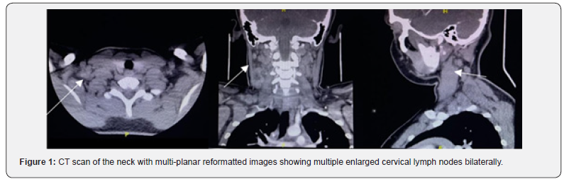
A 14-year-old male patient, previously healthy, presented to the out-patient clinic with 2 months history of neck swelling, that was gradually increasing. On Physical examination, he was conscious, alert and fully oriented. His vitals were within normal range. He had multiple enlarged cervical lymph nodes of 2x1 cm. Laboratory studies showed white count level of 5.4x10⁹ mmol/L with high lymphocyte level of 55.6% and monocyte level of 13.2%, a hemoglobin level of 140 g/L, a platelet level of 325 g/L, a creatinine level of 58 mmol/L, a sodium level of 138 mmol/L and potassium level of 4.6. CT scan showed multiple bilateral discrete and amalgamated cervical lymph nodes at multiple levels with mild stranding of the surrounding fat planes, the largest lymph node amalgam is noted on the right-side measuring about 16 x 13 x 23 mm at level III and 12 x 11 x 25 mm at level V (Figure 1). No significant related overlying skin thickening, no evidence of areas of cystic degenerations. No abscess formation at time of examination. No calcifications or areas of abnormal post contrast enhancement. No significant mediastinal or hilar lymph nodes enlargement at time of examination. Ultrasound of neck showed thyroid gland of average size and echo pattern no definite focal lesion or nodule. Bilateral multiple cervical lymph node more prominent on the right side posterior cervical space, largest about 2.2x0.8 cm at that region, with attenuated fatty hilum. Bilateral axillary small lymph node with attenuated fatty hilum on each side measuring 0.8x0.4 cm and 0.8x 0.6 cm on the right side, with attenuated fatty hilum. Guided fine needle aspiration was attempted from right posterior cervical lymph node measuring 1.5x1 cm reduced fatty hilum. Smears show polymorphous population of lymphoid cells with few plasma cells and lymphohistiocyst entangling tangible body macrophages.
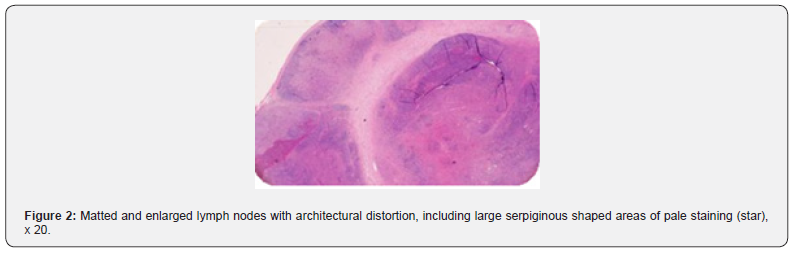
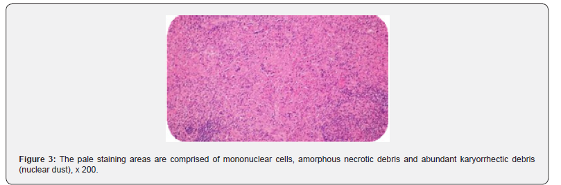
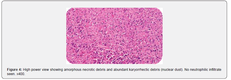
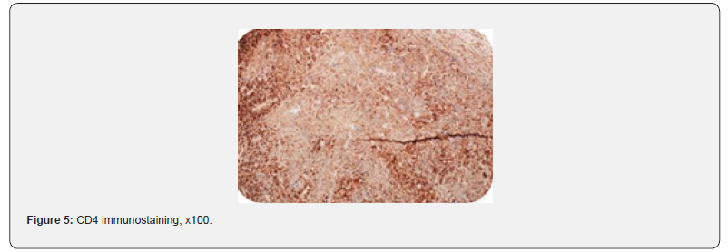
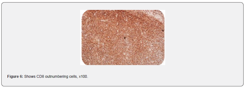
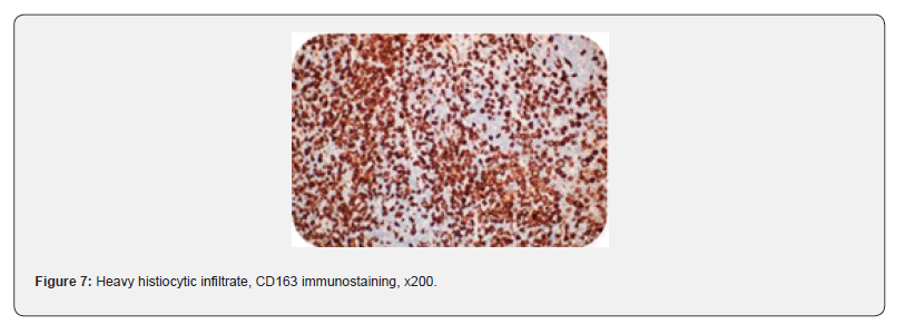
So, the patient underwent lymph node excisional biopsy under general anesthesia. The operation started with palpation which identified multiple lymph node in posterior triangle followed by skin incision along skin crease line. Dissection of subcutaneous fat or layer until lymph node was identified, Spinal accessory nerve was identified and preserved. Excision of multiple lymph nodes was done and were sent for histopathology. Post operatively, patient was doing well and was discharged with oral analgesia and Augmentin for 7 days. The histopathology result showed multiple matted lymph nodes featuring partially effaced architecture. The paracortex is expanded by numerous immunoblots admixed with T-lymphocytes (CD8+>CD4+), some of which features crescentic nuclei or xanthomatous morphology. The lymph nodes exhibit patchy large foci of necrosis within the paracortex. The necrosis contains no neutrophils, has abundant karyorrhectic nuclear debris, and is surrounded by a mixture of histiocytes, immunoblots, and T-lymphocytes as well as scattered foamy histiocytes. No hematoxylin bodies are seen. Special stains used showed negative result for bacteria, fungi, or acid-fast bacilli, respectively. By immunohistochemistry, the infiltrate is composed mainly of CD3 positive T cells, with CD8 positive cells outnumbering CD4 positive cells. CD30 highlights immunoblots and is negative for atypical cells. CD163 highlights abundant histiocytes. CD21 and CD23 highlight focally preserved follicular dendritic cell meshwork. MPO highlights histiocytes only. ISH for EBV and CMV is negative (Figure 2-7).
Discussion
Anatomy
Lymph nodes are kidney-shaped and receive lymph via multiple afferent vessels, and filtered lymph then leaves via one or two efferent vessels. Nodes typically have an associated artery and vein, which terminates into a high endothelial venule (HEV). The HEV is the site of trans-endothelial migration of circulating lymphocytes due to T and B-cell endothelial surface receptors. Lymph nodes usually range in size from 1 to 2cm and are enclosed in an adipose tissue capsule. Normal size depends upon location, as well as the axis which is being measured. The long axis should be 1 cm or less. They are considered pathological if they lose their oval shape, if there is a loss of the hilar fat, if there is an asymmetrical thickening of the cortex and if they are persistently enlarged. Lymph node structure consists of capsule, subscapular sinus, cortex and medulla [2].
Physiology
The primary function of lymph nodes is filtering interstitial fluid collected from soft tissues and eventually returning it to the vascular system. Filtering this exudative fluid allows for exposure of T-cells and B-cells to a wide range of antigens. For antigenspecific B and T cells to activate, they must first suffer exposure to antigens with the aid of antigen-presenting cells, dendritic cells, and follicular dendritic cells. These form part of both the innate immune response and play a role in adaptive immunity.
Pathogenesis
The lymphatic system is involved in infective, inflammatory, and malignant diseases, and as such, enlargement of lymph nodes can be attributed to multiple causes. In the case of lymphadenopathy of unclear origin, endoscopic ultrasound (EUS) with or without fine-needle aspiration (FNA) can aid in diagnosis [3]. For Kikuchi- Fujimoto disease, histology following lymph node dissection is critical to diagnosis: typical paracortical lesions with confluent zones of necrosis, atypical mononuclear cells, T-lymphocytes and histiocytes without granulocytes are exhibited. The lymph nodes are usually affected bilaterally, located in the posterior cervical triangle and are hard and tender [1,4].
Etiology and differential diagnosis
Causes can differ from Infectious, Immunologic to malignancies. Infectious could be: Viral, bacterial or Fungal. Viral like CMV, hepatitis or influenza. Bacterial: could be acute case like staphylococcus, Beta hemolytic group A streptococcus or Hemophilus influenza type B; or chronic case like in tuberculosis or brucella (brucellosis). Fungal like Histoplasmosis, coccidioidomycosis, blastomycosis and tinea are fungal infections caused by environmental contamination through the skin or the respiratory tract. Could be Immunologic in cases of granulomatous diseases like sarcoidosis, hyper -IgM syndrome or chronic granulomatous diseases. Could also be seen in rheumatic diseases like juvenile idiopathic arthritis or SLE. Could be seen in lymphoproliferative and histiocytic disorders like sinus histiocytic, Langerhans cell histiocytosis, hemophagocytic syndrome, Kikuchi-Fujimoto disease or PFAPA syndrome (a periodic fever, aphthous stomatitis, pharyngitis and cervical (lymph) adenitis). In addition to metabolic cases like in storage diseases including Neimann-Pick disease or Gaucher’s disease; or in cases of hypersensitivity such as serum sickness. Lastly, it could be in neoplastic cases which includes malignant lymphomas such as non- Hodgkin or Hodgkin’s lymphoma, and cases of leukemia such as case of acute lymphoblastic leukemia or AML. It could also be metastasis from nearby carcinoma like nasopharyngeal carcinoma or neuroblastoma [3].
Clinical features and presentation
Depending on the cause of lymph node enlargement, clinical features may present with differences and similarities. Similarities seen in most cases are fever, swelling/enlarged lymph nodes and pain. Similarities seen in infectious diseases such as viral, bacterial or fungal includes diarrhea, fever, or upper tract symptoms. Similarities seen in majority of malignant cases are B symptoms including fever, drenching night sweats, and unintended weight loss. The clinical manifestations of KFD in children are diverse. In some studies, fever, tenderness in the lymph node and leukopenia were more common in children with KFD. Other uncommon manifestations included rash, splenomegaly, hepatomegaly, oral ulcer and joint pain. Most children have lymphadenopathy localized in the cervical region. Extra-cervical lymphadenopathy was not uncommon [3,4].
Treatment
For Kikuchi-Fujimoto disease, therapeutic options are limited to symptomatic treatment (lowering fever, analgesia, intake of fluids), or surgical excision for biopsy. Since treatment guidelines for KFD have not been developed and recommendations are mostly derived from experts’ opinions, case reports, and case series. Most important step is to eliminate the underlying infection which is the mainstay of the treatment. The treatments are mainly adapted to the relevant pathogens. Viral infections mainly get supportive therapy, while bacterial infections are given antibiotics based on the pathogen; but if patients are immunocompromised treatment should be given, antiviral/ antibiotics. For immunologic cases, therapy depends on the clinical symptoms and predominantly treats the associated autoimmune or malignant disorders. Usually, first choice of therapy is corticosteroids or immunoglobulins. While for malignant cases, Chemotherapy or radiotherapy is given according to the stage and entity of the disease [3].
Conclusion
This case highlights the importance of proper examination, especially of lymph node and use of different diagnostic modalities for the exact diagnosis of disease. KFD is a rare disease that can occur in various ethnic groups, typically presenting with cervical lymphadenopathy and fever. While its treatment is still not developed, it’s important to identify it to treat symptoms [5].
References
- Vikram K Mahajan, Vikas S, Neeraj S, Ritu R (2023) Kikuchi-Fujimoto disease: A Comprehensive Review. World J Clin Cases 11(16): 3664-3679.
- Bujoreanu I, Gupta V (2023) Anatomy Lymph Nodes.
- Stephan L, Benjamin K (2014) Cervical lymph node diseases in children. GMS Curr Top Otorhinolaryngol Head Neck Surg 13: 8.
- Liu B, Sun Y, Shi WY, Hu B, Guo LY, et al. (2025) Clinical characteristics and risk factors for recurrence of Kikuchi-Fujimoto disease in children: a case-control study. BMC Pediatr 25(1): 485.
- Pitchaporn Y, Nattanicha C, Miriam P, Alexandra H, Kuldeep L, et al. (2025) Case Report: Two Cases of Kikuchi Disease in West Texas With Non-Self-Limited Disease. J Investig Med High Impact Case Rep.

























