Overlooked, Rare, but Curable Cause of Severe Snoring for Decades in a Sexagenarian
Sangeet Kumar Poddar1*, John Victor2 and Panna P Shetty3
1ENT Consultant, NMC Speciality Hospital, Abu Dhabi, UAE
2ENT Consultant, NMC Speciality Hospital, Abu Dhabi, UAE
3Pathology Specialist, NMC Royal Hospital, Abu Dhabi, UAE
Submission: January 12, 2024; Published: February 08, 2024
*Corresponding author: Sangeet Kumar Poddar, ENT Consultant, NMC specialty Hospital, Abu Dhabi, UAE
How to cite this article: Sangeet Kumar Poddar*, John Victor and Panna P Shetty. Overlooked, Rare, but Curable Cause of Severe Snoring for Decades in a Sexagenarian. Glob J Oto, 2024; 26 (4): 556193. DOI: 10.19080/GJO.2024.26.556193
Abstract
Purpose: To report a case of rarely encountered but curable cause of severe snoring overlooked by various physicians and the patient herself.
Case report: An elderly female patient complaining of severe snoring and choking in sleep was evaluated and was found to be suffering from a rare pathology - Epidermoid cyst of floor of mouth. She underwent surgical excision and was completely cured of her complaints.
Conclusion: Early diagnosis and surgical intervention would have cured this patient many years back from a rare cause of severe snoring.
Keywords: Snoring; Cyst; Epidermoid; Neck swelling; MRI
Introduction
Obstructive pathologies of the upper airway leads to snoring and if severe, sleep apnea. The obstruction can be at multiple levels in the upper airway tract necessitating a thorough evaluation to localize the site of obstruction. Mild or occasional snoring usually isn’t a cause for concern unless associated with excessive daytime sleepiness, choking in sleep and gasping for breath with sudden wakefulness - a condition known as obstructive sleep apnea. In obstructive sleep apnea, the risk of certain serious diseases like metabolic disorders, stroke and myocardial infarction are high. Ignoring it as just a trivial symptom and not seeking medical consultation is very common in society.
We present here a case where the concerned patient in her 60’s and probably the oldest patient suffering from this rare condition did not seek medical attention for decades and unfortunately whenever she went for any medical consultation for other conditions, a significant submental swelling was overlooked. The patient also thought of it as double chin and ignored it. Only when her snoring was getting louder, and she started facing difficulty in sleeping due to choking in sleep did she come to us for consultation.
Case Report
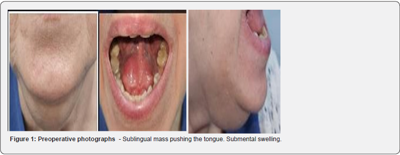
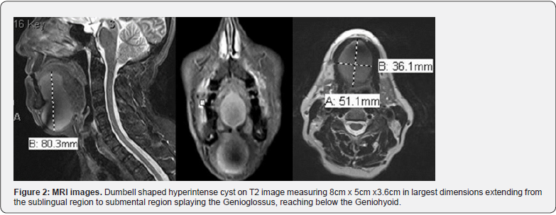
A 63-year-old female patient presented with complains of snoring for decades, worsening over time and past few months history of choking in sleep. She didn’t have any other complaints like nasal obstruction, recurrent cold, Post nasal drip etc. On examination a significant submental lump was seen. It was soft in consistency and non-tender. Oral examination revealed the tongue pushed supero-posteriorly by a huge soft, non-tender midline sublingual swelling (Figure 1). She didn’t give any history of previous surgery, trauma in mouth or neck region. There were no other neck lumps. MRI neck was advised which is of utmost importance [1] not only for the differential diagnosis but also for the surgical planning. MRI revealed- Midline thin-walled, well-defined dumbbell shaped cystic lesion in sublingual space splaying genioglossus muscle of tongue and extending into floor of mouth measuring 5.1 x 3.6cm. It was hypointense on T1, hyperintense in T2 and stir sequences (Figure 2). Differential diagnoses: 1)Ranula 2) Sublingual epidermoid cyst.
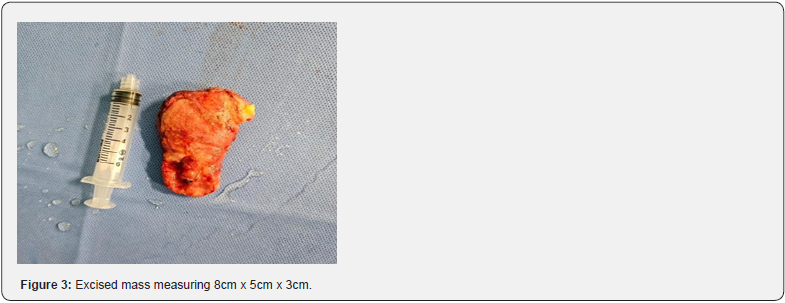
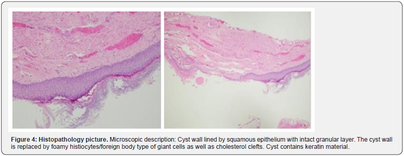
Sleep study was not considered as the diagnosis was straightforward about the obstruction of the oropharyngeal region due to, we removed the tumor through external route by a 5cm transverse submental incision exposing the tumor capsule and then dissecting all around it followed by finger dissection all around. It was a huge oblong mass (Figure 3) extending from the Mylohyoid muscle layer upward into the sublingual space between the genioglossus where there was narrowing giving the dumbbell appearance on MRI. The wound was closed in three layers after placing a corrugated rubber drain. The drain was removed on 3rd post op day and the sutures removed after 8 days. Histopathology reported the lesion as Epidermoid cyst (Figure 4). Healing was uneventful (Figure 5) and the patient’s snoring and choking in sleep has completely stopped.
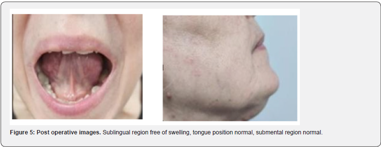
The present case report is made in view of mindset of ignoring health conditions in society unless it becomes really serious, rarity of the Epidermoid cyst in the floor of mouth, its huge size mimicking plunging Ranula and for surgical planning.
Discussion
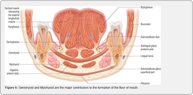
The floor of the mouth, also called “diaphragma oris,” includes the soft tissues between the medial aspect of the mandibular body and the hyoid bone. As such, it represents the inferior border of the oral cavity. Four of the six large salivary glands are associated with the floor of the mouth, i.e., the sublingual and submandibular glands. The main muscles contributing to the floor of the mouth include the mylohyoid and the geniohyoid muscles (Figure 6).
Dermoid cysts are slow-growing, benign developmental cysts that arise from ectodermal tissue and can occur anywhere in the body. Less than 7% of these cysts involve the head and neck region, with only 1.6% of cases presenting in the oral cavity [2-4]. They often remain asymptomatic for years until they grow enough to interfere with speech, deglutition and less often with breathing which can pose a critical risk to the airway and require immediate surgery [5]. The spectrum of Teratomas includes True dermoid cysts, epidermoid cysts, and teratoid cysts. The differentiation is on the basis of histopathological examination of the contents of the cyst. True dermoid cysts are cavities lined with epithelium showing keratinization and with identifiable skin on the cyst wall. Epidermic cysts are lined with simple squamous epithelium with a fibrous wall and no attached structures. The lining of teratoid cysts varies from simple squamous to a ciliate respiratory epithelium containing derivatives of ectoderm, mesoderm, and/or endoderm [6]. Treatment consists in complete surgical removal, trying not to rupture the cyst, as luminal contents may act as irritants to fibrovascular tissues, producing postoperative inflammation with excellent prognosis in cases free of complications [7]. The intraoral approach is preferred in sublingual located cysts, whereas the extraoral approach is preferred in submental and submandibular region located cysts [8].
Conclusion
Although rare, Epidermoid cyst should be considered as a differential diagnosis for sublingual swelling in midline. MRI is the most important investigation for extent of the lesion and surgical planning. Complete excision is the curative treatment and histopathology confirms the diagnosis.
References
- TJ Vogl, W Steger, S Ihrler, P Ferrera, G Grevers (1993) Cystic masses in the floor of the mouth: value of MR imaging in planning surgery. AJR Am J Roentgenol 161(1): 183-186.
- HB Santos, LS Rolim, CC Barros, IL Cavalcante, RD Freitas, et al. (2020) Dermoid and epidermoid cysts of the oral cavity: a 48-year retrospective study with focus on clinical and morphological features and review of main topics. Med Oral Patol Oral Cir Bucal 25(3): e364-e369.
- Koca H, Seckin T, Sipahi A, Kazanc A (2007) Epidermoid cyst in the floor of the mouth: Report of a case. Quintessence Int 38(6): 473-477.
- Surekha RP, Rudrayya SP, Satya Prakash, M Bimba (2016) Epidermoid cyst: Report of two cases. J Oral Maxillofac Pathol 20(3): 546.
- Brunet-Garcia A, Lucena-Rivero ED, Brunet-Garcia L, Faubel-Serra M (2018) Cystic mass of the floor of the mouth. J Clin Exp Dentistr 10(3): e287-e290.
- Meyer I (1955) Dermoid cysts (dermoids) of the floor of the mouth. Oral Surg Oral Med Oral Pathol 8(11): 1149-1164.
- Dimov Z, Dimov K, Krstev N, Krstev D, Baeva M, et al. (2000) Dermoid, epidermoid and teratoma cysts of the tongue and oral cavity floor. Khirurgiia (Sofiia) 56(2): 30-32.
- Akao I, Nobukiyo S, Kobayashi T, Hitoshi K, Izumi K (2003) A case of large dermoid cyst in the floor of the mouth. Auris Nasus Larynx 30: S137-S139.


