Clarification for the Differences Between Microfat and Nanofat
David Alessi*, Katrin Salehi and Ava Simpson
Alessi Institute Bevery Hills, Cedars Sinai Medical Center, USA
Submission:September 27, 2022;Published:October 07, 2022
*Corresponding author: David Alessi, M.D, Alessi Institute Bevery Hills, Cedars Sinai Medical Center, USA
How to cite this article: David A, Katrin S, Ava S. Clarification for the Differences Between Microfat and Nanofat.Glob J Oto, 2022; 25 (3): 556164. DOI: 10.19080/GJO.2022.25.556164
Abstract
Microfat and Nanofat are both autologous adipose-derived grafting methods that have clear differences in characteristics. As later discussed, we mention why the terms should be used in a standardized fashion with each term reserved for that specific fat grafting process. Nanofat has no live adipose cells and no volumizing qualities and Microfat the opposite.
Keywords: Newborn hearing; Screening; Hearing loss; Children; Disability
Introduction
Fat grafting for cosmetic filling and facial reconstruction has been around since the late 1800s. In recent years, the process has been refined by using finer and more processed adipose injections in multiple tissue layers if possible [1]. This has been associated with an increase of survival rates. This is due to the surgical excision and low negative-pressure aspiration with large-bore cannulas minimizing adipocyte damage during fat grafting [2]. It is generally accepted that a less traumatic approach to harvesting and processing the fat will lead to better outcomes. The use of stem cells in cosmetics and reconstruction is rapidly becoming popular in fat grafting. More specifically, the use of adipose stem cells for the treatment of scars [3]. Obtaining regenerative cells from adipose tissue can be done in two ways: Enzymatic and mechanical [4].
Copcu makes an argument for abandoning the use of Nanofat. We disagree with this for the aforementioned reasons. Moreover, the use of Nanofat is becoming popular and use of the Nanofat/ Microfat terminology is widespread. The issue is the occasional misunderstanding of the terms in the literature. However, there seems to be confusion with the use of Nanofat versus Microfat as seen in the paper by Verpaele for the use to fill in a lip scar defect [5]. We use both adipose-derived stem cells (ADSC) and extensively processed fat frequently in our clinic. It appears the use of Nanofat isn’t standardized. The highly processed fat can be injected with fine cannulas for small corrections which we call Micro Fat injections. Our use for the term Nanofat is for the injection of ADSC. The following will describe the differences between Nanofat and Microfat and the reasoning for the standardization of nomenclature.
Materials and Methods Micro fat
Microfat is small clumps of fat that are derived from lipoaspirate. The technique used for Microfat in cosmetic filling or facial reconstruction can be used as an isolated procedure or combined with another one [6]. Survival rates and cosmetic outcomes for Microfat injections is better with multiple layers of fat injection as opposed to a large clump of Microfat in a single location. It is broken down so that it may be injected with fine cannulas. Our process does not use collagenase but is simply a method to mechanically break down the aspirate into usable material the particles injected are well above the upper limit of Nanoparticles (100 Nanometers in diameter).
Our method is as follows:
a) 200cc to 500cc of tumescent solutions (50cc of lidocaine with epinephrine in 500cc of normal saline) is infiltrated into the anterior abdominal fat pad or the thighs.
b) 50 to 150cc of lipoaspirate is removed using a 1.5mm Byron harvesting cannula under hand pressure. Mechanical liposuction is avoided.
c) Gravity drainage is used to remove the tumescent fluid and oily layer (occasionally the aspirate is centrifuged if there is not adequate gravity separation).
d) The fat aspirate is broken down with Tulip sizers (sequential fragmenters) until it can pass through a 22-gauge blunt tip needle. (Figure 1) shows the appearance of the Microfat before injection.
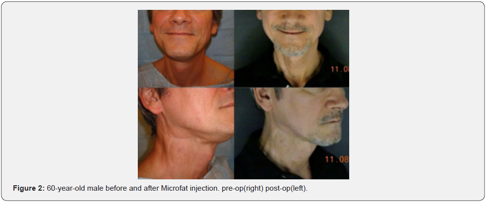
e) The fat is then injected into the predetermined area (soft tissue defect). (Figure 2) shows a patient treated with Microfat.


Nanofat
Nanofat is made when the ADSC is processed from the lipoaspirate with all the adipose cells removed. Using condensed Nanofat combined with fat grafts in a novel technique to improve atrophic facial scars by raising both the surface and the bottom of the affected area is the best way to use nanofat [7]. The use of Nanofat on scars has both a cosmetic improvement but has been show histologically to improve the dermal layer. It has no filling qualities and is useful for the stimulation of collagen and elastic and useful in scar treatment and facial rejuvenation. It is the remnant of the lipoaspirate consisting of the stromal vascular fraction along with cytokines, epidermal growth factors, collagen stim, and intracellular hormones [5].
Our method is as follows:
a) 200cc to 500cc of tumescent solutions (50cc of lidocaine with epinephrine in 500cc of normal saline) is infiltrated into the anterior abdominal fat pad or the thighs
b) 50 to 150cc of lipoaspirate is removed using a 1.5mm Byron harvesting cannula
c) Gravity drainage is used to remove the tumescent fluid and oily layer (occasionally the aspirate is centrifuged if there is not adequate gravity separation).
d) The fat aspirate is broken down with Tulip sizers. (Sequential fragments) until it can pass through a 22-gauge blunt tip needle.
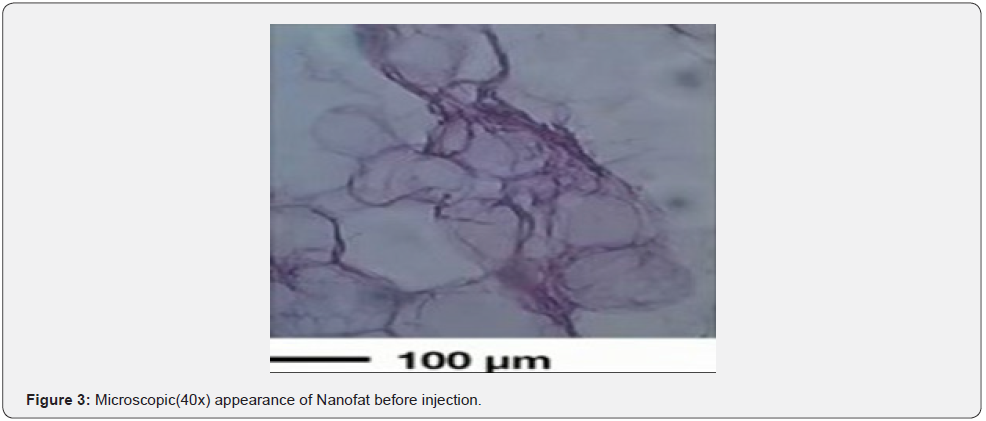
e) The processed liquid Microfat is then placed through a filter device (Tulip) to generate a Nanofat liquid. (Figure 3) shows the appearance of the Nanofat before injection.
f) The Nanofat is then injected into the predetermined area via a 27-gauge needle in a mesotherapy technique. (Figure 4) shows a picture of a patient with burn scars before and after Nanofat.


Discussion
Microfat is a term that should only be reserved for adipocytecontaining volumizer. This lipoaspirate is broken down into a fairly thin liquid that can be injected via a 22-gauge needle or smaller. Micro fat is a filler and contains live adipose cells. Nanofat is a term that should only be reserved for lipoaspirate broken down into a fairly thin liquid that has NO adipose cell and is the stromal vascular fraction of the lipoaspirate. It has NO volumizing qualities and serves as a regenerative modality. Nanofat is the residual of processed lipoaspirate that is broken down into Microfat. The Nanofat is isolated when the adipocytes are removed from Microfat. The resultant stromovascular fraction contains different types of cells such as endothelial cells, monocytes, macrophages, granulocytes and lymphocytes.
The stromal vascular fraction also includes a substantial amount of mesenchymal stem cells (adipose-derived stem cells). These multipotent stem cells have the ability to form fibroblastlike colonies in vitro. Unlike other sources of multipotent stem cells, these multipotent stem cells are found in high quantities in lipoaspirate. Adipose-derived stem cells have an extensive proliferative capacity and the ability to differentiate into other mesenchymal cells. Other terms use for adipose-derived non-augmenting biologics include adipose-derived stem cells ADSC and adipose-derived exosomes. True nanoparticles have sizes from 1 to 100 nanometer portions of the stromal vascular fraction fit that size criteria [8]. For example, enzymes typically range from 100 to 1000nm.
Many peptides fit into this nanometer range. With Nanofat, even though all of the components are not nanoparticles, several are. Therefore, we feel comfortable with the term Nanofat [4]. The impact of Nanofat on skin rejuvenation is not entirely understood. Several studies imply the potential effectiveness of Nanofat [4,9,10]. Long-term follow-ups on multiple patients that have undergone autologous fat injections showed that the absorption rate varies considerably in each individual case but was estimated to be 40-60% of the injected fat. Furthermore, the long-term follow-ups proved that with final corrections after two or more repetitions of fat injections the longevity of the injections persisted for many years with one of the longest being proved to be more than 12 years [11,12].
Additionally, many issues come to play a role in the clinical longevity of correction after autologous fat transfer/injections and it is less dependent on the harvesting and reinjection methods. The degree of augmentation is a determining factor which results from the amount of fibrosis induced along with the number of viable fat cell grafts. That number also depends on the anatomic site, the mobility and vascularity of the recipient tissue or any underlying causes and diseases [13].
Conclusion
We propose that the terms Nanofat and Microfat be used in a standardized fashion. Moreover, we anticipate further studies will delineate the effectiveness of these modalities.
References
- Riccardo F, Isabella C (2013) The Fascinating History of Fat Grafting. J Craniofac Surg 24(4): 1069-1071.
- Simonacci F, Bertozzi N, Grieco MP, Grignaffini E, Raposio E (2017) Procedure, applications, and outcomes of autologous fat grafting. Ann Med Surg 20: 49-60.
- Stachura A, Paskal W, Pawlik W, Mazurek M, Jawoeowski J (2021) The Use of Adipose-Derived Stem Cells (ADSCs) and Stromal Vascular Fraction (SVF) in Skin Scar Treatment-A Systematic Review of Clinical Studies. J Clin Med 10(16): 3637.
- Zhang B, Lai CR, Wei KS, Andre BHC, Ellen BL, et.al. (2021) Topical Applications of Mesenchymal Stem Cell Exosomes Alleviates the Imiquimod Induced Psoriasis-Like Inflammation. Int J Mol Sci 22(2): 720.
- Verpaele A, Tonnard P, Jeganathan C, Ramaut L (2019) Nanofat Needling: A Novel Method for Uniform Delivery of Adipose-Derived Stromal Vascular Fraction into the Skin. Plast Reconstr Surg 143(4): 1062-1065.
- Trepsat F (2009) Midface reshaping with micro-fat grafting. Ann Chir Plast Esthet 54(5): 435-443.
- Gu Z, Li Y, Li H (2018) Use of condensed nanofat combined with fat grafts to treat atrophic scars. JAMA Facial Plast Surg 20(2): 128-135.
- Verma D, Gulati N, Shreya K, Mukherjee S, Nagaich U (2018) Protein Based Nanostructures for Drug Delivery. J Pharm (Cairo) 2018: 9285854.
- Zhou Y, Bo Zhao, Xin-Liao Z, Yi-Jun L, Shou-Tao L, et. al. (2021) Combined topical and systemic administration with human adipose-derived mesenchymal stem cells (hADSC) and hADSC-derived exosomes markedly promoted cutaneous wound healing and regeneration. Stem Cell Res Ther 12(1): 257.
- Tonnard P, Verpaele A, Geert P, Moustapha H, Maria C, et. al. (2013) Nanofat Grafting Basic Research and Clinical Applications: Plast Reconstr Surg 132(4): 1017-1026.
- Dasiu-Palkida D (2003) Fat injections for facial rejuvenation: 17 years experience in 1720 patients. J Cosmet Dermatol 2(3-4) :119-125.
- Copcu H, Oztan S (2021) Not Stromal Vascular Fraction or Nanofat: Total Stromal Cells (TOST): A New Definition. Systemic Review of Mechanical Stromal-Cell Extraction Techniques. Tissue Eng Regen Med 18(1): 25-36.
- Sommer B, Sattler G (2000) Current concepts of fat graft survival: histology of aspirated adipose tissue and review of the literature. Dermatol Surg 26(12): 1159-1166.


