A Rare Case of Recurring Ossifying Fibroma of the External Auditory Canal
Sarthak Sachdeva1*, Manisha Yadav1*, Vinod Kumar Arora2, Richa Gupta2, Vipin Arora1 and Akanksha Agarwal2
1Department of Otorhinolaryngology, University College of Medical Sciences and GTB Hospital, India
2Department of Pathology, University College of Medical Sciences and GTB Hospital, India
Submission:April 22, 2021; Published: May 14, 2021
*Corresponding author:Sarthak Sachdeva and Manisha Yadav, Junior resident, Department of Otorhinolaryngology, University College of Medical Sciences and GTB Hospital, Delhi, India
How to cite this article: Sarthak Sachdeva, Manisha Yadav, Vinod Kumar Arora, Richa Gupta, Vipin Arora, Akanksha Agarwal. A Rare Case of Recurring Ossifying Fibroma of the External Auditory Canal. Glob J Oto, 2021; 24 (3): 556138. DOI: 10.19080/GJO.2021.24.556138
Abstract
Fibro-osseous lesions are a group of disorders that can arise from any part of the facial skeleton and include ossifying fibroma, fibrous dysplasia, giant cell lesions etc. Almost 70 percent of these lesions arise in the head and neck. The most common sites affected are maxilla and mandible. Occasionally, they have also been reported in orbitofrontal bone, paranasal sinuses, nasopharynx, and skull base. We report a rare case of recurring ossifying fibroma of the external auditory canal.
Keywords: Benign fibro osseous lesion; EAC; Ossifying fibroma
Introduction
Ossifying lesions in the External auditory canal (EAC) are most commonly Exostosis and Osteoma. Exostoses comprise the most common neoplasms of the EAC and are mainly caused by local irritation in the ear [1]. Although exostosis is most common ossifying lesion of EAC, but its overall incidence is merely 0.6%. Osteomas, which are rarer than exostosis, are usually single, unilateral, squamous bone lesion located laterally with respect to isthmus [2]. Apart from these, exceedingly rarely ossifying fibromas and monostotic fibrous dysplasia have also been reported and are collectively termed as Benign fibro-osseous lesions of the EAC [3]. Here, we describe a rare case of ossifying fibroma of the EAC which has some histopathological and clinical features differentiating it from the earlier reported cases, and difficulty encountered while managing it because of the high incidence of recurrence after primary excision.
Case Presentation
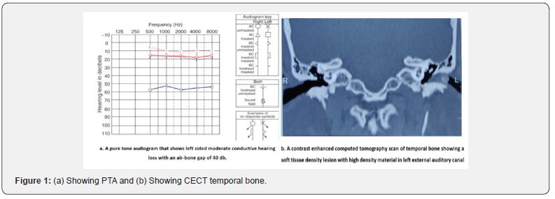
A 50-year-old male presented with occasional otalgia and persistent hearing impairment for six months. Patient had a history of head trauma 1.5 years back. Otoscopy revealed an extensive skin covered firm mass completely filling his left EAC. Pure tone audiogram (PTA) showed a 60 dB hearing loss and an air–bone gap of 40dB in the left ear (Figure 1a).

High-resolution CECT of the temporal bone showed a minimally enhancing soft-tissue density lesion in the EAC measuring 1.7x0.7x0.3cm (tr*cc*ap) with central osteoid differentiation measuring 8mm. This pedunculated lesion was seen arising from the antero-inferior wall of the lateral most point of the bony EAC medially reaching up to the tympanic membrane and laterally up to the external orifice of the bony external auditory canal. A small lytic defect was also noted in the anterior bony auditory canal. No e/o periosteal reaction was seen. The ossicle chains were intact, and the middle ear cavity was free (Figure 1b).
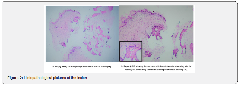
An excisional biopsy of the mass was carried out under local anesthesia, leaving the skin under the mass and the eardrum intact. Histopathological examination revealed osteoma like bone formation with islands of bony trabeculae and sparse osteoblastic areas in a cellular fibrous stroma (Figure 2a). Minimal ‘osteoblastic rimming’ was also seen at the advancing front of the bony islands in surrounding soft tissues and dermis (Figure 2b). Subsequently, there was an improvement in hearing loss postoperatively, but the hearing loss recurred over the next few months. An endoscopic examination of the EAC showed recurrence of similar mass which was further confirmed by radiological examination suggesting the need of a more robust procedure with complete removal and large canaloplasty for this patient.

Discussion
Fibro-osseous lesions form a diverse group of conditions with similar histology but varying etiologies. They are lesions with misshaped trabeculae of compact bone embedded in the surrounding connective tissue. They seem to abruptly arise over the healthy bone [4]. Benign fibro-osseous lesions include non-neoplastic lesions such as fibrous dysplasia and neoplastic lesions such as ossifying fibroma, giant cell tumor, osteoma, osteoblastoma, and aneurysmal bone cysts [3]. These lesions need to be differentiated so that the aggressiveness of the lesion can be anticipated, and the surgeon can choose from a plethora of treatment options ranging from simple enucleation to complete excision.
Biopsy done from our case shows islands of bony trabeculae in a fibrous stroma with infiltration into surrounding soft tissue and dermis with minimal activity and occasional ‘osteoblastic rimming’ over the margins of advancing bony trabeculae (Figure 2a & 2b). These findings are consistent with benign fibro-osseous lesions, possibly, ossifying fibroma. Pertaining to the rarity of ossifying fibroma, it may be misdiagnosed as a more common condition like osteoma. This is because they share similar histopathological features with very minute differentiating characters and their diagnosis requires clinical and radiological correlation as well as good knowledge of subtle histological differences (Table 1).

Ossifying fibroma is a benign fibro-osseus lesion that consists of conventional ossifying fibroma and juvenile ossifying fibroma. Conventional ossifying fibroma could be ossifying, cementifying or storiform form. Juvenile ossifying fibromas are aggressive and further divided into trabecular and psammomata varieties [5,6]. Our histopathological findings show greater similarity to conventional ossifying fibroma which in literature has been known to show irregular lamellar bony trabeculae with hypercellular fibrous stroma. There is no atypia of fibroblastic cells and there is no mitosis. Mature lesions show minimal osteoblastic rimming [7].
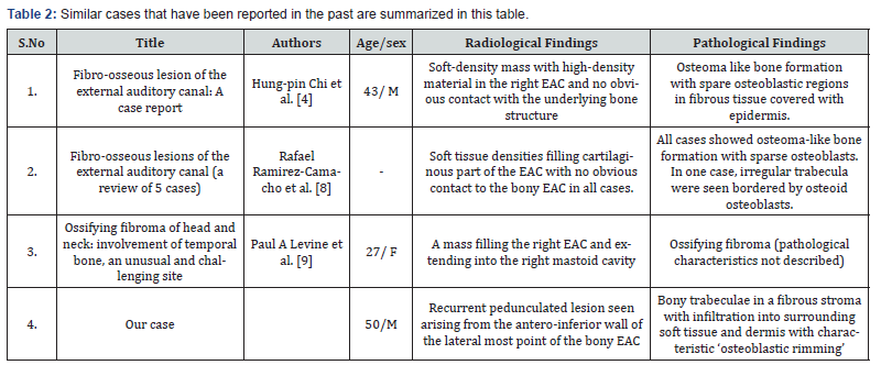
Similar cases that have been reported in the past are summarized in Table 2. The current case is unique as it was observed that the pedunculated mass occupied the bony part of the EAC only rather than cartilaginous EAC which is reported by Ramirez et al. [8] in his series of 5 cases. Our case histopathology also showed characteristic ‘osteoblastic rimming’ which has not been described in previous studies. Although benign but if left untreated they may locally destruct the bony EAC, temporomandibular joints, middle ear, mastoid cavity etc. and manifest in the form of various symptoms associated with it [9]. Malignant transformation is not reported in the literature. The various treatment modalities available for such lesions are enucleation, curettage, and resection with reconstruction. Recurrence rates of up to 28% have been reported in literature [10]. Recurrent cases of ossifying fibroma have been found to be larger in size and with an extensively altered radiographic appearance as it is believed that surgery can reactivate the growth of lesion [10]. Hence the preferred modality of treatment in such lesions should be complete excision, canaloplasty and reconstruction.

Conclusion
This is a rare case report of recurring ossifying fibroma of EAC which probably developed from a progressive inflammatory reaction with fibrosis, osseous metaplasia, and eventual calcification. Such patients require a complete removal of all tissue along with a large canaloplasty and reconstruction to correct the conductive hearing loss. Also, a long-term follow-up of these patients is essential as recurrences can occur up to 10 years following treatment.
References
- Graham MD (1979) Osteomas and exostoses of the external auditory canal: A clinical, histopathologic and scanning electron microscopic study. Ann Otol Rhinol Laryngol 88(4): 566-572.
- Kline OR PR (1954) Osteoma of External Auditory Canal. Arch Otolaryngol Head Neck Surg (59): 588-593.
- Atilla S, Pabuscu Y, Celasun B, Gerek M, Pocan S (1998) Radiology of an atypical fibro-osseous lesion confined to the nasal cavity. Neuroradiology 40(11): 752-754.
- Chi HP, Ho KY, Tsai KB, Lee KW, Ta CF, et al. (2004) Fibro-osseous Lesion of the External Auditory Canal: A Case Report. Kaohsiung J Med Sci 20(1): 31-34.
- Slootweg PJ, Panders AK, Koopmans R, Nikkels PGJ (1994) Juvenile ossifying fibroma. An analysis of 33 cases with emphasis on histopathological aspects. J Oral Pathol Med 23(9): 385-388.
- Williams HK, Mangham C, Speight PM (2000) Juvenile ossifying fibroma. An analysis of eight cases and a comparison with other fibro-osseous lesions. J Oral Pathol Med 29(1): 13-18.
- Slootweg PJ (1996) Maxillofacial fibro-osseous lesions: classification and differential diagnosis. Semin Diagn Pathol 13(2): 104-112.
- Liu Y, You M, Wang H, Yang Z, Miao J, et al. (2010) Ossifying fibromas of the jaw bone: 20 cases. Dentomaxillofacial Radiol 39(1): 57-63.


