Extranasopharyngeal Angiofibroma of Nasal Septum: A Rare Clinical Presentation at an Unusual Site
Humra Shamim, Kamlesh Chandra and Mehtab Alam*
Department of ENT, JNMCH, AMU, India
Submission: March 2, 2020; Published: March 17, 2020
*Corresponding author:Mehtab Alam, Assistant professor, Department of ENT, JNMCH, AMU, Aligarh, UP, India
How to cite this article: Mehtab Alam, Humra Shamim, Kamlesh Chandra. Extranasopharyngeal Angiofibroma of Nasal Septum: A Rare Clinical Presentation at an Unusual Site. Glob J Oto, 2020; 21(5): 556075 DOI: 10.19080/GJO.2020.21.556075
Abstract
Aim: Extranasopharyngeal angiofibroma is very rare diagnosis, it should be kept in mind as a differential diagnosis with any unilateral nasal mass causing nasal obstruction and epistaxis
Background: Nasopharyngeal Angiofibroma (NAs)is a benign tumor, consisting of fibrous tissue with varying degrees of vascularity, characterized by a proliferation of stellate and spindle cells around the blood vessels. It has been hypothesized that NAs is a testosterone-dependent tumor that arises from a fibrovascular nidus in the nasopharynx that lies dormant until the onset of puberty; hence the incidence is more in males with peak incidence between the ages of 14 and18 years. The term extranasopharyngeal angiofibroma (ENA) has been applied to vascular, fibrous nodules occurring outside the nasopharynx like invoving the septum, inferior turbinate’s, and maxillary sinus. The maxillary sinus is the most common site involved, while the nasal septum represents an extremely rare localization with only 6 cases having been reported till now
Case Description: We present a case of extranasopharyngeal angiofibroma originating from the nasal septum. A young male presented to the with an insidious onset and gradually progressive right sided nasal obstruction and recent presentation of a nasal mass along with few episodes of medically controlled minor nasal bleeds. After careful evaluation the case was taken up for surgery and was the mass was found out to be consistent with angiofibroma of the nasal septum.
Conclusion: Clinically, extranasopharyngeal angiofibroma’s, such as the one described in this case, fall into that category of odd presentations that do not fit within the accepted clinical parameters that one always expects to find in cases of NAs. Clinical Significance: Although extranasopharyngeal angiofibroma is very rare diagnosis, it should be kept in mind as a differential diagnosis with any unilateral nasal mass causing nasal obstruction and epistaxis.
Keywords: Extranasopharyngeal Angiofibroma of Nasal Septum: A Rare Clinical Presentation at an Unusual Site
Background
Nasopharyngeal Angiofibroma (NAs)is a benign tumor, consisting of fibrous tissue with varying degrees of vascularity, characterized by a proliferation of stellate and spindle cells around the blood vessels. It is originating from the pterygoid plate and the region of the sphenopalatine foramen. There are many theories trying to explain the pathology underlying this tumor, like genetic, hormonal, and developmental ones, but none of them had general acceptance. It has been hypothesized that NAs is a testosterone-dependent tumor that arises from a fibrovascular nidus in the nasopharynx that lies dormant until the onset of puberty; hence the incidence is more in males with peak incidence between the ages of 14 and18 years [1].
The term extranasopharyngeal angiofibroma (ENA) has been applied to vascular, fibrous nodules occurring outside the nasopharynx like invoving the septum, inferior turbinate’s, and maxillary sinus. The maxillary sinus is the most common site involved, while the nasal septum represents an extremely rare localization with only 6 cases having been reported in the international literature to date [2]. Besides the different location, typical clinical characteristics of ENA such as, symptoms, age, gender, do not conform to a great extent with that of NAs. These factors led to presumption whether ENA, though structurally similar, should be considered as being different from NAs [3]. Due to these differing features, ENA can present a diagnostic challenge and a meticulous evaluation with a high index of suspicion is essential in establishing the correct diagnosis and treatment. We present a case of extranasopharyngeal angiofibroma originating from the nasal septum which is a rare entity.
Case Description
A 38yr old male presented to the ENT OPD of JNMCH, AMU, Aligarh with an insidious onset and gradually progressive right sided nasal obstruction and recent presentation of a nasal mass. He also had few episodes of medically controlled minor nasal bleed from the same nostril. The patient had no previous history of trauma or infection. Anterior rhinoscopy showed a reddish mass filling the right nasal cavity and nasal septum deviation towards the left side. The mass could be probed all around except the medial side. Nasal endoscopy which revealed a reddish, well circumscribed non friable mass with no areas of surface bleed, adhering to the anterior part of septum. A contrast enhanced computerized tomography (CT) scan of the nose and paranasal sinuses showed a mild contrast enhancing mass arising from the septum and extending backwards up to the posteroinferior border of middle turbinate on right side of the nasal cavity. The mass was well circumscribed (Figure 1).


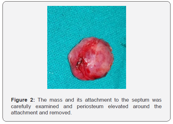
There was no extension beyond the nasal cavity into the nasopharynx or any paranasal sinuses and no bony erosions were noted. After careful evaluation the case was taken up for surgery (Figures 1 & 2) and block with minimal intra-op bleed and the excisional margin was cauterized. The rhinotomy incision was repaired with nonabsorbable sutures. The immediate and late post-operative recovery was uneventful.
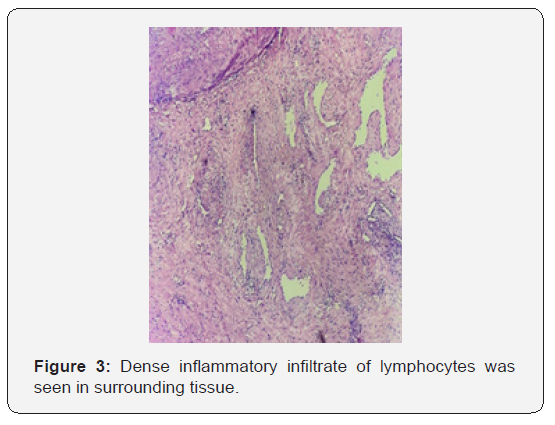
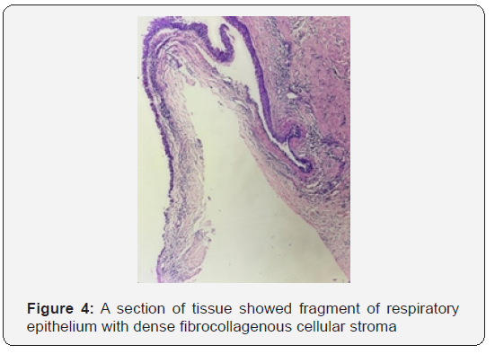
Histopathological examination was consistent with angiofibroma which revealed dense fibro collagenous stromal tissue with variable caliber of vessels, few vessels showed staghorn shape and were lined by flat endothelial cells (Figures 3 & 4).


Discussion
Angiofibroma was first described by Hippocrates in 5th century BC and Friedberg first used the term angiofibroma in 1940 [4]. The tumours virtually always arises from the nasopharynx and only later may extend into the nasal cavity [5]. It is histopathologically benign but potentially locally destructive vascular tumor. It is an un-encapsulated neoplasm composed of a rich vascular network lined by flat endothelial cells within a fibrous stroma [6]. In a review of 704 cases of nasopharyngeal angiofibroma by Tasca and Compadreti (2008), only 13 cases presented outside the nasopharynx suggesting that Extranasopharyngeal location is a rare possibility [2]. Compared to NAs, ENA have fewer features in common and the use of the term angiofibroma for these lesions may therefore be confusing. In fact, these rare, benign, neoplasms characterized by a different biological history and clinical features with respect to nasopharyngeal angiofibroma and, for these reasons, they should be regarded as a separate clinical entity.
Compared to NAs or juvenile nasopharyngeal angiofibroma’s (JNA), in ENA patients affected are older, may present in females, symptoms develop more quickly, and hypervascularity is less common [7]. The clinical manifestations depend on the site and extent of tumor. ENA of nasal septum commonly presents with nasal obstruction and nasal bleeding. The case described above presented with several atypical features like presentation in the late third decade of life, milder epistaxis episodes and lesser hypervascularity. The administration of a contrast agent in nasopharyngeal angiofibroma leads to a strong and usually homogeneous enhancement on CT and MRI T1 sequences [8]. On the other hand, ENA usually have mild enhancement of contrast, due to the frequent poor vascularity of the tumor [9]. In our case, the poor vascularity of the lesion allowed for easy removal without any significant intra operative blood loss. Surgical excision of the mass is the treatment of choice, and recurrence is rare [10].
Conclusion
Clinically, extranasopharyngeal angiofibroma’s, such as the one described in this case, fall into that category of odd presentations that do not fit within the accepted clinical parameters that one always expects to find in cases of NAs. Although extranasopharyngeal angiofibroma is very rare diagnosis, it should be kept in mind as a differential diagnosis with any unilateral nasal mass causing nasal obstruction and epistaxis.
Clinical Significance
The case presented here draws attention to the point that every case of nasal obstruction with epistaxis should be dealt with high index of suspicion and rare causes must be kept in mind while managing such patients.
Acknowledgement
The author is sincerely thankful to senior faculty members of the department for their support and valuable suggestions.
Ethical Approval
All procedures performed in the case reported were in accordance with the ethical standards of the institutional and/or research committee and with the 1964 Helsinki declaration and its later amendments or comparable ethical standards. The case report does not contain any studies with animals performed by any of the authors.
References
- Pillenahalli Maheshwarappa R, Gupta A, Bansal J, Kattimani MV, Shabadi SS, et al. (2013) An Unusual Location of Juvenile Angiofibroma: A Case Report and Review of the Literature. Case Rep Otolaryngol 2013.
- Tasca I, Ceroni Compadretti G (2008) Extranasopharyngeal angiofibroma of nasal septum. A controversial entity. Acta Otorhinolaryngol Ital 28(6): 312-314.
- Lucas RB (1976) Pathology of tumours of the oral tissues. 3rdEdn, Edinburgh: Churchill Livingstone, US.
- New GB, Erich JB (1941) Tumors of the nose and t Arch Otolaryngol 34(5):1039-1064.
- Harrison DF (1987) The natural history, pathogenesis, and treatment of juvenile angiofibroma. Personal experience with 44 patients. Arch Otolaryngol Head Neck Surg 113(9): 936-942.
- Huang RY, Damrose EJ, Blackwell KE, Cohen AN, Calcaterra TC (2000) Extranasopharyngeal angiofibroma. Int J Pediatr Otorhinolaryngol 56(1): 59-64.
- Akbas Y, Anadolu Y (2003) Extra Nasopharyngeal angiofibroma of the head and neck in women. Am J Otolaryngol 24: 413-416.
- Maroldi R, Borghesi A, Maculotti P (2005) CT and MR Anatomy of Paranasal Sinuses: Key Elements. In: Maroldi R, Nicolai P, editors. Imaging in Treatment Planning for Sinonasal Diseases. Berlin, Heidelberg: Springer Berlin Heidelberg9–27, Germany.
- Alvi A, Myssiorek D, Fuchs A (1996) Extranasopharyngeal angiofibroma. J Otolaryngol 25(5):346-348.
- Sarpa JR, Novelly NJ (1989) Extranasopharyngeal angiofibroma. Otolaryngol--Head Neck Surg Off J Am Acad Otolaryngol-Head Neck Surg101(6):693-697.


