Involvement of the External Auditory Canal by an Otherwise Discreet Nasopharyngeal Carcinoma Via A Spread by the Eustachian Tube
Ardhaoui H, Khdim M*, Douimi L, Abada R, Rouadi S, Roubal M and Mahtar M
ENT Department and Head and Neck Surgery, Ibn Rochd Teaching Hospital, Casablanca, Morocco
Submission: November 02, 2019; Published: November 14, 2019
*Corresponding author: Khdim M, ENT Department and Head and Neck Surgery, Ibn Rochd Teaching Hospital, Casablanca, Morocco
How to cite this article: Ardhaoui H, Khdim M, Douimi L, Abada R, Rouadi S, Roubal M, Mahtar M. Involvement of the External Auditory Canal by an Otherwise Discreet Nasopharyngeal Carcinoma Via A Spread by the Eustachian Tube. Glob J Oto, 2019; 21(2): 556057. DOI: 10.19080/GJO.2019.20.556057
Abstract
Nasopharyngeal tumors (NPT), mostly dominated by undifferentiated squamous cell carcinomas: UCNT type, have a variable incidence. NPT can extend within or out of the nasopharynx to the other lateral wall or to the oropharynx or postero-superiorly to the base of skull or to the nasal cavity. It then typically metastases to cervical lymph nodes. Cervical lymph adenopathy is the most common sign of call, beside otological or rhinological symptoms such as: otitis media, nasal obstruction, hearing loss, epistaxis or outright cranial nerve palsies. The following report is about an unusual clinical presentation of nasopharyngeal tumor. It describes a case of 25-year-old woman consulting for a tight and severe trismus, for whom clinical examination found a mass coming out of the external auditory canal (EAC) and further evaluation reveal a nasopharyngeal tumor.
Keywords: UCNT; Nasopharyngeal tumors; External auditory canal; Eustachian tube; Trismus
Introduction
Undifferentiated nasopharyngeal carcinoma (UCNT) is an epidermoid head and neck tumor related to the Epstein Barr Virus (EBV). It has a geographical endemic distribution among well-defined ethnic groups [1]. The classic triad of symptoms is a neck mass, nasal obstruction with blood tinged sputum, tinnitus or serous otitis. However, these symptoms rarely occur simultaneously [2]. Yet, because of its deep basicranial topography not very accessible to the examination, and despite a symptomatology rich but misleading, this cancer remains of late diagnosis.

This article describes a nasopharyngeal tumor revealed by a trismus and a mass of the external auditory meatus; a clinical presentation extremely rare or even unheard of in the literature. We report the clinical and histological data, CT scan findings, and highlight its management.
Clinical Report
A 25-year-old patient was complaining of a tight trismus, for which, she consulted a dentist who treated her symptomatically. Because of the non-clinical improvement and the appearance of otalgia and pain facing the temporo mandibular joint, she was referred to our department for a specialized medical care. Physical examination after admission has shown a severe tight trismus, and no sign of cranial nerve palsies. Trans nasal flexible laryngoscopy showed a nasopharyngeal mass, and at the otoscopy a mass coming out of the external auditory canal. Further investigation has found a medical history of intermittent and occasional epistaxis and a hearing loss. Cervical examination doesn’t find any lymph node.
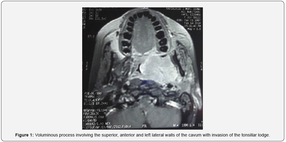
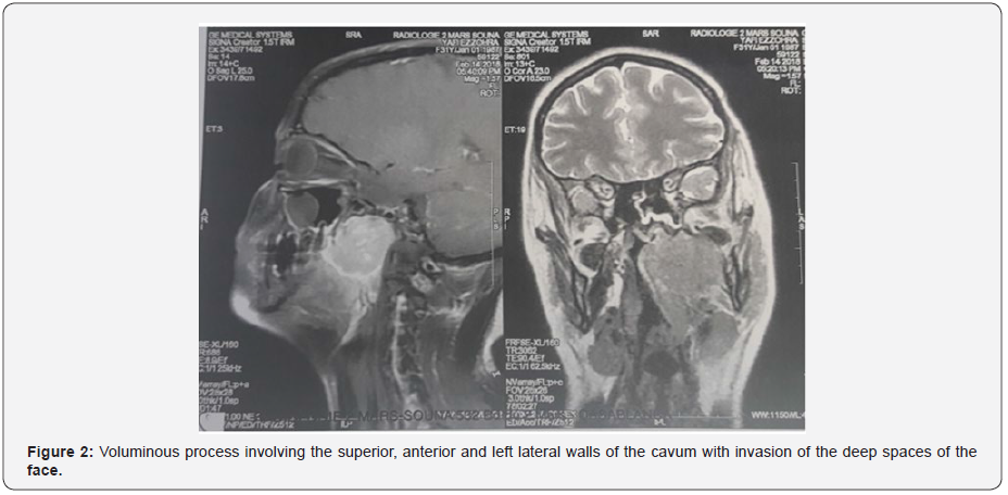
The patient has undergone a rhinocavoscopy in order to take biopsies of the lesion, for which the anatomopathological study revealed an UCNT. Then, the patient has benefited from Cervicofacial MRI to study the extension of the tumor. It has revealed voluminous process involving the superior, anterior and left lateral walls of the cavum with invasion of the tonsillar lodge and deep spaces of the face (Figures 1 & 2). The patient has received a cure of radiotherapy, and evolved well, with a rhinocavoscopy of control showing disappearance of the tumor.

Discussion
Nasopharyngeal cancers tend to invade adjacent structures. It can spread along the pharyngeal muscles down to the oropharynx responsible of trismus [3]. It can also grow through the choanes into the nasal cavity leading to nasal obstruction. The parapharyngeal space is also a frequent site of tumor invasion since the pharyngeal wall around the Eustachian tube is not very resistant [2]. Moreover, cranial invasion is not uncommon, likewise for cranial nerve palsies [4]. In fact, the lymphatic drainage of the nasopharynx is very extensive draining through three pathways: the internal jugular vein, the posterior cervical and retropharyngeal chains; which explains the very high frequency of nodal involvement [2]. Because the nasopharynx is a deep anatomical region, NPT are often discovered at a locally advanced stage, when it has already spread to the lymph nodes. This explains the presence almost constant of cervical lymphadenopathy at the time of diagnosis. Yet, tumors limited to the nasopharynx and without clinical lymph nodes account only 9% [5].
Otherwise; middle ear or external auditory canal invasion is uncommon and unusual. In fact, the Eustachian tube (ET) can be used by NPC as a lane to invade the middle ear and the mastoid [6]. Bulent Satar et Al. described two NPC cases with middle ear and outer ear involvements through the ET [7]. Other studies have been made for this purpose. We mention the case of three patients among 214 having NPC with middle ear invasion via the ET according to çuhruk et al. in Turkey [8].
Another theory might explain the spread of tumor through the outer ear canal. That is the erosion of anterior wall of the outer ear canal bypassing the middle ear; which can be confirmed by CT scan [7]. JWY Lai et Al. rapported a case of a locally advanced NPC with left ET, middle ear, and EAC involvements, documented by MRI. His CT scan showed a little erosion of the surrounding temporal bone; which could point to true mucosal or submucosal invasion along the Eustachian tube, rather than a locally advanced NPC invading the ET and middle ear externally through extensive skull bone involvement [9]. However, to date invasion mechanism of the EAC is still unclear and not very elucidated. Patients having NPC with middle ear and EAC involvements, whether it was primary or recurrence, present otological symptoms such as: hearing loss, otalgia, otitis media, tinnitus, vertigo, EAC polyps, and sometimes facial paralysis.
Conclusion
Nasopharyngeal tumors can spread via direct extension, perineural invasion, or via lymphatic or hematogenous mode into anatomical sites classified as high, medium and low risk [10]. However, direct invasion to the external auditory canal is unusual and rarely seen, but possible. This extension may imitate ordinary auditory symptoms such as otalgia, hearing loss, otitis media, facial paralysis, for which we should be aware and vigilant.
References
- H Boussen, N Bouaouina, A Gamoudi, N Mokni, F Benna et al. (2007) Cancers du nasopharynx. Oto-rhino-laryngologie 2(1): 1‑23.
- M Altun, A Fandi, O Dupuis, E Cvitkovic, Z Krajina, et al. (1995) Undifferentiated nasopharyngeal cancer (UCNT): Current diagnostic and therapeutic aspects. International Journal of Radiation Oncology Biology Physics 32(3): 859‑877.
- K Ichimura, Tanaka T (1993) Trismus in patients with malignant tumours in the head and neck. J Laryngol Otol 107(11): 1017‑1020.
- Brennan (2006) Nasopharyngeal carcinoma. Orphanet Journal of Rare Diseases 1(1): 23.
- A W Lee (1992) Retrospective analysis of 5037 patients with nasopharyngeal carcinoma treated during 1976–1985: Overall survival and patterns of failure. International Journal of Radiation Oncology Biology Physics 23(2): 261‑270.
- R L Cundy, I Sando, W G Hemenway (1973) Middle Ear Extension of Nasopharyngeal Carcinoma via Eustachian Tube: A Temporal Bone Report. Archives of Otolaryngology - Head and Neck Surgery 98(2): 131‑133.
- F A Tosun, Y Özkaptan (2008) Nasopharyngeal Carcinoma A Report of Two Cases with Unusual Extension. T Klin J E N T 1:45-50
- Çuhruk Ç, Aktürk T, Bacacı K (1979) Tüba Östaki Yoluyla Dış Kulak Yoluna Yayılım Gösterilen Nazofarenks Kanseri (3 olgu bildirimi). En ligne 4(3): 267-274.
- J Lai, A Cheng, M S Tong, C Yau (2015) Sneaking Its Way Up: External Auditory Canal Involvement by an Otherwise Inconspicuous Nasopharyngeal Carcinoma via Spread Through the Eustachian Tube. Hong Kong J Radiol 18: e1-6.
- W F Li, Ying Sun, Mo Chen, Ling Long Tang, Li Zhi Liu, et al. (2012) Locoregional extension patterns of nasopharyngeal carcinoma and suggestions for clinical target volume delineation. Chin J Cancer 31(12): 579‑587.


