Middle Turbinate Saving Technique in Cribriform Plate CSF Leak Repair
Pankaj Srivastava*
Director, ENT Hospital, Alambagh, Lucknow, India
Submission: July 24, 2019; Published: August 26, 2019
*Corresponding author:Pankaj Srivastava, Director, ENT Hospital, Alambagh, Lucknow, India
How to cite this article: Pankaj Srivastava. Middle Turbinate Saving Technique in Cribriform Plate CSF Leak Repair. Glob J Oto, 2019; 20(5): 556047. DOI: 10.19080/GJO.2019.20.556047
Abstract
Introduction: Middle Turbinate is an important structure of lateral nasal wall for air-conditioning of inhaled air, more importantly we need Middle turbinate as a landmark for revision surgeries if any. Saving Middle Turbinate would be a great Idea if we can perform surgery with same good exposure and not sacrificing one structure.
Method: In cases planned for CSF repair from Cribriform plate, usual investigations and surgical procedures were followed. We cut the necessary anterior portion of Middle turbinate and push it downwards, keeping its posterior attachment intact. After the surgery and sealing the Cribriform area with Fascia, the Turbinate is brought to its normal position and Merocel packing is done below turbinate.
Results: Three patients were done CSF repair having leak from one side Cribriform plate. Middle Turbinate saving procedure was followed. Per operative and post-operative results noted
Conclusion: We can save Middle turbinate for normal Rhinological function, as surgical landmark and for better post-operative result in CSF leak repairs.
Keywords: Middle Turbinate; CSF leak; Cribriform plate; Sphenopalatine; Lamina Paparacia
Introduction
Middle Turbinate is an important structure of lateral nasal wall for air-conditioning of inhaled air, more importantly we need Middle turbinate as a landmark for revision surgeries if any. Saving Middle Turbinate would be a great Idea if we can perform surgery with same good exposure and not sacrificing one structure
Review of Literature
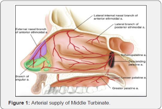
Middle Turbinate is supplied mainly by branch of Sphenopalatine artery which enters it from posterior end and also a branch of anterior ethmoidal [1]. The Blood supply to the middle turbinate arises from the proximal portion of the posterior lateral nasal artery just after sphenopalatine foramen. The middle turbinate can be preserved in almost all, endoscopic skull base surgery while providing good exposure for surgery and skull base reconstruction. Postoperative sinonasal function may be better preserved with this technique [2,3]. The endoscopic transsphenoidal approach to the midline skull base structures has been increasing in popularity as it provides a minimally invasive yet maximally aggressive therapeutic treatment option. Short-term outcomes compare favorably to more traditional open or microscopic approaches [4-7]. The turbinates are important nasal structures that help humidify, filter, andregulate temperature of nasal airflow before entering the lower airways. They also provide nasal resistance and contain sensory fibers essential for the perception of nasal airflow. In addition, the middle and superior turbinate contain olfactory fibers in the superior region. Finally, the middle turbinate serves as an important surgical landmark for the skull base, frontal sinus, and orbit [8].
The indications for partial or total middle turbinectomy in the setting of sinusitis are controversial. Most surgeons agree that a compromised middle turbinate secondary to polypoid degeneration, concha bullosa, or a paradoxical middle turbinate contributing to nasal obstruction or sinus disease is an acceptable reason to remove a portion of the turbinate. However, routine turbinectomy for surgical access to the paranasal sinuses has become less favorable amongst surgeons in recent years. Many surgeons describe the routine sacrifice of one or both middle turbinates in endoscopic transsphenoidal skull base surgery, whereas other authors report adequate visualization and surgical access with preservation of this structure. Saving middle turbinates in nasal surgery is needed to save nasal functions. Concerns about partial or total middle turbinectomy include alteration of nasal function, synechia formation withobstruction of sinonasal outflow tracts, promotion of frontal sinusitis, development of hyposmia, formation of excessive scar tissue, intra- and postoperative epistaxis, increased crusting postoperatively, loss of anatomic landmarks for revision surgery, and development of atrophic rhinitis or empty nose syndrome [9-13].
Materials and Methods
In cases planned for CSF repair from Cribriform plate, usual investigations and surgical procedures were followed. We cut the necessary anterior portion of Middle turbinate and push it downwards, keeping its posterior attachment intact. After the surgery and sealing the Cribriform area with Fascia, the Turbinate is brought to its normal position and Merocel packing is done below turbinate
Results
Three patients were done CSF repair having leak from one side Cribriform plate. Middle Turbinate saving procedure was followed. Per operative and post-operative results noted. In all three no difficulty in operative field was felt. Saved and displaced Middle turbinate did not come in operative field in any way. No problem in instrumentation was noted. Middle turbinate was easily replaced back in all three and Merocel packing done. No problem in pack removal was felt
In all three uneventful CSF leak stopped.
a) Patients were followed for maximum 12 months. No dryness change in smell, empty nose syndrome or any complaint was present.
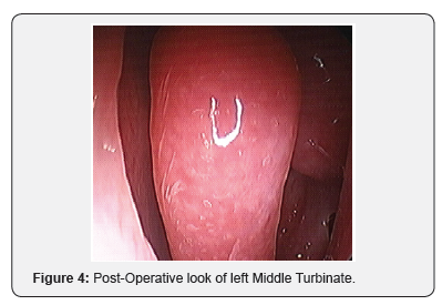
b) Post-operative endoscopic picture was as natural as normal nasal study in all three.
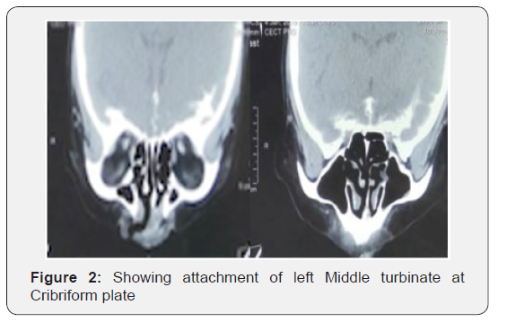
c) Post-operative CT Scan PNS revealed full length normal Middle turbinate in all three but the of left Middle Turbinate at Lamina Paparacia
Discussion
As arterial supply of Middle turbinate comes from both the ends, while detaching Middle turbinate in Cribriform plate CSF leak cases we preserve the posterior attachment of Turbinate and keep it downwards with its intact posterior Sphenopalatine supply (Figure 1). As it is pushed down there is no hindrance in surgical field. After the surgery Turbinate is brought to its possible natural position. Giving normal looking anatomy. Per operative it gives a good live tissue support and pressure to the material kept for sealing like Fascia and may help in early vascularization of the graft (Figure 2). All the functions of Middle turbinate like humidification of inhaled air, filtration and temperature controls are unaffected. Chances of empty nose syndrome is avoided. Post operatively in case of any revision surgery Middle turbinate being important landmark is saved. The replaced Middle turbinate is attached at different place from its natural attachment in post-operative CT Scan PNSSo, if revision surgery is planned and done for any reason, we should keep in mind that its changed attachment may misguide as landmark (Figures 3 & 4).


Conclusion
We can save Middle turbinate for normal Rhinological function, as surgical landmark and for better post-operative result in CSF leak repairs.
Summary
Middle Turbinate is an important structure of lateral nasal wall for air-conditioning of inhaled air.
ii. We need Middle turbinate as a landmark for revision surgeries if any.
iii. Saving Middle Turbinate in cribriform plate CSF Leak surgery would be a great Idea if we can perform surgery with same good exposure.
iv. Thus, we are not sacrificing one structure (Turbinate) and doing surgery without compromising exposure.
References
- Lee HY, Kim HU, Kim SS, Son EJ, Kim JW (2002) Surgical anatomy of the sphenopalatine artery in lateral nasal wall. Laryngoscope 112(10): 1813-1818.
- Luk LJ, Ikeda A, Wise SK, DelGaudio JM (2019) Middle Turbinate Friendly Technique for Cribriform Cerebrospinal Fluid Leak Repair. Skull Base 20(5): 343-347.
- Gurston G, Nyquist, Vijay K, Anand, Seth Brown, et al. (2010) Middle Turbinate Preservation in Endoscopic Transsphenoidal Surgery of the Anterior. Skull Base 20(5): 343-347.
- Frank G, Pasquini E, Farneti G (2006) The endoscopic versus the traditional approach in pituitary surgery. Neuroendocrinology 83(3-4): 240-248.
- Tabaee A, Anand VK, Barrón Y (2009) Endoscopic pituitary surgery: a systematic review and meta-analysis. J Neurosurg 111(3): 545-554.
- Schwartz TH, Fraser JF, Brown S, Tabaee A, Kacker A, et al. (2008) Endoscopic cranial base surgery: classification of operative approaches. Neurosurgery 62(5): 991-1005.
- Leopold DA, Hummel T, Schwob JE, Hong SC, Knecht M, et al. (2000) Anterior distribution of human olfactory epithelium. Laryngoscope 110(3 Pt 1): 417-421.
- Kennedy DW (1998) Middle turbinate resection: Evaluating the issues-should we respect normal middle turbinates Arch Otolaryngol Head Neck Surg 124(1): 107.
- Rice DH, Kern EB, Marple BF, Mabry RL, Friedman WH (2003) The turbinates in nasal and sinus surgery: a consensus statement. Ear Nose Throat J 82(2): 82-84.
- Cappabianca P, Cavallo LM, De Divitiis O, Solari D, Esposito F, et al. (2008) Endoscopic pituitary surgery. Pituitary 11(4): 385-390.
- Kassam A, Snyderman CH, Mintz A (2005) Expanded endonasal approach: The rostrocaudal axis. Part I. Crista galli to sella turcica. Neurosurg Focus 19(1): E3 1-12.
- De Divitiis E, Cavallo LM, Cappabianca P, Esposito F (2007) Extended endoscopic endonasal transsphenoidal approach for the removal of suprasellar tumors: Part 2. Neurosurgery 60(1): 46-59 discussion 58-59.
- Frank G, Pasquini E, Doglietto F (2006) The endoscopic extended transsphenoidal approach for craniopharyngiomas. Operative Neurosurgery 59(1 Suppl): S75-S83.


