Clinicopathological Profile of Aural Polypoidal Masses: A Retrospective Study of 41 Cases
Gabriel Toye Olajide1*, Olagoke Olaseinde Erinomo2, Timothy Adewale Olajide3, Mathew Segun Agboola4 and Waheed Atilade Adegbiji5
1Department of Ear, Nose and Throat, Federal Teaching Hospital, Ido Ekiti /Afe Babalola University, Ado Ekiti, Nigeria
2Department of Histo & Anatomic Pathology, Federal Teaching Hospital, Ido Ekiti/Afe Babalola University, Ado Ekiti, Nigeria
3Department of Surgery, Federal Teaching Hospital, Ido Ekiti /Afe Babalola University, Ado Ekiti, Nigeria
4Department of Family Medicine, Federal Teaching Hospital, Ido Ekiti /Afe Babalola University, Ado Ekiti, Nigeria
5Department of Ear, Nose and Throat, Ekiti State Teaching Hospital, Ado Ekiti, Nigeria
Submission:June 10, 2019;Published:June 21, 2019
*Corresponding author:Olajide Toye Gabriel, Department of Ear, Nose and Throat Surgery, Federal Teaching Hospital, Ido Ekiti/College of Medicine and Health Sciences, Afe Babalola University, Ado Ekiti (ABUAD), Nigeria
How to cite this article:Gabriel T O, Olagoke O E, Timothy A O, Mathew S A, Waheed A A. Clinicopathological Profile of Aural Polypoidal Masses: A Retrospective Study of 41 Cases. Glob J Oto, 2019; 20(3): 556036.DOI: 10.19080/GJO.2019.20.556036
Abstract
Background/Aim: Several diseases can affect the external auditory canal (EAC) which may cause conductive hearing loss among other things. However, some of them are locally invasive and if they are not diagnosed early may lead to morbidity with low quality of life. The aim of this study was to determine the clinicopathological profile of aural polypoidal masses as well as their treatment and outcome.
Methodology: An 8 – year retrospective clinicopathologic analysis of patients with aural mass seen and treated at the ENT clinic of Federal Teaching Hospital, Ido Ekiti and Group Consultant Clinic (Private Clinic), Ado Ekiti, Nigeria between July 2007 and June 2015 was carried out. Information on patient’s biodata, clinical and histological diagnosis, treatment and outcome were retrieved from their case notes.
Results: Forty one (41) patients had complete data for analysis. Their age ranged from 6 to 63 years. Majority (34.1%) of the patients falls within the age group of 20 – 30 years. Thirty (73.2%) were males with male: female ratio of 2.7:1. Right ear were more affected than the left in 65.9 % of the patients. The major complaints they presented with was otalgia 30 (73.2%). The commonest histological diagnosis was aural polyp in 17(41.5 %) of the patients after surgical excision (simple aural polypectomy).
Conclusion: Aural masses of different forms are commonly present to Otolaryngologist. Otalgia was the commonest symptom leading to early presentation in our study. All the patients had simple aural polypectomy. Commonest histological diagnosis was aural polyps. Histopathology remains the key tool in differentiating aural masses. Early diagnosis will prevent morbidity and mortality.
Keywords:Clinicopathological; Aural; Profile; Polypoidal Masses
Introduction
Mass lesions within the external auditory canal (EAC) may cause progressive conductive hearing loss, otalgia, or otorrhea [1]. Such mass usually bulges or projects downwards or outward from the normal mucosa surface. Although primary tumors involving the EAC are extremely rare, early and accurate diagnosis through histopathologic examination is important for their proper treatment [1]. Such mass may be due to the most frequently occurring simple polyps or other pathological entities likely to be infective process to polypoidal neoplasms including the malignant ones. Failure to diagnose some of them early may cause morbidity and mortality. Majority of the aural masses presenting as aural polyps can be managed by simple aural polypectomy, while others may need extensive surgical intervention. Those that arise from inflammatory ear diseases are more difficult to manage and are also associated with grievous consequences [2]. The aim of this study was to determine the clinicopathological profile of aural polypoidal masses as well as their treatment and outcome This type of study has not been carried out in our locality; the result will serve as a base line for further studies as well as compared with similar research in other centers.
Methodology
We carried out an 8 - year retrospective study and analysis of patients with aural masses seen and managed at the Ear, Nose and Throat (ENT) clinic of Federal Teaching Hospital, Ido Ekiti and Group Medical Clinic (Private Clinic), Ado Ekiti, Nigeria between July 2007 to June 2015. Case notes of all the patients with clinical diagnosis of aural polyps that had simple excision biopsies (aural polypectomies) done was retrieved from the hospital medical record department. The information that was extracted from the case notes included their biodata, presenting symptoms, duration of symptoms, affected ear, otomicroscopic findings including location of the mass, state of tympanic membrane, clinical diagnosis, their histopathology report, treatment and outcome. Tuning fork tests and their radiological investigations (X- ray mastoids) were also taken into consideration. The information retrieved was entered into a data sheet that was designed for the study. Inclusion criteria include all patients with a mass in the external auditory canal and with a complete records/data. Exclusion criteria include any patients with mass arising from pinna or patients that had documented tumour in the ears with evidence of local and regional aggression. Ethical approval to conduct this study was obtained from the hospital ethics and research committee.
Data Analysis
A simple descriptive analysis of the data obtained was carried out using SPSS version 20.0 and the result was presented in simple tables and charts.
Results
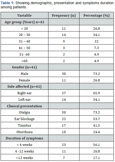
NB: Some patients presents with more than one symptom.
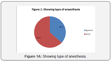
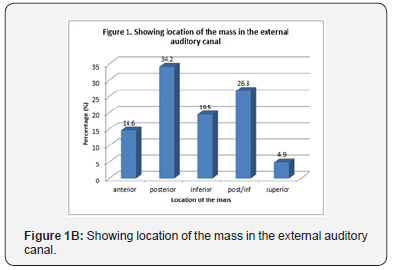
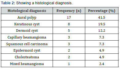
Out of 46 patients studied only 41 had complete data for analysis. Their ages ranged between 6 to 63 years with a mean of 28.63 ± 14.47 years. Majority (34.1%) of the patients falls within the age group of 20 – 30 years. Thirty (73.2%) were males while 11(26.8%) were females given a male: female ratio of 2.7:1. Right ear were more affected than the left in 65.9 % of the patient (Table 1). The major complaints the patients presented with was otalgia 30 (73.2%), followed by ear blockage in 22 (53.7%) patients, tinnitus in 17 (41.5%) patients while 10 (24.4%) patients had otorrhoea. Duration of the symptoms was less than 4 weeks in 23(56.1%) of the patients, 4–12 weeks in 11(26.8 %) and more than 12 weeks in 7(17.1%). This is also shown in Table 1. The commonest histological diagnosis was aural polyp 17 (41.5 %), keratinous cyst 8 (19.5%), dermiod cyst 5(12.2%). Others are shown in Table 2. Majority 14 (34.2%) of the masses were located at the posterior wall of the EAC. Other locations are shown in Figure 1A. Treatment given include surgical excision under general anesthesia in 15 (36.6%) patients while 26 (63.4%) had the excision under local anesthesia (Figure 1B).
Discussion
Several diseases can affect the external ear canal with it associated problems. In this study males were more affected than females, this is similar to study carried out by other authors [3,4]. However it was in contrast to study done by Sogebi [5] in which higher number of female patients was recorded. Majority of our patient presented with otalgia and that also corroborated earlier presentation of our patients in the hospital. In our study more than half of our patient presents within four weeks. Majority of patients with aural mass usually has underlying chronic ear infections inform of otitis external or media. With otitis media it may be characterized by copious blood ear discharge at times. These findings are not unexpected as the mass tend to cause acute exacerbation of the symptoms with increasing discomfort from earache [6]. Our patient also presented with ear blockage which in laymen to some of them will be equated to inability to hear very well on the affected ear. This may be due to the mass effect of the aural mass in the EAC which impaired sound conduction into the inner ear [5]. Right ear was mostly affected in this study as few studies also demonstrated differential lateralization of pathologies to particular ears [7,8].
An aural polyp which is a benign lesion was the most common histopathological diagnosis of aural mass in our study. Baylan et al 4 recorded similar finding in their study. Most of the ear mass had attachments to the posterior wall of the EAC. For proper management of a mass in the EAC, histopathological analysis of the mass is the key factor so as to arrive at appropriate diagnosis and also assists in the final management since such mass may be difficult to distinguished clinically from each other. In our study some of our patients had simple aural polypectomy under general anesthesia (light sedation) while majority had it under local anesthesia. Some of our patient had cauterization to the base of the lesion after polypectomy. We lost some of our patients to follow up, this is one of the challenges occasionally faced with as a clinician in our setting, where patient tend to default once they have relief from their symptoms. For our patients that was followed up (70.7%) of them for more than five years, none had recurrence of their mass. Although it is difficult to find a time scale for recurrence in the literature, general agreement was that long – term monitoring is needed [9]. Williams et al emphasized that simple aural polypectomy may be relevant in initial diagnosis, and often times in the treatment of aural masses, it may not be adequate in treating extensive disease associated with cholesteatoma [10]. The Limitation of this study was that audiometric tests were not conducted on some of the patients which could have given the state of hearing status. Although the tuning fork tests that was performed on them showed some clinical evidence of conductive hearing loss.
Epidermal Cyst
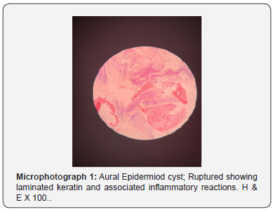
Epidermal inclusion cyst refers to those cysts that are the result of the implantation of epidermal elements in the dermis and proliferation of the epidermal cells within a circumscribed space of the dermis [11]. They are rare tumours that may occur anywhere in the body. About 7 % of them are found in head and neck region [12]. It usually arises on hair-bearing areas including scalp, face, neck, trunk, extremities and scrotum [13]. Their appearance in the EAC is very rare. They are slowly growing, elevated, firm, round intradermal or subcutaneous tumours that cease to grow after reaching a size of 1 to 5 cm in diameter [11,14]. In our study only 2 cases were seen and treated. No recurrences of the lesion after following up (Microphotograph 1).
Haemangioma
It is a vascular lesion usually found in the dermis. Commonly seen in young children and it progressively disappears before the ages of 5-6 years [15]. Haemangioma can be either capillary type when vessels are organized in capillary structures or cavernous type when presenting large vascular spaces, which is more common in the skin and in the mucosa. We lost 3 out of 4 of our patients to follow up.
Cholesteatoma
Cholesteatoma is a serious disorder of the middle ear cleft that is comprised a sac filled with keratin and lined by keratinizing squamous epithelium [4]. Cholesteatomas can be congenital (primary) or acquired. The acquired ones are much more common, and they occur in older children and young adult 3. External auditory canal cholesteatoma is a rare disease entity with uncertain aetiology. The condition is not often diagnosed and may be merely an incidental finding [16-18]. Some authors have reported that the EAC cholesteatoma was as a result of trauma to EAC, chronic inflammation, ear canal stenosis, or that it arises spontaneously [19,20]. They are not true neoplasm, but clinically they can mimic malignant neoplasm because of their propensity to destroy surrounding tissue and recur after excision [21].
Squamous Cell Carcinoma
Squamous cell carcinoma of the external auditory canal is a rare tumour that can occur from chronic irritation of the ear canal from chronic external otitis [22]. It may also be as a result of long history of chronic otitis media followed by bloody discharge and hearing loss [23]. It may occur occasionally for unknown reason. Early tumor is highly treatable while a somewhat more advanced tumour can be incurable. Three of our patients have histological diagnosis. Two was lost to follow up and one referred to other centre for further treatment.
Aural Polyp
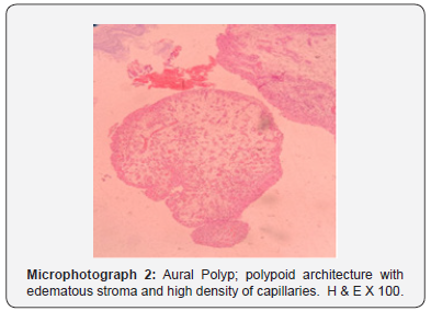
Aural polyps are fleshy and oedematous growth in the external auditory canal (EAC) of the ear. At times it may arise from middle ear. It occurs worldwide; although it tends to be more common in the developing countries due to its association with chronic inflammatory diseases of the ear which are more prevalent in these places [23,24]. It is a condition that can affect any group without any gender predisposition. It can affect any individual irrespective of the age or sex of the individual. It affects all races and ethnicities equally. This condition is usually associated with blood stained ear discharge with earaches [24]. In our study, they constituted major histological diagnosis (41.5%). No recurrence was recorded among those that came for regular follow up (Microphotograph 2).
Conclusion
Aural masses in different forms are commonly present to otolaryngologist. Otalgia was the commonest symptom leading to early presentation in our study. All the patients had simple aural polypectomy. Commonest histological diagnosis was aural polyps. Histopathology remains the key tool in differentiating aural masses. Early diagnosis and creating awareness will prevent morbidity and mortality.
References
- Chang-Hee Kim, Hye Seung Lee, Sung-Yong Kim, Jung Eun Shin (2019) Unusual Tumors Obstructing the External Auditory Canal: Report of Two Cases. J Audiol Otol 23(1): 59-62.
- Prasannaraj T, De NS, Narasimhan I (2003) Aural polyps: safe or unsafe disease? Am J Otolaryngol 24(3): 155-158.
- Agarwal NM, Popat VC, Traviad C, Srivastava A (2013) Clinical and Histopathological Study of Mass in Ear: A Study of Fifty Cases. Indian J Otolaryngol Head Neck Surg 65(3): 520-525.
- Baylan MY, Koc EE, Kilic N, Meric F (2010) Histopathological evaluation of the polyps with and without the presence of Cholesteatoma. Int Adv Otol 6(1): 201-205.
- Sogebi OA (2012) Clinicopathological Characteristics of Aural Polyps. East and Centr Afr J of Surg 17(3): 58-66.
- Chen B, Ye F, Wang L (2011) The clinical characteristics and the treatment of external auditory canal cholesteatoma. Lin Chung Er Bi Yan Hou Tou Jing Wai Ke Za Zhi 25(19): 868-870.
- Yoon YH, Park CH, Kim EH, Park YH (2008) Clinical characteristics of external auditory canal cholesteatom in children. Otolaryngol Head Neck Surg 139(5): 661-664.
- Olusesi AD (2008) Otitis media as a cause of significant hearing loss among Nigerians. Int J Pediatr Otorhinolaryngol 72(6): 787-792.
- Garin P, Degols JC, Delos M (1997) External Auditory canal cholesteatoma. Arch Otolaryngol Head Neck Surg 123: 62-65.
- Williams SR, Robinson PJ, Brightwell AP (1989) Management of the inflammatory aural polyp. J Laryngo Otol 103(11): 1040-1042.
- Kenneth AB, Isabella T (1997) Epidermoid inclusion cyst. E-medicine-specialties dermatology benign neoplasm.
- Ali I, Ahmad R, Iqbal I (2011) Epidermal cyst of the bony external auditory canal in adult. Journal of Clinical Immunology and Immunopathology Research 3(2): 20-21.
- Kim HK, Kim SM, Lee SH, Racadio JM, Shin MJ (2011) Subcutaneous epidermal inclusion cysts. Ultrasound (US) and MR imaging findings. Skeletal Radiol 40(11): 1415-1419.
- Toshihiro S, Masakatsu T, Toshiaki S, Tatsuya M (2007) Epidermal cyst of the bony external auditory canal. Otolaryngology head and neck surgery 136(1): 155-156.
- Neto JFL, Miura MS, Saleh C, Andrade M, Assmann M (2007) Hemangioma as a mass of External Ear Canal. Intl. Arch. Otorhinolaryngol 11(4): 498-500.
- Heilbrun ME, Salzman KL, Glastonbury CM, Harnsberger HR, Kennedy RJ, et al. (2003) External auditory canal cholesteatoma: clinical and imaging spectrum. AJNR Am J Neuroradiol 24(4): 751-756.
- Choi JH, Woo HY, Yoo YS, Cho KR (2011) Congenital primary cholesteatoma of external auditory canal. Am J Otolaryngol 32(3): 247-249.
- Darr EA, Linstrom CJ (2010) Conservative management of advanced external auditory canal cholesteatoma. Otolaryngol Head Neck Surg 142(2): 278-280.
- Naim R, Linthicum F, Shen T, Bran G, Hormann K (2005) Classification of the external auditory canal cholesteatoma. Laryngoscope 115(3): 455-460.
- Holt JJ (1992) Ear canal cholesteatoma. Laryngoscope 102(6): 608-613.
- Silverberg SG, DeLellis RA, Frable WJ, Virginia AL, Wick MR (2006) Silverberg’s principles and practice of surgical pathology and cytopathology, New York: Churchill Livingstone 2(4): 2267–2284.
- Djalilian HR (2016) Clinical Consultation. Symptom: Ear canal mass and pain. The hearing Journal p. 39-42.
- Monasta L, Ronfani L, Marchetti F, Montico M, Vecchi Brumatti L, et al. (2012) Burden of disease caused by otitis media: systematic review and global estimates. PLoS One 7(4): e36226.
- Kerkar P (2018) What are Aural Polyps: Causes, Symptoms, Treatment, Prognosis, Complications, Risk Factors.


