Bilateral Maxillary Mucocoele: A Rare Case Report
Snigdha1, Karthikeyan R1*, Alexander2, Saxena3, Sivaraman4, Subhashini P5, Vignesh K5 and ReddyC1
1 Junior Resident, Department of ENT, Jawaharlal Institute Postgraduate Medical Education and Research (JIPMER), India
2Additional Professor and Head, Department of ENT, JIPMER, India
3Professor, Department of ENT, JIPMER, India
4Associate Professor, Department of ENT, JIPMER, India
5Senior Resident, Department of ENT, JIPMER, India
Submission: August 25, 2018; Published: September 04, 2018
*Corresponding author: Karthikeyan Ramasamy,Department of ENT,Jawaharlal Institute PostgraduateMedical Education and Research (JIPMER), India.
How to cite this article: Snigdha, Karthikeyan R, Alexander, Saxena, Sivaraman, Subhashini P, Vignesh K, Reddy C. Bilateral Maxillary Mucocoele: A Rare Case Report. Glob J Oto, 2018; 17(3): 555964. DOI: 10.19080/GJO.2018.17.555964
Abstract
Mucocoele of paranasal sinus occurs commonly due to ongoing inflammation against closed drainage pathway of the sinus. Incidence of mucocoele is most common in frontal sinus followed by ethmoid sinus, less common in maxillary sinus. Moreover, incidence of bilateral maxillary mucocoele is of rare occurrence, has been classically described in children with cystic fibrosis or following open sinus surgery. Other causes of mucocoele being chronic sinusitis, allergic rhinosinusitis, trauma, previous surgery. Here we present a case report of a 26-year-old adult who was found to have bilateral maxillary mucocoele of uncertain aetiology, probably secondary to chronic sinusitis and further predisposed by anatomical variations in the sinus. Presentation, differential diagnosis and management by combined sublabial and endoscopic approach is discussed.
Keywords: Bilateral Maxillary Mucocoele; Maxillary Sinus Mucocoele; Combined Approach
Abbreviations: CT: Computed Tomography
Bilateral Maxillary Mucocoele: A Rare Case Report
Mucocoeles are benign expansile cyst like lesions of paranasal sinuses. They occur because of an ongoing inflammatory process in an obstructed sinus.Mucocoeles are most commonly encountered in frontoethmoid region, and their occurrence in maxillary sinus is uncommon, found to be less than 10%[1]. Overall, less than 5% were noted to be bilateral/ multiloculated[2]. A Bilateral maxillary mucocele is rare clinical entity that has been reported only in infants with cystic fibrosis or following sinus surgeries such as Caldwell Luc operation[3-6]. In this case report, we thread through a case of bilateral maxillary mucocoele in a 26 year old adult of unknown etiology. Possible etiology, differential diagnosis, and optimal management modalities are discussed.
Case Report
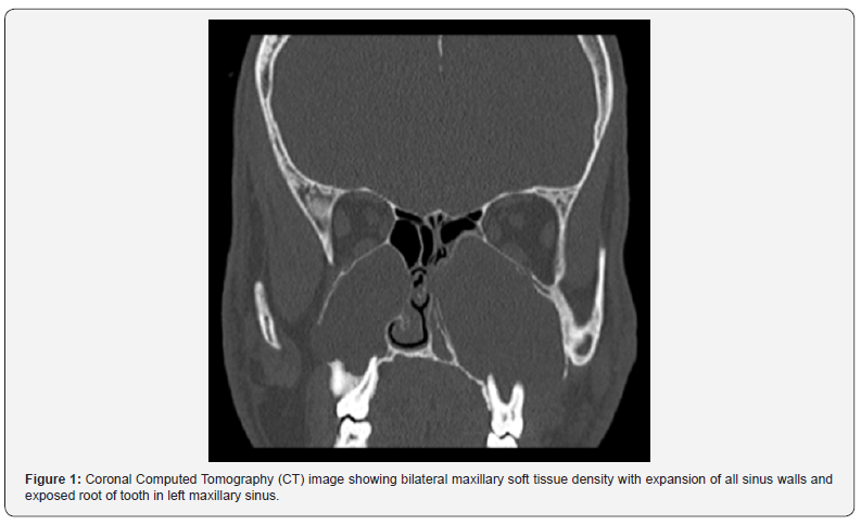
26-year-old adult male presented with complaints of purulent nasal discharge from bilateral nasal cavity for one year. He also had one episode of purulent discharge from left upper first molar region. He had no dental procedure or nasal surgery or trauma prior to these symptoms. There was no loosening of teeth, headache, epistaxis, displacement of eye or visual disturbances. Nasal endoscopy shows bulging lateral nasal wall with polypoidal mucosa prolapsing in left middle meatus, purulent nasal discharge from bilateral middle meatus. Contrast enhanced Computerised Tomography (CT) was done which showed non-enhancing homogenous density filling bilateral maxillary sinus with remodelling of surrounding bone leading to thinning out and ballooning of all walls of maxillary sinus. There was extreme thinning of all walls including floor of sinus exposing root of tooth in the sinus. Biopsy of polypoidal mucosa at middle meatus showed inflammatory mucosa.Based on the clinical features and imaging, a working diagnosis of bilateral maxillary mucocoele or dental cyst is made. Patient was planned for bilateral Caldwell Luc approach under general anaesthesia due to suspected bone erosion of left maxillary sinus floor. Anterior wall of sinus was thinned out on both sides. On performing antrostomy, rupture of cyst wall noted with mucinous fluid filling the cyst in right maxillary sinus, and purulent fluid noted in the left one. Entire cyst wall removed, and no bony defect was noted along the floor of maxillary sinus on either side intraoperatively. Middle meatal antrostomy was done endoscopically to improve ventilation to the sinus. Combined approach i.e., canine fossa antrostomy by sublabial approach with endoscopic middle meatal antrostomy was thus done. Postoperative period was uneventful. No cheek anaesthesia or loosening of teeth was noted. sublabial wound healed well.Diagnosis was made based on findings on imaging and confirmed intraoperatively and histologically as mucocoele and mucopyocoele. A five-month follow-up showed no recurrence of symptoms.
Discussion
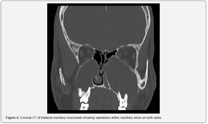
Mucocoele are cystic expansible lesions lined by secretory respiratory epithelium filled with mucus and characterized by expansion and thinning of sinus walls. They are believed to arise due to accumulation of mucus secondary to obstructed sinus outflow while some believe that it may arise from an enlarging mucous retention cyst filling entire sinus[4]. While mucocoeles are most common in frontal sinus followed by ethmoid sinus, they are of rare occurrence in maxillary sinus accounting to 3-10% of mucocoeles[7,8]. Maxillary mucocoelesare more common in Japan as a long-term sequel after Caldwell Luc surgery, presumed to be due to entrapped sinus mucosa[1]. Apart from these, common causes of maxillary sinus mucocoele are chronic sinusitis, allergic rhinosinusitis, benign or malignant tumours occluding ostiomeatal complex[1]. Bilateral maxillary mucocoele presentation is even rarer, and is classically seen in children with cystic fibrosis, rarely even as a presenting feature[4]. Mucocoele can remain asymptomatic for a long time. Progressive expansion of maxillary mucocoele leads to displacement of surrounding structures and can cause proptosis, loosening of teeth, cheek swelling[1]. Our patient here presented with purulent discharge from bilateral nasal cavity, for an year and there was no history of allergy, trauma or previous surgery.Discharge from upper molar may be secondary to sinus infection or vice versa. The cause of mucocoele remains uncertain, possibly due to chronic sinusitis. Mucocoeles are best diagnosed by computed tomography (CT) that shows homogenous non-enhancing soft tissue density filling entire sinus with surrounding bony remodelling in terms of thinning out and expansion. Differential diagnosis of soft tissue density filling maxillary sinus includes, mucus retention cyst, antrochoanal polyp, although, bony remodelling is not classically seen in these. Surrounding bone destruction and erosion should lead towards diagnosis of benign or malignant tumours of sinonasal origin[1]. In this case, CT showed expansile lesion filling bilateral maxillary sinus with all walls ballooned out with prominent medial extension on the left and focalerosion of floor of maxillary sinus exposing root of teeth (Figure 1). Bone erosion in mucocoele has been attributed inflammatory cytokines, which trigger prostaglandins and collagenases which in turn results in bone resorption[1,9]. Periapical cyst, odontogenic cyst remained to be differential diagnosis. Caldwell Luc approach was chosen to proceed with.Cystic lesion filling maxillary sinus and filled mucus/ mucopus noted in maxillary sinus. Although, floor of sinus was found intact during surgery and septations were noted in maxillary sinus, as also seen in preoperative CT (Figure 2), which might have led to localised accumulation of mucus progressively and resulted in loculated mucocoele formation.


Bilateral maxillary mucocoeles are rare, have been classically described in children with cystic fibrosis, or following Caldwell Luc surgery[4,5]. This case we believe may be secondary to chronic sinusitis, drainage in turn affected by multiple septations in maxillary sinus predisposing to loculated mucocoele formation.Mucocoele was managed by historically by Caldwell Luc operation and removal of mucocoele lining. Now the preferred treatment is endoscopic marsupialization and wide middle meatal antrostomy[3,4]. Although, when there is significant extension outside the sinus like facial tissue, pterygopalatine fossa, open approach by Caldwell Luc surgery is preferred for complete removal of mucocoele lining[1,3]. In this case, combined approach was used, where canine fossa antrostomy was done to confirm diagnosis , to rule cyst from dental origin, followed by middle meatal antrostomy for ventilation of sinus. A five-month follow-up showed no recurrence of symptoms or complications.
Conclusion
Bilateral maxillary mucocoele is of rare occurrence in adults secondary to chronic sinusitis, and it may be predisposed by intrasinus septations. Close differential diagnosis should be considered based on symptomatology and imaging while planning management. Endoscopic marsupialisation has good outcome for maxillary mucocoele management, can be combined with open technique by antrostomy to rule out close differential diagnosis, and ensure complete removal of pathology.
References
- Caylakli F, Yavuz H, Cagici AC, Ozluoglu LN(2006) Endoscopic sinus surgery for maxillary sinus mucoceles. Head & face medicine 2(1): 29.
- ScottBrown W, Gleeson M (2008) Scott-Brown’s Otorhinolaryngoloy, head and neck surgery. (7th edn), [England], pp.1531-1538.
- Busaba NY, Salman SD(1999) Maxillary sinus mucoceles: clinical presentation and long-term results of endoscopic surgical treatment. The Laryngoscope 109(9): 1446-1449.
- Qureishi A, Lennox P, Bottrill I (2012) Bilateral maxillary mucoceles: an unusual presentation of cystic fibrosis. The Journal of Laryngology & Otology 126(3): 319-321.
- Tunkel DE, Naclerio RM, Baroody FM, Rosenstein BJ (1994) Bilateral maxillary sinus mucoceles in an infant with cystic fibrosis. OtolaryngologyHead and Neck Surgery 111(1): 116-120.
- Hasegawa M, Saito Y, Watanabe I, Kern EB (1979) Postoperative mucoceles of the maxillary sinus. Rhinology 17(4): 253-256.
- Natvig K, Larsen TE (1978) Mucocele of the paranasal sinuses: A retrospective clinical and histological study. The Journal of Laryngology & Otology 92(12): 1075-1082.
- Christensen JR,HouckLP (1954)Mucocele of the maxillary sinus. AMA archives of otolaryngology 59(2): 147-151.
- Lund VJ, Milroy CM (1991) Frontoethmoidal mucocoeles: a histopathological analysis. The Journal of Laryngology & Otology 105(11): 921-923.


