Deep Circumflex Iliac Artery Flap for Reconstruction of Mandibular Defects Exceeding 9 cm
Almamidou Assoumane1,2, K Liu2, Zhe Shao1 and ZJ Shang1,2*
1The State Key Laboratory Breeding Base of Basic Science of Stomatology (Hubei-MOST) and Key Laboratory for Oral Biomedicine Ministry of Education, Wuhan University, China
2Department of Oro-maxillofacial - Head and Neck Oncology, School and Hospital of Stomatology, Wuhan University, China
Submission: June 10, 2017; Published: June 21, 2017
*Corresponding author: Zheng Jun Shang, Department of Oro-maxillofacial - Head and Neck Oncology School and Hospital of Stomatology, Wuhan University, 237 Luoyu Road, Wuhan 430079, China Tel: +862787686129; Fax: +862787873260; Email: shangzhengiun@hotmail.com
How to cite this article: Almamidou A, K Liu, Zhe S, ZJ Shang. Deep Circumflex Iliac Artery Flap for Reconstruction of Mandibular Defects Exceeding 9 cm.Glob J Oto 2017; 8(4): 555742. DOI: 10.19080/GJO.2017.08.555742
Abstract
The deep circumflex iliac artery flap (DCIAF) or vascularized iliac crest flap is widely used for mandibular reconstruction. However despite the large mandibular defect recorded in our developing environment, DCIAF continu! es to play a role in its used for anterior mandibular defects(S1), lateral mandibular defects (L) and posterior mandibular defects (S2). The aim of this study is to show the clinical outcomes and complications of using deep circumflex iliac artery flap in immediate reconstruction of mandibular defects according to the site and sizes of defects. We retrospectively reviewed records of sixty seven (67) patients (35 males and 32 females) with mandibular tumor and traumatic defect, who underwent resection of anterior mandibular defects (S1:n=7); lateral mandibular defects (L: n=54) and posterior mandibular defects (S2: n=35). All of them were reconstructed using deep circumflex iliac artery flap with the bony defect less or exceeding 9 cm in length. All of 67 patients achieved optimal functional results and clinical outcomes postoperatively. Despite our clinical data , it is suggestible that the deep circumflex iliac artery be considered for anterior, lateral and posterior mandibular defects reconstruction, considering the size of defect less or exceeding 9 cm.
Keywords: Deep circumflex iliac artery flap; Mandibular defects exceeding 9 cm; Reconstruction
Abbreviations: S1: Anterior Mandibular Defect; L: Lateral Mandibular Defect; S2: Posterior Mandibular Defect; KCOT: Keratocystic Odontogenic Tumor; SCC: Squamous Cell Carcinoma; RA: Recurrence of Ameloblastoma; OCT: Odontogenic Calcifying Tumor; DCIAF: Deep circumflex iliac artery flap
Introduction
Mandibular defect resection is the most important decision to be made in the treatment of patients with tumor, traumatic defect and others. Reconstruction of mandibular defects continues to pose a challenge to surgeons and patients because the bony defect compromising functional results and aesthetic [1,2]. Indication for reconstruction type was based on type of mandibular defect, anterior includes both canines, lateral mandibular defect here defined as region from distal surface of central incisors to its ipsilateral condyle, posterior mandibular defect (Hemimandibulectomy and condyle) and other analysed variables were defect in size and dimension of bone ( length, width). However, deep circumflex iliac artery flap is a reconstruction option with many potential applications because its offers a large amount of bone complex reconstruction in the dentate mandible [3], provides a large segment of vascularised bone , was popularized in association with an inconspicuous donor scar and low donor site morbidity [4,5]. It may be the first choice for mandibular defect less nine (9 cm) in size because the shape of the vascularised iliac crest bone is similar to the mandible angle and has abundant bone tissue. In addition, the deep circumflex iliac artery flap is used in cases of defect more than 9 cm and is not the only technique available for immediate mandibular reconstruction, the fibula flap [6] has been equally successful. Although fibula and scapula free flaps remains the best-suited vascularised bone flap. These debates have mainly centred on donor site morbidity, length of surgery and donor bone adequacy [7,8]. The aim of this study is to show the clinical outcomes and complications of using deep circumflex iliac artery flap in immediate reconstruction of mandibular defects according to the site and size of defects less or exceeding 9 cm in length and 2.4 cm in width.
Materials and Methods
Patients
The present study included sixty seven (67) patients who underwent immediate reconstruction of mandibular defects using deep circumflex iliac artery flap at the department of oromaxillofacial and Head Neck Oncology School and Hospital of Stomatology from July 2014 to august 2016 following mandibular surgery for malignant, benign tumors and traumatic defect. The study was conducted in accordance with the principles of the declarations of Wuhan University School and Hospital of Stomatology, the ethics committee for clinical study had reviewed and approved the study protocol. We retrospectively reviewed records of sixty seven patients (35 males and 32 females) with mandibular tumor and traumatic defect. Clinical data were collected by demographic information (ages, gender), mandibular defects diagnosis loading resection of tumors, sizes of bone harvest, clinical outcomes and complication encountered. Patients were grouped by bone defect type and size according to the new classification for mandibular defects after oncological resection described by James S Brown and Colleagues [9].
It was based on the four corners of mandible (two angles and two canines) from class I to class IV. Class I lateral mandibulectomy (includes the angles, or one vertical corner but not the condyle), Class Ic lateral defect including condyle, Class II hemimandibulectomy ( includes the angles vertical corner and the ipsilateral canine but not the contralateral canine, Class IIc hemi mandibulectomy including condyle, class III hemimandibulectomy (includes both canines but neither angle), class IV extensive mandibulectomy (includes both canines and one or both angles) and Class IVc extensive anterior mandibulectomy including both canines and one or both angles. Of 67 patients, fifty four (54) defects as type class I with size less or 9 cm; seven (7) defects were classified as type Class III with sizes of bony 10 cm, thirty five (35) defects as type class IIc with size 12 cm and no defects were classified as type class IV because all the patients including in this study have the defects sizes ranging from 4.5 to 12cm.
Surgical Procedures
All surgical procedures were performed using a two - tea m approach: an extirpative surgery team and a reconstructive surgery team. Tumors resections along with modified radical neck dissection or selective neck dissection were done simultaneously in all patients and transmandibular approach with or without a partial mandibulectomy. After completion of the tumor resection and harvesting of the deep circumflex iliac artery flap, appropriate preparation of both ends of vessels was performed for microsurgery show in ( Figure 1). The GEM microvascular anastomotic coupler system (synovis-microcampanies Alliances. In) was used for the microanastomoses in the standard fashion in accordance with the manufacture instructions. In brief the anastomosis was performed in the following steps:
- Placement of vessels in proximity to the recipient vessels occluded using vascular clamps,
- Determination of the coupler size using a coupler measuring gauge.
- Pulling and Finning of vessels to coupler,
- Rotation of instrument knob to mate the vessel ends , and
- Securing the two rings in opposition using a fine hemostat before separation of the instrument from rings.
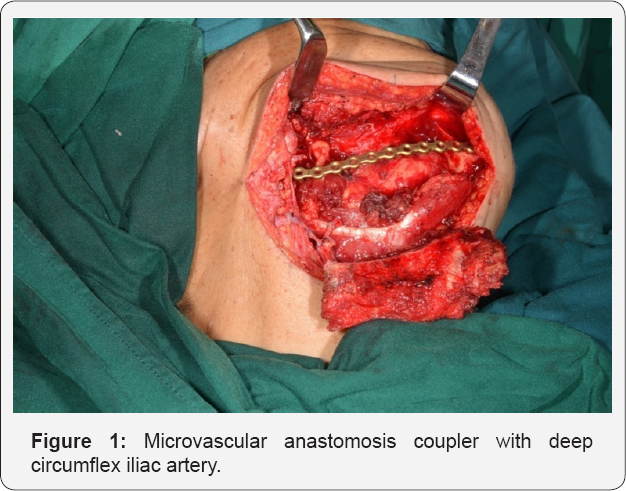
During the vascular coupling, the vessels were rinsed repeatedly with heparinized saline .After a careful inspection of patency and appropriate drainage placement, autograft fixation and subsequent suture of the incision was finally performed. Postoperative conventional techniques for flap monitoring method were used including clinical examination and observation, supplemented beside external Doppler every 30 minutes for the first 24 h, every one hour for the next 24h and every 4h in the following days. Postoperative pharmacological therapy was used when necessary, low dose and low molecular weight heparin. All patients staying three (3) days in intensive care unit (ICU) and the length of all patients in hospital ranged from 8 to 30 days (average 18.70).
Postoperative follow-up
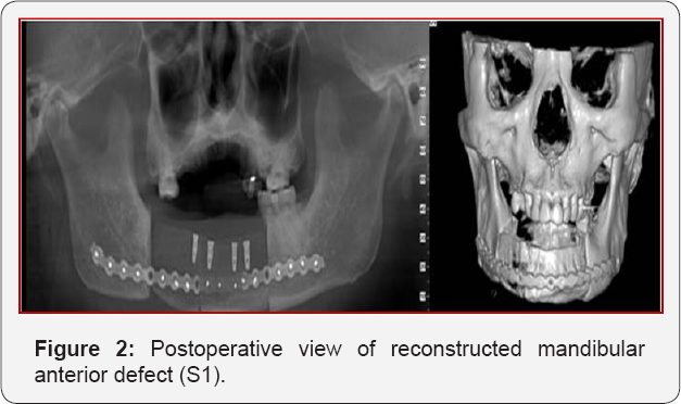
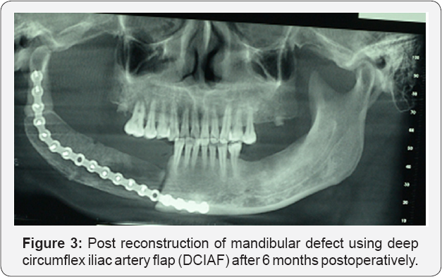
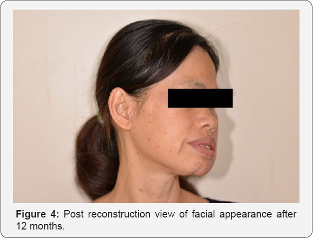
The deep circumflex iliac artery flap outcome was evaluated with CBCT computed tomography Scan images over 6-12 months post reconstruction of mandibular defects (Figures 2 & 3). When postoperative images showed complete consolidation and continuity of the bone graft with residual mandible it means, the outcome was successful. Clinical outcome was evaluated in terms of function and aesthetics. Standardized questionnaires were used to assess the outcomes: pain, mouth opening, recipients' site alterations and facial appearance/aesthetics (Figures 4 & 5). To assess the functional outcomes three points scale were used in all patients: Excellent, acceptable and poor. It was defined as "Excellent" when the patient no feeling pain, no alteration of mouth opening and recipient site. "Acceptable" when the patient feeling moderate pain in mouth opening and " poor" when the patient feeling pain, limitations of mouth opening and alteration of the recipient site were significant. Aesthetic or facial appearance outcomes were evaluated as "good" when the patient and surgeon did not find any alteration to site of reconstruction, "acceptable if alterations were moderate and "poor" when alterations were significant [10]. Postoperative complications related to the donor site were not find, excepted one (1) patient had fistula in the recipient site due to radiotherapy before reconstruction.

Statistical Analysis
Data were performed using SPSS for window version 17.0 (SPSS. Inc. Chicago, II USA), means with standard deviation (S.D) and variables analyses were obtained for ages, gender, site of defects, bone harvest dimension, clinical outcome and complication encountered postoperatively. A test of significance was set at P value<0.05. P values were calculated and there was not significant difference between the independent variables (ages, gender , sites of defects, bone harvest dimensions, clinical outcomes and complications encountered) and dependent variables (days in staying in hospital).
Results
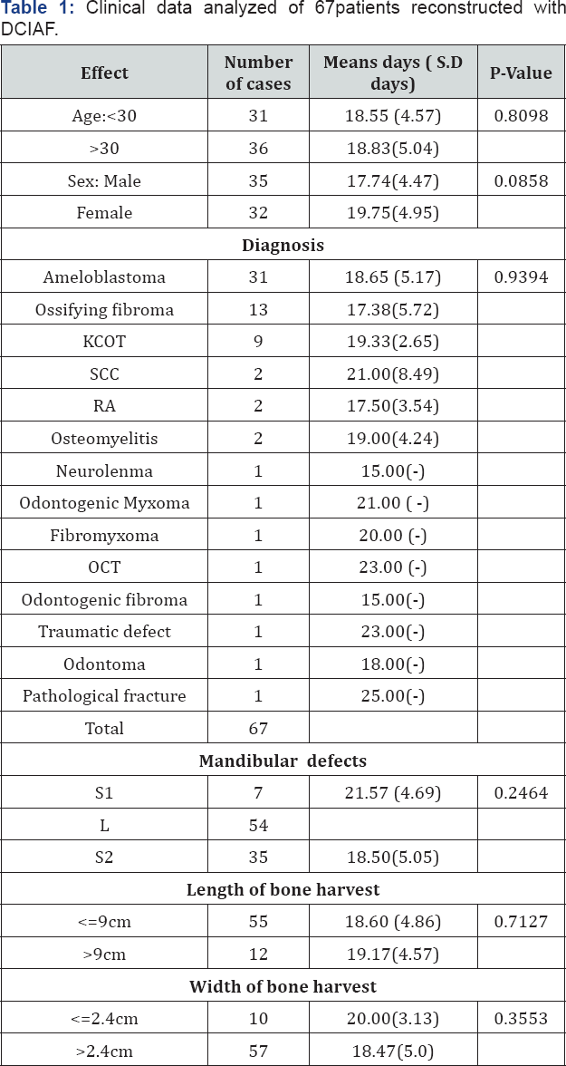
Between July 2014 and August 2016, we treated sixty seven (67) patients with tumor diseases and traumatic defect in mandible. Mean age of patients was 36.6 years (ranging from 14 to 63 years) with males (n=35) and females(n=32). The average (standard deviation) of males was 17.74(4.47) while for females was 19.75(4.95) days in staying in hospital with P value (P<0.08). Of sixty seven (67) patients indicated for reconstruction due to malignant tumors (n=2); benign tumors (n=63) and others (n=2), were treated successfully using deep circumflex iliac artery flap. Reconstruction of mandibular defects based on the new classification for mandibular defects after oncological resection described by James S Brown was used. Reconstruction sites involved fifty four (54) cases as type class I; seven (7) cases as type class III and thirty five(35) cases as type class IIc.
Harvesting bone as donor site, the length less than nine (9) cm, thirty (30) males and 25 females , while the length of bone harvest greater than nine( 9) cm , males (n=5) and females( n=7). The average( standard deviation ) of days staying in hospital in both males were 18.60(4.86) and females were 19.17(4.57). Ten (10) cases with width of bone harvest less than 2.4 cm with average (S.D) was 20(3.13) days ; while fifty seven(57) cases greater than 2.4 cm with average (S.D) was 18.47(5.0). Functional and aesthetic or facial appearance outcomes were mostly "good" in fifty four cases as type class I and " acceptable" thirty five(35) cases as type class IIc . Only one of seven (7) cases as type class II had fistula due to radiotherapy before reconstruction, no donor site complication was recorded in all patients. Reported clinical data analysis with mandibular defects P value (P<0.246) and dimension of bone harvest (length of bone P<0.712 and width of bone P<0.355) indicated to be statistical not significant as show in (Table 1).
Discussion
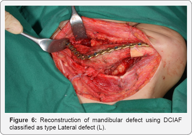
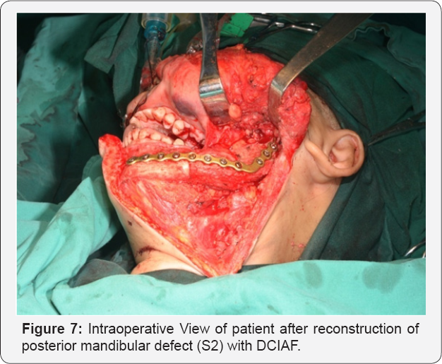
Advances in microsurgical technique free vascularised bone grafts have become the preferred method for reconstructing mandibular defects. However multiple variety of donor sites have been introduced, such as the fibular flap, scapular, rib, radius, and deep circumflex iliac artery flap. In this study; the choice of vascularised deep circumflex iliac artery flap (DCIAF) was based on the site and sizes of defects of mandibular and the population was relatively young with ages less or greater than 30 years. Based on type of defects, sizes; all patients were selected in this study with emphasis on lateral side of mandibular defects(L); posterior mandibular defects (S2) and anterior mandibular defects ( S1) show in (Figures 6-8).

We treated all patients in this clinical study according to the new classification for mandibular defects after oncological resection described by James S Brown [9]. The introduction of this new classification is a step forward because existing suggestions of classification have not been widely adopted. Urken and Colleagues classification is purely descriptive and does not suggest a method of reconstruction for mandibular defects. In additional Jewer et al. [11] or HCL classification defined as follows: H: hemi mandible segment that includes the condyle; C: bone defect involving the entire central segment between 33 and 43 and L: lateral segment excluding the condyle. Based on this classification, most of the oncological patients underwent mandible resections of types L, H, CH, HC, LC, LCL and HH. This classification was not used in this retrospective study of sixty seven (67) patients. However, all the patients were treated according to the classification described by James S Brown, of 67 cases, 54 cases as class I, 7 cases as class III and 35 cases as class IIc, no class IV and IVc (no extensive resection).
Both sizes of each defect encountered were less than 9 cm, 10 cm and 12cm in length. Functional outcomes and facial appearance were observed in Class I and Class IIc, accepted one case in class III had fistula in the recipient site, who underwent radiotherapy before surgical reconstruction of defect, after 36 days the wound was healed. The site of the mandibular defect seemed highly predictive with the highest failure rate (35%) in the anterior region reported by Boy JB et al [12]. Radiotherapy was associated with recipient site complication and outcome in this study, although Clark et al. [13] found significantly more surgical complications in patients who had undergone irradiation previously. Controversy exists as to the effect of radiotherapy on surgical complications and success after free flap reconstruction. Cloke et al. [14] did not find more wound infections in oral cancer patients with free flap reconstruction who had undergone irradiation previously.
In the present study, we successful used the new classification for mandibular defects after oncological resection using deep circumflex iliac artery flap to reconstruct the recipient site with functional and aesthetic outcomes as " good" in fifty four cases for Class I defect, thirty five cases as " acceptable" for class IIc . However for extensive resection, this study did not show good results because for limitations of size of defect less or exceeding 9 cm in length. The study findings by Ho Yi et al. [15] showed that the deep circumflex iliac artery flap (DCIAF) is easy to contour, has minimal complication at the donor site and achieved good functional and cosmetic repair of the mandibular defect, and the use of DCIAF as the flap of choice for reconstruction of mandibular defects.
The disadvantages of a new classification for mandibular defects after oncological resection are that soft tissue defects and type of mandible in terms of dentate status have not been incorporated. The principle concerns in mandibular reconstruction are restoration of the occlusion in a dentate cases and obtaining a functional and aesthetic/or facial appearance. Immediate implant might need to be placed in cases of class II or Class III directly into the grafted bone which has been reported in the medical literature [16]. Despite of the typical factors like defects sizes, location and location condition of the recipient site can be a factor influencing the donor site. In this retrospective study, any complication of donor site did not encountered and showed the new classification for mandibular defects after oncological resection is useful in clinical practice with acceptable outcomes of patients treated with deep circumflex iliac artery flap. This study has some limitation because for retrospective study and the number of cases are few to show the statistical significant and need in the future the prospective study with more patients treated.
Conclusion
Despite our clinical data, it is suggestible that the deep circumflex iliac artery be considered for anterior, lateral and posterior mandibular defect reconstruction, considering the size and defect less or exceeding 9 cm.
References
- Chandu A Smith AC, Rogers SN (2006) Health related quality of life in Oral Cancer: a review J Oral Maxillfac Surq 64(3): 495-502.
- Murphy BA, Ridmer S, Wells N (2007) Quality of life research in head and Neck cancer: a review of the current state of the science. Crit Rev Oncol Hematol 62(3): 251-267.
- Brown JS (1996) Deep circumflex iliac artery free flap with internal oblique muscle as a new method of immediate reconstruction of maxillectomy defect: Head Neck 18(5): 412-421.
- Kudo K Injioka Y (1978) Review of bone grafting for reconstruction of discontinuity defects of the mandible. J Oral Surg 36(10): 791-793.
- Bitter K, Schlesinger westerman U (1983) The iliac bone or osteocutaneous transplant pedicled to the deep circumflex iliac artery J Maxillofac Surg 11:241-247.
- Hidalgo DA(1989) Fibular Free Flap: A New method of mandible Reconstruction. Plast Reconstr Surg; 84:71-79
- Van Gemsert IT, Van Es RJ, Van Cann EM, Koole R (2009) Nonvascularised bone grafts for segmental reconstruction of the mandible a reappraisal. J Oral Maxillfac Surg 67(7):1446-1452.
- Yu DD, Schmidt BL (2008) Quality of life evaluation for patients receiving vascularized versus nonvascularized bone graft reconstruction of segmental mandibular defects. J Oral Maxillofac Surg 66: 1856-1863.
- James S Brown, Canor Barry, Michael Ho, Richard Shaw (2016) A new classification for mandibular defects after oncological resection. Lancet Oncol 17: 23-30.
- Van Gemert JT, Van Es RJ, Rosenberg AJ, Van derBilt A, Koole R, et al. (2012) Free vascularized flaps for reconstruction of the mandible: Complications, success, and dental rehabilitation J Oral Maxillofac Surg 70: 1692-1698.
- Jewer DD, Boyd JB, ManKteLow RT, ZuKer RM, Rosen IB (1989) Orofacial and mandibular reconstruction with the iliac crest free flap: A review of 60 cases and a new method of classification. Plast Reconstr Surg 84: 391-403.
- Boyd JB, Mulholland RS, Davidson J (1995) The free flap and plate in oromandibular reconstruction : Long-term review and indications. Plast Reconstr Surg 95: 1018-1028.?
- Clark JR, Mc Cluskey SA, Hall F (2007) Predictors of Morbidity following free flap reconstruction for cancer of head and neck Head Neck 29: 1090-1101.
- Cloke DJ, Green JE, Khan AL (2004) factors influencing the development of wound infection following free flap reconstruction for intra-oral cancer. Br J Plast Surg 57: 556-560.
- Ho Yi, Korea Lee JH, YiH, LeeI W (2007) Mandibular reconstruction using the DCIA Composite free flap.
- Zenha HA, Rios L, Pinto L (2011) The application of 3-D Biomodeling technology in complex mandibular reconstruction - experience of 47 clinical cases. Eur J Plast Surg 34(4): 257-265.


