Influence of Tympanic Membrane Perforation on Hearing Loss
Teja Deepak Dessai1* and Rhea Philip2
1Lecturer, Nitte Institute of Speech and Hearing, India
2Undergraduate student, Nitte Institute of Speech and Hearing, India
Submission: March 04, 2016; Published: March 23, 2017
*Corresponding author: Teja Deepak Dessai, Lecturer, Nitte Institute of Speech and Hearing, Nithyananda Nagara, Derelakatte, Mangalore, Dakshina Kannada, India, Tel: +91 824 220 4069; Email:dessaiteja@gmail.com
How to cite this article: Teja D D, Rhea P. Influence of Tympanic Membrane Perforation on Hearing Loss. Glob J Oto 2017; 5(5): 555673. DOI: 10.19080/GJO.2017.05.555673
Abstract
Ear is the most pressure sensitive organ of the body and is susceptible for rupture with altered pressure. Tympanic membrane (TM) perforation reduces the total ratio of surface area, allowing the sound waves to directly pass through the middle ear However, implications of the tympanic membrane perforation on sound transmission in the middle ear is not well characterised. The study was undertaken to correlate size and site of TM perforation with the hearing loss. Participants of the study involved both the sexes with dry ear. Individuals below 15years of age, active ear discharge, tympanosclerosis, ossicular chain fixation, presence of mixed and sensori-neural component were excluded from the study. A total of 50 patients within 15 to 50years with tympanic membrane perforation were recruited for the study. The participants were classified in 5 groups based on the size and site of tympanic membrane perforation.
Pure tone Audiometry was performed on the participants who complained of ear discharge 5-6days before testing. Three frequency (500Hz, 1000Hz and 2000Hz) air conduction thresholds were used to determine the hearing level. One-Way ANOVA test and Duncan post hoc test was applied to obtain a statistically significant difference with p>0.05 to obtain a significant difference between the groups and hearing loss across 250Hz, 500Hz, 1000Hz, 2000Hz, 4000Hz frequencies using SPSS software version 16. Results revealed that the degree of hearing loss increases as the size of TM perforation increases. Clinically, prediction of the hearing loss would be easy based on the site and size of TM perforation.
Keywords: Tympanic Membrane; Hearing Loss; Perforation
Introduction
Since the prehistoric times, Chronic Supperative Otitis Media (CSOM) has played a key role in causing the middle ear disease [1] and is considered as the main cause for hearing loss in most of the developing countries [2]. In India, overcrowd and poor living environment with reduced hygiene and nutrition is commonly observed; thereby, adding to the widespread prevalence of chronic supperative otitis media [1]. Ear is the most pressure sensitive organ of the body and is susceptible for rupture with altered pressure [3]. Tympanic membrane (TM) perforation reduces the total ratio of surface area, allowing the sound waves to directly pass through the middle ear.
CSOM is further divided into two types; tubotympanic and Atticoantral type [4]. Tubo-tympanic kind of CSOM is most commonly characterised with parse tensa perforations. Size of TM perforation may define the severity of hearing loss. With larger perforation size, severity of the hearing loss increases. The site of perforation is in correlation with the degree of hearing loss. Similarly, with total loss of TM, absence of transformer action is noted [5]. Presence of tympanic membrane perforation due to CSOM leads to conductive hearing loss ranging from a mild to moderate loss [4]. Pattern of hearing impairment is in concordance with the site of tympanic membrane perforation and has a quantitative relation with the surface area of TM perforation and hearing impairment [6]. Greater the perforation more number of sound waves pass through it causing a nullifying effect. Occurrence of greater hearing loss is more when round window directly gets exposed to the sound waves, specifically when the tympanic membrane perforation is in the posterior quadrant in comparison to the anterior quadrant. In addition, blow on hearing is more when the perforation is near the attachment of the manubrium [7].
Need for the study
WHO observed overall CSOM prevalence of 7.8 % and CSOM with hearing impairment prevalence of 77% in Indian population. Global burden of CSOM is borne in South East Asian countries like India with regional burden of hearing impairment with CSOM of 57,900,000 out of the 115800 000 total population with CSOM. Around 164million subjects with hearing impairment are the cause of CSOM and 90% of it develops in the developing countries worldwide [4]. South India (Tamil Nadu) has reported CSOM prevalence of 6% in children with 5.2% in preschool children and 6.2% in primary school going children [8].
As Audiologist, we evaluate the tympanic membrane on the daily basis; however, exact prediction of hearing impairment in correlation with TM perforation is still not a straight factor. Anthony and Harrison [9] verified the importance of surface area and site of perforation to conclude on the degree of hearing impairment. However, implications of the tympanic membrane perforation on sound transmission in the middle ear is not well characterised. A superior report on effects of perforation on transmission of middle ear sounds is needed so that the audiologist knows the frequencies and its magnitude of hearing loss to be expected in subjects with various tympanic membrane perforations. Knowledge of such information will allow the audiologist to conclude on the presence of any additional factor leading to hearing impairment. Thus the current study was undertaken to correlate the TM perforation and the frequencies involving the hearing impairment.
Aim of the study
To correlate size and site of TM perforation with the hearing loss
Method and Materials
The present study was carried out on first 50samples with TM perforation in the department from Jan-July 2015. Participants of the study involved both the sexes with dry ear and who gave a verbal consent of participation in the study. Individuals below 15years of age, active ear discharge, tympanosclerosis, ossicular chain fixation, presence of mixed and sensori-neural component were excluded from the study. Pre-audiological assessment included detailed history taking, visualisation of the ear and the tympanic membrane in the department of Oto- Rhino-Laryngology to describe the site and size of perforation. Individuals were further divided in 5 groups depending on the site and size of perforation.
- Large Central perforation
- Small Central perforation
- Pin hole perforation
- Posterior superior/inferior quadrant perforation
- Anterior superior/inferior quadrant perforation
Audiometric evaluation was carried out with a clinical calibrated audiometer calibrated ISO standards in a sound treated room setup. Pure tone Audiometry was performed on the participants of the study who complained of ear discharge 5-6days before testing. Both air and Bone conduction thresholds were determined at frequencies 250Hz, 500Hz, 1000Hz, 2000Hz 4000Hz and 8000Hz using Hughson-Westlake procedure [10]. Three frequency (500Hz, 1000Hz and 2000Hz) air conduction thresholds were used to determine the hearing level. Data analysis was done using SPSS software version 16. One-Way ANOVA test and Duncan post hoc test was applied to obtain a statistically significant difference with p>0.05 to obtain a significant difference between the groups and hearing loss across 250Hz, 500Hz, 1000Hz, 2000Hz, 4000Hz frequencies.
Results & Discussion
Total of 50 individuals were enrolled in the study. The result of the study is as below. Individuals below 15years were excluded from the study due to the possible difficulty in testing. Similarly, individuals above 50years were excluded because they may exhibit presence of presbycusis. For calculation of the hearing level three (500Hz, 1000Hz and 2000Hz) frequencies were considered. Pure tone audiometry is considered as a golden tool in assessing the thresholds of the individual; thus, it was implemented in the study.
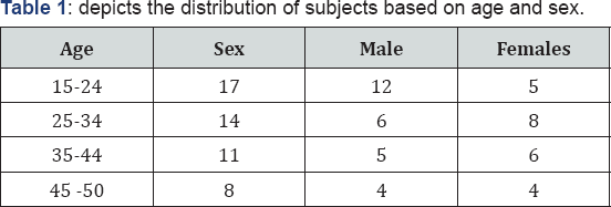
Age and Sex distribution

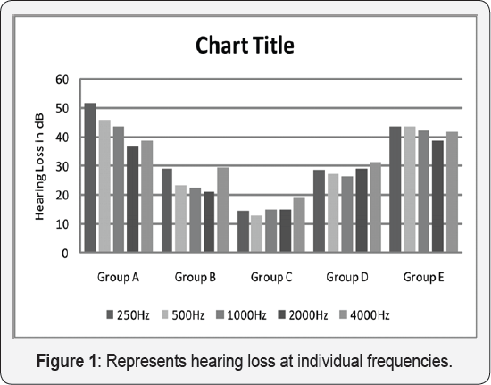
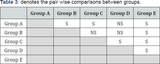
In the current study, most commonly affected age group was 15-24years with 17 individuals. The possible reason could be that this group is socially active and is more health conscious. This result is similar to the finding of 15 individuals between the age ranges of 13-25years [11] (Figure 1). With increasing size and site of TM perforation hearing loss also increased. The data revealed a positive relationship with size and site of TM perforation. Maximum hearing loss (41.6dB) was observed in A group and minimum (14dB) in C group. One-way ANOVA with group as between subject factor revealed significant main effect of group [F(4,45)=10.65, p<0.001]. Duncan's post-hoc comparison was done to resolve main effect of group. These results are given in Tables 1-3.


S: Significant; NS: Non-Significant
These findings are in similar to the findings of Ahmad 8Ramani [7]. The study reported 30dB hearing loss in above 40% TM perforation and 8dB in 10% of TM perforation. In addition, Austin [12] also reported similar findings. Hearing loss steadily increases with increasing size of TM perforation. Pressure sensitive transducer tympanic membrane performs conduction of sound waves across the middle ear, acts as a shield to the round window preventing the direct exposure of sound waves called as 'round window baffle' and also aids in protection mechanism of the middle ear clefts. Loss of Round window baffle that creates phase difference and prevents direct impinging of sound on the oval and round window simultaneously and dampens the unilateral flow of sound waves to the oval window would have been the possible understanding of 41.3dB loss in E group [13]. Literature reveals a strong correlation between site and size of TM perforation with the hearing loss in animals and human [7,9]. Current study confirmed the above findings that the site and size of TM perforation has a major role on hearing. However, more research is called for as the sample size used in the study is limited.
Conclusion
The current study attempted to correlate the degree of hearing loss to the different site and size of perforation. From the present study we can infer that the degree of hearing loss increases as the size of TM perforation increases. Clinically, prediction of the hearing loss would be easy based on the site and size of TM perforation.
References
- Booth JB (1997) Management of chronic suppurative otitis media. Scott-Brown's Otology 3: 10-11.
- Shrestha R, Baral K, Neil W (2001) Community ear care delivery by community ear assistants and volunteers: a pilot study. J Laryngol Otol 115(11): 869-873.
- Patow CA, Bartels J, Dodd KT (1994) Tympanic membrane perforation in survivors of a SCUD missile explosion. Otolaryngol Head Neck Surg 110(2): 211-221.
- Child and Adolescent Health and Development Prevention of Blindness and Deafness World Health Organization Geneva, Switzerland 2004, Chronic suppurative otitis media Burden of Illness and Management Options ISBN 92-4-159158 7.
- (2005) American Academy of Otolaryngology-Head and neck. Perforated Ear Drum. US: American Academy of Otolaryngology-Head and neck.
- Payne MC, Githler fJ (1951) Effects of Perforations of the tympanic membrane on cochlear potentials. Arch. Otolar 54(6): 666-674.
- Ahmad SW, Ramani GV (1979) Hearing loss in tympanic membrane perforations. JLO 93(11): 1091-1098.
- Rupa V, Jacob A, Joseph A (1999) Chronic suppurative otitis media: prevalence and practices among rural South Indian children. Int J Pediatr Otorhinolaryngol 48(3): 217-221.
- Anthony WP, Harrison CW (1972) Tympanic membrane perforation effects on audiogram. Archives of Otolaryngology 59(6): 506-510.
- Carhart R, Jerger J (1959) Preferred Methods for Clinical Determination of Pure-Tone Thresholds. J Speech Hear Res 24: 330-345.
- Prasansuk S, Hinchcliffe R (1982) Tympanic membrane perforation discriptors and hearing levels in otitis media. Audiology 21(1): 43-51.
- Austin DF (1990) Mechanism of hearing. In: Glasscock ME, Shambough GE, (Eds). Surgery of the ear. (4th edn). WB Sounders, Philadelphia, USA, pp. 247-248.
- Ogisi FO, Adobamen P (2004) Type I Tympanoplasty in Benin: A 10 year review. The Nigerian Postgraduate Medical journal 11(2): 84-87.


