




 ARR Home




Abstract
Local ocular anesthesia remains an essential component of modern ophthalmic surgery, providing effective analgesia and, when required, akinesia while avoiding many of the systemic risks associated with general anesthesia. Although topical anesthesia is widely used for cataract surgery, orbital regional anesthesia continues to offer important advantages in selected patients. Conventional injection techniques, however, are associated with potentially significant complications, including retrobulbar hemorrhage, chemosis, conjunctival hemorrhage, eyelid ecchymosis, and inadvertent globe penetration. This study describes and evaluates sopra-bulbar anesthesia, a modified orbital regional anesthetic technique developed to provide effective ocular anesthesia and akinesia while potentially reducing complications associated with traditional orbital injections.
Keywords: Sopra-orbital anesthesia; Ophthalmic anesthesia; Regional ocular anesthesia; Cataract surgery; Orbital block; Local anesthesia; Ocular akinesia; Ophthalmic surgery; Retrobulbar anesthesia; Peribulbar anesthesia
Abbreviation: SOA: Sopra-Orbital Anesthesia; LA: Local Anesthesia; GA: General Anesthesia; RB: Retrobulbar Block; PB: Peribulbar Block
Introduction
The use of local ocular anesthesia is vital to the current practices of ophthalmologists worldwide. Local ocular anesthesia has been implemented for the past 140 years starting with Karl Koller in 1884, revolutionizing the ability to perform ophthalmic surgery without the risks of general anesthesia. Common eye procedures can often successfully avoid general anesthesia through localized nerve blocks and topical anesthetics. Most ophthalmic surgeries can be undertaken using local anesthesia, which is administered either topically or via orbital regional techniques. Over recent years, topical anesthesia has gained popularity as a preferred mode, notably for cataract surgery. However, many ophthalmologists favor orbital regional anesthesia. Recent studies suggest that a substantial number of patients exhibit a preference for orbital regional anesthesia due to its advantages. The utilization of local anesthetic techniques is favored as they provide orbital analgesia and/or akinesia with a reduced likelihood of systemic side effects compared to general anesthesia. These attributes are particularly pertinent for elderly and frail patients undergoing ocular surgery. There are risks associated with orbital regional techniques, including retrobulbar hemorrhage, chemosis with conjunctival hemorrhage, lid ecchymosis, and penetration of the globe. However, there is reasonable evidence to conclude that a different approach to localized injections may minimize this risk. This modification involves the creation of an innovative technique called sopra orbital anesthesia, was developed by Charles Zwerling, MD Goldsboro Eye Clinic, N.C. The study explores the innovative technique of sopra orbital anesthesia injection in the context of cataract surgery. The objective is to compare this approach's complications, costs, surgery duration, and success rates against conventional anesthesia methods frequently discussed in the literature for cataract surgery, including retrobulbar, peribulbar, and topical approaches.
Patients & Methods
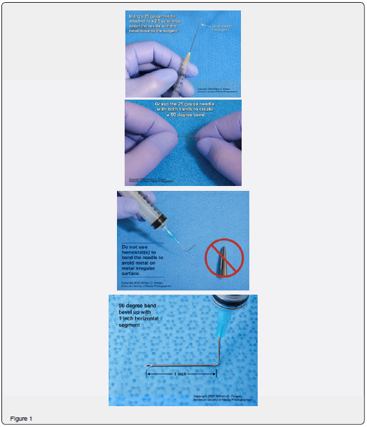
In the past 20 years from 2001-2020, a superior bulbar injection method of local anesthesia was perfected to maximize the effects of local anesthesia for ophthalmic surgery while minimizing the side effects and complications of such a procedure. After the patient has been prepped for eye surgery, the surgeon prepares the injection method as follows. A 1.5” 22-gauge needle is attached to a 5cc syringe which has been filled with the appropriate anesthetic fluids. In this series a 1:1 ratio of 0.5% Marcaine was mixed with 2.5% Lidocaine. The needle is bent to a 90-degree angle so that approximately 0.5” extends directly from the syringe and the remaining 1” of the needle is at 90 degrees with the bevel facing towards the surgeon (Figure 1).
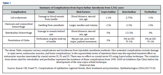
With the patient in a reclined position the surgeon then presses their index finger in the upper eyelid crease displacing the globe inferiorly (a reverse approach to the Schneider method for retrobulbar elevation injection technique) and thus creates a virtual space between the globe and the middle of the supraorbital bone. The 1” shaft of the needle is then introduced into this space and advances slowly until the needle contacts the supraorbital bone. Then the needle is carefully moved posteriorly in a stepwise fashion until the surgeon can’t feel any contact with the supraorbital bone. The fluid is injected slowly filling the sopra orbital space. In most cases only 2.0 cc of anesthetic fluid is needed. The needle assembly is now gently removed.
Clinical Note: There exists no one perfect method for local anesthesia of the globe and /or surrounding tissues. Complications may arise with any technique. Fortunately, no serious injection complications have emerged from this sopra-orbital technique. However, the authors would caution other surgeons that this technique might create a higher risk in highly myopic eyes. Unless the surgeon can create a safe space for needle entry, this technique could cause global perforation which has been documented with retro bulbar injections in highly myopic eyes. Moreover, this technique of sopra-orbital injections would present a higher risk for patients with poor blood coagulation.
Results
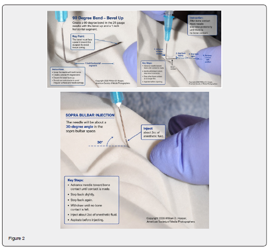
A total of 5,542 cases were performed under local MAC anesthesia at Wayne Memorial Hospital in Goldsboro NC (rebranded as Wayne UNC since 2019). Complications are listed in the table below and consist of intra-operative lid ecchymosis and post-operative lid edema with ecchymosis, ptosis, subconjunctival hemorrhage. Although similar to peri-bulbar anesthetic method, the number of conjunctival hemorrhages was significantly less. Additionally, there is a definite benefit of reduced extra-ocular muscle movement from anesthesia. Of the 5,542 patients in this series, (61.2%) were females and (38.8%) were males. Patients ranged in age from 28 years to 96 years. The average age was 76 years. No allergic reactions during surgery. There were minimal morbidity and no mortality. Vital signs were stable in all cases except for patients with unrelated preexisting physical conditions. No cardiac arrests. Four patients with previous cardiac history exhibited multiple premature ventricular contractions (PVCs) during surgery. All these patients responded as would be expected with routine administration of intravenous lidocaine. No cases of intraoperative nausea or vomiting. 311 patients in the series described some degree of transient, minor nausea after surgery. Minor vomiting episodes occurred in the recovery room in only 5 cases, and no case of severe vomiting occurred. All responded to intravenous metoclopramide. 79 patients reported a moderate postoperative headache. No severe headaches post-operatively reported (Figure 2) & (Table 1).
Discussion
Orbital & Eyelid Anatomy
A thorough working knowledge of the anatomy of the ocular adnexa lids, and orbit is essential in performing both routine and complex procedures. Ophthalmic and anatomical textbooks should be consulted for fine details of lid and adnexal anatomy. The following summary is an overview and is not intended as a comprehensive presentation of orbital and eyelid anatomy. The outline should serve as a guide to the practitioner who is not well acquainted with these areas and a reminder of the most important anatomical landmarks [1-10].
Bony Orbit of the Eye – Summary
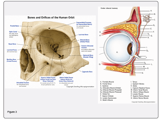
The orbits are bilateral, pyramidal skeletal cavities that house and protect the eyes. Each orbit is formed by seven bones: sphenoid, frontal, zygomatic, ethmoid, lacrimal, maxilla, and palatine, and consists of a roof, floor, medial wall, and lateral wall. The eyeball occupies ~1/5 of the orbital volume, with the remaining space filled by extraocular muscles, neurovascular structures, fat, and connective tissue.
Roof
a) Formed by the orbital plate of the frontal bone and lesser wing of the sphenoid.
b) Separates the orbit from the cranial cavity.
c) Houses the orbital part of the lacrimal gland, frontal sinus, and trochlear fovea (attachment for superior oblique tendon).
d) The supraorbital notch/foramen transmits the supraorbital nerve.
e) Generally thin; thins further with age, requiring care during surgery to avoid trochlear injury (risk of Brown’s syndrome).
Medial Wall
i. Formed by the orbital plate of the ethmoid (lamina papyracea) and sphenoid body.
ii. Lamina papyracea is very thin, prone to fracture or surgical breach, allowing orbital infection spread (e.g., subperiosteal abscess).
iii. Ethmoid foramina transmit anterior and posterior ethmoidal nerves and vessels. Injury can cause orbital bleeding or epistaxis.
iv. Maxillo-ethmoid suture marks a thick inferomedial structure preserved during decompression to prevent globe displacement.
v. Medial wall articulates with roof at frontal-ethmoid suture; excessive bone removal risks cerebrospinal fluid leakage [11-14].
Floor
a) Composed of the orbital plate of the maxilla, articulated with zygomatic (anterolateral) and palatine (posteromedial) bones.
b) Roofs the maxillary sinus; infraorbital groove/canal transmits infraorbital nerves and vessels.
c) Frequently involved in blowout fractures, causing diplopia, enophthalmos, restricted ocular motility, and infraorbital sensory deficits.
d) Posterolaterally, separated from the lateral wall by the inferior orbital fissure, which transmits V2 (maxillary nerve) and is vulnerable during surgery.
Lateral Wall
i. Formed by the greater wing of the sphenoid and frontal process of the zygomatic bone.
ii. Thickest orbital wall, particularly posteriorly, separating the orbit from the middle cranial fossa.
iii. Anteriorly, separates the orbit from the infratemporal fossa.
iv. Zygomatic portion is thin; sphenoid portion thickens near the superior orbital fissure.
v. The bony orbit provides structural protection, defines orbital shape, and houses key neurovascular pathways critical for ocular function and surgical planning (Figure 3).
Eyelid Anatomy – Summary
The upper and lower eyelids are anatomically similar structures. The primary upper eyelid retractor is the levator palpebrae superioris muscle and its aponeurosis, while the primary lower eyelid retractor is the capsulo-palpebral fascia, a fascial extension of the inferior rectus muscle. Embryologically, the levator develops from the superior rectus muscle.
The eyelid consists of six principal layers:
i. Skin
ii. Orbicularis oculi muscle
iii. Levator aponeurosis
iv. Müller's muscle
v. Tarsal plate
vi. Palpebral conjunctiva
Skin
The eyelid skin is among the thinnest in the body, facilitating eyelid mobility and blinking. It is composed of:
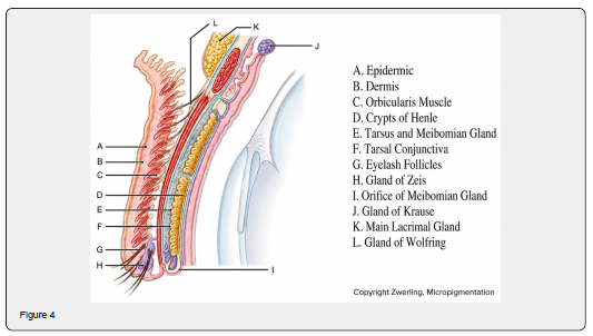
a) Epidermis, consisting of five layers: stratum corneum, lucidum, granulosum, spinosum, and basale. The stratum spinosum and basale form the germinative layer responsible for continual epithelial cell production.
b) Dermis, which contains blood vessels, nerves, connective tissue, hair follicles, sweat glands, sebaceous glands, and smooth muscle fibers.
Unlike the brow and temple, the eyelid contains minimal subcutaneous fat and loose connective tissue (Figure 4).
Eyelashes
Upper eyelid eyelashes are more numerous, thicker, and longer than those of the lower eyelid, typically measuring 7–9 mm in length and arranged in three to five irregular rows.
Upper Eyelid Surface Anatomy
The upper eyelid is divided into:
a) Orbital (pre-septal) portion – between the orbital rim and superior border of the tarsus.
b) Tarsal portion – containing the tarsal plate.
The upper eyelid crease, typically located 7–12 mm above the eyelid margin, is formed by attachments of the levator aponeurosis to the skin. This crease may be absent or less prominent in some Asian individuals.
Lateral periocular skin folds, commonly known as crow’s feet, develop near the lateral canthus. Natural eyelid creases often serve as preferred locations for surgical incisions due to their ability to conceal scars.
Aging Changes
The pre-septal skin lacks firm attachments to deeper tissues and tends to stretch with age, resulting in redundant skin folds. Excess upper eyelid skin, termed dermatochalasis, may overhang the lashes and impair the superior visual field.
Eyelid Glands
The eyelids contain:
i. Glands of Zeis – modified sebaceous glands associated with eyelash follicles.
ii. Glands of Moll – specialized sweat glands located along the eyelid margin.
These glands contribute to maintenance of the eyelid margin and tear film.
Eyelid Margin and Subcutaneous Anatomy – Summary
Lid Margin
The lid margin is the 2 mm transitional zone between eyelid skin and palpebral conjunctiva. Key features include:
a) Puncta: Located 6–7 mm from the medial canthus, dividing the horizontal lid into medial 1/6 and lateral 5/6. Oriented slightly inward toward the globe; marks the nasal end of the tarsus.
b) Gray line: Slightly anterior to the mid-lid margin; anterior border of the tarsus. Meibomian gland orifices lie posterior to it (~30 in upper lid, slightly fewer in lower lid). Medial to puncta, only a mucocutaneous junction is present; the pilar epithelium marks the keratinizing-to-nonkeratinizing transition.
c) Canthi: Lateral canthus ~2 mm higher than medial. Lymphatics drain primarily to parotid nodes.
Subcutaneous Tissue
Beneath the skin is loose areolar connective tissue with minimal fat, allowing free skin movement over orbicularis. Hair follicles originate in this layer; follicle bases lie ~2.5 mm below the epidermis. Eyelid hair follicles lack associated erector muscles.
Orbicularis Oculi Muscle
i. Function: Eyelid protractor; concentric sheet around palpebral fissure. Innervated by cranial nerve VII.
ii. Divisions: Orbital (thickest, strongest), pre-septal, and pretarsal portions.
iii. Attachments: Medial orbital margin, frontal process of maxilla, frontal bones, lateral raphe over lateral canthal tendon. Orbital portion connects with frontalis, procerus, corrugator muscles; lateral portion overlaps temporalis and cheek.
iv. Lacrimal function: Preseptal fibers over lacrimal sac assist lacrimal pump mechanism.
Medial Canthal Tendon
a) Pretarsal orbicularis condenses medially into two heads:
i. Anterior limb: in front of canaliculi
ii. Posterior limb (Horner’s muscle): behind canaliculi, inserts into lacrimal fascia and posterior lacrimal ampulla; aids lacrimal flow during blinking
b) Tendon structure: Superficial thick portion anterior to lacrimal fossa; thin deep portion posteriorly attaches to posterior lacrimal crest.
c) Trauma or excessive skin tension can displace the canthus, damage underlying tissue, cause edema or bleeding, and thin lid layers, increasing risk of deeper needle penetration into orbicularis.
Neurovascular Anatomy and Eyebrows – Summary
Motor Innervation
Motor control of the eye and eyelids is provided by the third (oculomotor), fourth (trochlear), and sixth (abducens) cranial nerves, which enter the orbit through the orbital apex and innervate the extraocular muscles. The seventh cranial nerve (facial nerve) supplies the muscles responsible for eyelid closure and facial expression, including the orbicularis oculi.
Sensory Innervation
Sensory innervation of the eye, eyelids, and surrounding tissues is supplied primarily by the ophthalmic (V1) and maxillary (V2) divisions of the trigeminal nerve (cranial nerve V). These nerves enter through the supraorbital and infraorbital regions and transmit sensation from the orbital and periorbital tissues to the brain.
Autonomic Innervation
The intrinsic ocular muscles and Müller’s muscle receive autonomic innervation. The ciliary ganglion, located near the optic nerve, serves as the synaptic relay for parasympathetic fibers and also transmits sympathetic and sensory fibers. Short ciliary nerves emerge from the ganglion and enter the globe around the optic nerve.
Arterial Supply
The primary blood supply to the eye and adnexa arises from the ophthalmic artery, a branch of the internal carotid artery. After entering the orbit through the optic canal, it courses within the muscle cone and gives rise to several important branches, including:
a) Central retinal artery
b) Ciliary arteries
c) Lacrimal artery
d) Ethmoidal arteries
e) Muscular arteries
f) Supratrochlear artery
Numerous anastomoses exist between the internal and external carotid circulations.
Venous Drainage
Orbital venous drainage occurs primarily through the superior ophthalmic vein and secondarily through the inferior ophthalmic vein.
a) The superior ophthalmic vein drains into the cavernous sinus.
b) The inferior ophthalmic vein drains into the pterygoid venous plexus.
Because orbital and facial veins lack valves, infections can spread between the face, orbit, paranasal sinuses, and intracranial structures through extensive venous communications.
Eyebrows
The eyebrows are crescent-shaped hair-bearing structures located along the superior orbital rim. They overlie an area where the frontalis and orbicularis oculi muscles interdigitate.
i. Frontalis muscle contraction elevates the eyebrows.
ii. Orbicularis oculi, corrugator supercilia, and procerus muscle contraction depresses the eyebrows and contributes to frowning and facial expression.
Historical and Current Anesthetic Methods
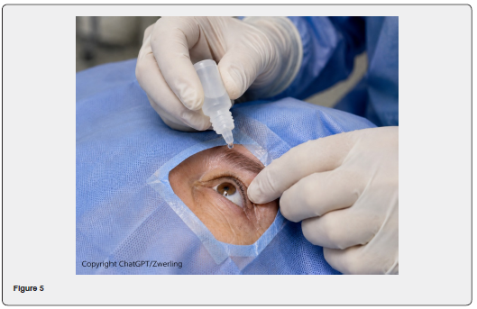
Topical Ocular Anesthesia- One type of ocular anesthesia that is commonly used today is topical ocular anesthesia. As the name applies, topical anesthesia for the eye is limited to cornea, conjunctiva, and anterior sclera only; thus, it avoids the use of anesthetic injections into deeper structures and risks commonly associated with them [15]. Topical ocular anesthesia avoids sutures, is affordable, and keeps the eye well lubricated [16]. It eliminates the need for a patch postoperatively and allows for early use of anti-inflammatory and anti-infective eye drops that prevent surgical complications [16]. The anesthetic agents used in topical ocular anesthesia are classified as either amide or ester compounds and include agents such as proparacaine, tetracaine, benoxinate (oxybuprocaine), cocaine and lidocaine. These agents work by blocking sodium channels inside nerves which in turn stops neural conduction along axons and decreases the perception of pain, however they have no effect on motor block & as such are the not ideal choice of anesthesia when dealing with unruly patients or very invasive eye procedures [15,17]. Additionally, topical anesthetic agent abuse has been associated with corneal defects, epithelial defects, conjunctival injection, & stromal edema [17]. Drawbacks of topical ocular anesthesia, particularly inadequate pain control and lack of motor block are addressed with injectable retrobulbar & peribulbar ocular anesthetic agents (Figure 5).
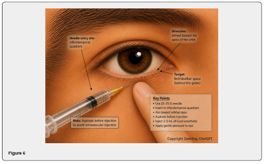
Retrobulbar Anesthesia- In 1884, the same year in which cocaine hydrochloride was discovered to be a useful topical ocular anesthetic, Herman Knapp, a German American ophthalmologist performed the first enucleation (removal of the eye) using a retrobulbar block with cocaine and went on to describe the technique in detail in his book [15]. Retrobulbar anesthesia is a nerve block behind the globe of the eye into the intraconal component [18]. It not only obtains anesthesia of the cornea, uvea, & conjunctiva, but also blocks motor function. Retrobulbar anesthesia inhibits the ciliary nerves, optic n, oculomotor n, & abducens n, which leads to akinesia of the extraocular muscles except for the superior oblique m as the trochlear n is not affected [18].
Retrobulbar anesthesia is a very invasive procedure & great care must be taken to protect sensitive underlying structures. In the modern technique a 31 mm long 23-gauge needle is inserted just above the junction of the inferior and lateral orbital wall and slowly directed upwards and posteriorly into the intraconal space [15]. Afterwards, aspiration is done to rule out damage to any blood vessels and if cleared about 5ml of anesthetic agent is inserted [15]. Some of anesthetic agents that can be used in a retrobulbar block are lignocaine, bupivacaine, ropivacaine, & levobupivacaine [15]. Although retrobulbar block has some advantages over topical ocular anesthesia it also can cause some serious complications such as retrobulbar hemorrhage, cardiorespiratory distress, optic n damage, myopathy secondary to trauma by needle injection, brainstem anesthesia leading to respiratory distress if it reaches the subarachnoid space [15,18]. These serious complications can be mitigated with the equally effective peribulbar anesthetic technique (Figure 6).
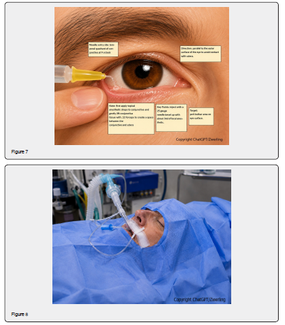
Peribulbar Anesthesia-In 1986, Davis and Mendel reported that peribulbar anesthesia was a safer technique than retrobulbar anesthesia [19]. Peribulbar injection is associated with a lesser chance of retrobulbar hemorrhage, optic nerve damage, ocular cardiac reflex, and brainstem anesthesia [15]. In peribulbar anesthesia, the anesthetic injection is administered extraconal or outside the muscle cone [19]. Complications such as myopathy secondary to injection trauma may be avoided, but a higher volume of anesthetic agent or supplemental injection may be needed to reach the intraconal region and anesthetize the nerves. Some minor complications of peribulbar anesthesia are more frequent conjunctival edema (chemosis) and subconjunctival hemorrhage (ecchymosis) than retrobulbar block, due to anterior spread of the local anesthetic agent and the damage of minor blood vessels with needle tip, but these resolve spontaneously [20] (Figure 7).
General Anesthesia- As an alternative to local anesthetic, general anesthesia should be considered if the potential risk factors of needle penetration of the globe and/or lack of patient cooperation outweigh the risks of general anesthesia. The decision to use general anesthesia can be made after consideration of the length and nature of the procedure and after discussion with the patient and the patient's personal physician. Although needle penetration of the globe during retrobulbar or peribulbar injections is uncommon, its occurrence can be reduced by using alternative methods in high-risk patients. If needle penetration of the globe occurs, management options and final visual acuities are dependent on the severity of the intraocular injury (Figure 8).
Future Use for Sopra-Bulbar Injection Technique
In addition to its potential application for regional ocular anesthesia, the sopra-bulbar injection technique may provide an alternative route for the targeted administration of selected non-anesthetic medications to the orbit. Traditionally, retrobulbar injections have occasionally been employed to deliver therapeutic agents directly into the posterior orbital space when localized drug delivery is clinically desirable. Potential applications include the administration of hyaluronidase in selected vascular emergencies associated with inadvertent intravascular injection of hyaluronic acid dermal fillers and resulting ocular vascular compromise. Retrobulbar administration of neurolytic or neuroleptic agents, including alcohol or chlorpromazine, has also historically been utilized in carefully selected patients with a painful blind eye or severe phantom eye pain when conventional medical therapy has been unsuccessful. In addition, targeted orbital administration of corticosteroids, antibiotics, or other therapeutic agents has been considered in selected inflammatory or infectious orbital disorders under specialist supervision.
The sopra-bulbar approach may therefore have potential applications extending beyond the administration of local anesthetics. By providing an alternative superior orbital route while avoiding the traditional inferior retrobulbar needle trajectory, this technique could potentially facilitate delivery of selected therapeutic agents while reducing some of the mechanical risks inherent to conventional retrobulbar injections. However, these potential non-anesthetic applications should presently be regarded as investigational extensions of the technique rather than established indications. Specific medications differ substantially in their pharmacologic properties, required site of delivery, and potential for ocular or orbital toxicity. Consequently, safety, efficacy, optimal needle position, drug selection, dosage, and clinical indications for non-anesthetic sopra-orbital administration would require prospective investigation before routine clinical use.
Conclusion
A novel technique of sopra-bulbar anesthesia injection has been used for ophthalmic surgery in the past 20 years from 2001-2020. This sopra-bulbar injection method of local anesthesia was developed to maximize the effects of local anesthesia for ophthalmic surgery while minimizing the side effects and complications for these procedures. Overall, the sopra-bulbar injection technique represents a potentially versatile modification of traditional orbital injection methodology. Its clinical value may ultimately extend beyond cataract anesthesia to selected situations requiring targeted orbital drug delivery, warranting further anatomical, pharmacologic, and clinical investigation.
References
- Brown G, Brown MM, Sharma S (2018) The Five Senses: A Patient Preference-Based Comparative Analysis. Clinical Research in Ophthalmology 1: 1-8.
- Scott WA, Bressler MN, Ffolkes S, Wittenborn SJ, Jorkasky J (2016) Public Attitudes About Eye and Vision Health. JAMA Ophthalmology pp. 1111-1118.
- Welp A, Woodbury RB, McCoy MA (2016) Making Eye Health a Population Health Imperative: Vision for Tomorrow. National Academies Press (US).
- Saaddine JB, Narayan KM, Vinicor F (2003) Vision loss: a public health problem? Ophthalmology Journal 110(2): 253-254
- Mundy KM, Nichols E, Lindsey J (2016) Socioeconomic Disparities in Cataract Prevalence, Characteristics, and Management. Seminars in Ophthalmology 31(4): 358-363.
- Lamoureux EL, Fenwick E, Pesudovs K, Tan D (2011) The impact of cataract surgery on quality of life. Current Opinion in Ophthalmology 22: 19-27.
- Boyd K (2022, September 6). What are cataracts? American Academy of Ophthalmology.
- Yu-Chi LM, Wilkins MM, Kim T, Malyugin B, Mehta SJ (2017) Cataracts. The Lancet pp. 600-612.
- Nizami AA, Gulani AC (2022) Cataract. [Updated 2022 Jul 5]. In: Stat Pearls [Internet]. Treasure Island (FL): Stat Pearls Publishing.
- Cataracts - Symptoms and causes. (2022, September 13). Mayo Clinic.
- Davis G (2016) The Evolution of Cataract Surgery. Mo Med 113(1): 58-62.
- Patel S (2023) Alpa Delmonte W, Derek. Houser, Kourtney. (2023, March 15). History of Cataract Surgery. American Academy of Ophthalmology.
- Ademola-Popoola DS, Owoeye JF (2004) Traditional Couching for Cataract Treatment: a cause of visual impairment. West African journal of medicine 23(3): 208-210.
- Kumar M, Chawla R, Goyal M (2015) Topical anesthesia. J Anesthesia Clin Pharmacol 31(4): 450-456.
- Sudarshan Khokhar, Sourabh Verma, Chitrakshi Dhull (2019) Anesthesia in Ocular Surgery. SL Journal of Anesthesia & Critical Care 2(1): 115.
- O'Brien TP (2002) Tips on Topical Anesthesia for Cataract Surgery. Cataract & Refractive Surgery Today.
- Bunya VY, Moon C, Ortiz-Morales G (2022) Topical Anesthetic Abuse Keratopathy. American Academy of Ophthalmology.
- Feldman, Brad H (2022) “Retrobulbar Anesthesia.” American Academy of Ophthalmology.
- Nomura R, Shimada Y, Sugimoto M, Tanikawa A, Mizuguchi T, et al. (2021) Comparison of analgesia and akinesia between sub-Tenon's capsule anesthesia and trans-Tenon's capsule retrobulbar anesthesia in vitrectomy. Fujita Med J 7(3): 105-109.
- Kumar, C M FRCA (2006) Orbital regional anesthesia: Complications and their prevention. Indian Journal of Ophthalmology 54(2): 77-84.