




 ARR Home



Abstract
Background: Background: Postpartum stroke is a rare but serious complication that can manifest with subtle and easily overlooked symptoms [1]. While haemorrhagic events are more common in the peripartum period, ischaemic infarcts, particularly those confined to the occipital lobe may present with visual disturbances such as a homonymous hemianopia where there is loss of the same visual field (either left or right) in both eyes.
Case Presentation: We report the case of a 33-year-old primigravida who developed an abrupt bilateral left-sided visual defect two days after an uncomplicated elective caesarean section. She exhibited no other neurological deficits, reported no history of migraines or seizure activity, and had had an otherwise normal pregnancy complicated only by transient, untreated hypertension towards term. Urgent MRI confirmed a right occipital lobe infarct. Extensive investigations excluded cardioembolic and thrombophilic disorders, arterial dissection, and venous sinus thrombosis. She was treated with antiplatelet and antihypertensive therapy, with full resolution of symptoms and no residual deficits.
Discussion: This case illustrates the diagnostic challenges posed by isolated cortical visual symptoms in the postpartum period [2]. The differential diagnosis includes eclampsia, posterior reversible encephalopathy syndrome (PRES) [3], migraine aura, and seizure-related phenomena all of which may unduly affect the maternal circulation at or around the time of birth. In this instance, the findings were consistent with an ischaemic infarct affecting the occipital lobe of the brain in the setting of mild gestational hypertension.
Conclusion: This report, along with other public narratives, highlights the neurovascular vulnerability of the post-partum state. It tasks clinicians to maintain a high index of suspicion for stroke during pregnancy and in the setting of neurological deficit no matter how subtle or atypical the symptoms may be.
Keywords:Postpartum stroke; Occipital lobe infarction; Homonymous hemianopia; Gestational Hypertension; Visual disturbance; Maternal cerebrovascular event.
Abbreviations: PRES: Posterior reversible encephalopathy syndrome; RCVS: Reversible cerebral vasoconstriction syndrome; PFO: Paradoxical embolus; CVST: Cerebral venous sinus thrombosis
Introduction
Stroke is a leading cause of severe maternal morbidity in high-income countries and carries a disproportionately high burden among postpartum women [4]. Although uncommon, its incidence appears to be increasing due to rising maternal age and hypertensive disorders of pregnancy [5]. The risk of maternal stroke peaks during the first few weeks postpartum, when the normal physiological changes of pregnancy are compounded by abrupt shifts in circulating blood flow, hormonal withdrawal, changes to vascular tone, and coagulation [6]. Although rare, cerebrovascular stroke is a serious and life-threatening complication of pregnancy which is made more likely by the unique susceptibility of the maternal brain and circulation after child birth [7]. In Camargo and Singhal’s 2023 epidemiological review, risk of adverse haemorrhagic or ischaemic events was found to peak in the peripartum period and remained elevated throughout the following six weeks [8]. This is a time of profound physiological instability, endothelial dysfunction, delayed autoregulation, and microvascular injury particularly in the posterior lobes where autoregulatory buffering is by default, relatively limited [9]. Hypertensive disorders such as pre-eclampsia, reversible cerebral vasoconstriction syndrome (RCVS) [10], arterial dissection, paradoxical embolus (PFO) and hypercoagulability, peripartum cardiomyopathy, and occult arrhythmia (paroxysmal AF) were all identified as potential provocateurs [11].
Posterior circulation infarcts are under-recognised in this population, due in part to their subtle presentation and the tendency to attribute early symptoms—such as headache, dizziness, and visual disturbance—to benign postpartum fatigue or migraine [12]. Homonymous hemianopia (HH), an indicator of retro-chiasmal injury, may be the only presenting feature of occipital infarction and is often missed without formal visual field testing [13]. The differential diagnosis is broad and includes eclampsia, posterior reversible encephalopathy syndrome (PRES) [14], reversible cerebral vasoconstriction syndrome (RCVS), migraine with aura, and multiple sclerosis. Rarer conditions include pituitary apoplexy, cerebral venous sinus thrombosis (CVST) [15], vasculitic processes such as primary angiitis of the CNS, or embolic phenomena secondary to peripartum cardiomyopathy or paradoxical embolus via patent foramen ovale. Metabolic and infectious causes, including hypoglycaemia, sepsisassociated encephalopathy, and varicella-zoster vasculopathy, should also be borne in mind in the postpartum population, where immune and vascular homeostasis remain in flux [16].
We report a case of isolated homonymous hemianopia caused by right occipital infarction in a postpartum woman with mild gestational hypertension. It lends weight to the understanding that postpartum cerebrovascular instability may permit adverse perfusion events even in the absence of significant antenatal or peripartum morbidity [17]. This highlights a call for greater vigilance of maternal wellbeing up to and beyond birth not only to mitigate the risk of stroke, but to manage it appropriately when it does occur [18].
Case Report
A 31-year-old primigravida presented postpartum following an uncomplicated elective caesarean section at term. Her initial postoperative course was unremarkable. However, on the second day, she reported a sudden onset of subtle visual loss, describing an inability to see objects in the left visual field of both eyes.
On examination, she was fully alert and orientated and had no symptoms of headache, nausea, or vomiting. Her blood pressure was noted to be 155/95 without significant proteinuria. Neurological findings were normal. Cranial nerve function, peripheral strength, sensation, coordination, and language were all intact. Pupillary responses and visual acuity were normal. Clinically, she presented with an isolated left homonymous hemianopia in the setting of good postpartum recovery. Her medical history was notable only for gestational hypertension, identified at routine antenatal assessment in late pregnancy with blood pressure recordings up to 140/90. Clinical and biochemical investigation excluded pre-eclampsia or renal dysfunction. She was managed conservatively with ongoing surveillance without need for antihypertensives. She had no personal or family history of migraine, thrombophilia, or autoimmune disease. She did not smoke or take illicit recreational drugs.
An urgent non-contrast CT brain with angiography revealed an acute infarct in the right occipital lobe. There was no evidence of haemorrhage, arterial stenosis, or dissection. MRI with MRA and MRV, performed on day five, confirmed an ischaemic infarct with no signs of venous sinus thrombosis, aneurysm, or posterior reversible encephalopathy syndrome (PRES). The infarct pattern was localised to posterior cerebral territory without large vessel occlusion, suggesting a microvascular or perfusion-based aetiology. Transthoracic echocardiography with bubble study was normal, showing no evidence of a cardiac thromboembolic source or shunt. Lower limb Doppler ultrasound excluded deep vein thrombosis. Thrombophilia screening—including anticardiolipin IgM/IgG, beta-2 glycoprotein antibodies, and lupus anticoagulant—was negative.
Initial management included commencement of amlodipine which was later transitioned to sustained-release nifedipine (30 mg daily), in combination with enalapril 5 mg. Antiplatelet therapy was commenced with aspirin 100 mg daily and a short course of clopidogrel 75 mg. During her remaining hospital stay, the patient had full resolution of visual deficit. She was discharged on Day 5 by which time there had been complete resolution of her visual defect. She remained on oral treatment and her BP was stable at 120 to 140mmHg systolic and 80 – 85 diastolic. She was booked for a repeat MRI and outpatient follow-up arranged with neurology and cardiology units.
Discussion
Cerebral Autoregulation and the Postpartum Brain: A System on the Edge
A thunderclap headache in the postpartum period is often a harbinger of acute cerebrovascular crisis—typically described as a sudden, severe headache that reaches maximal intensity within seconds. Though the differential diagnosis may include reversible cerebral vasoconstriction syndrome (RCVS), posterior reversible encephalopathy syndrome (PRES), and cerebral venous sinus thrombosis (CVST), the most alarming associations remain acute cerebral haemorrhage or ischaemia. Such presentations underscore the inherent vulnerability of maternal cerebral circulation during pregnancy and the puerperium. Under normal conditions, cerebral blood flow remains stable due to autoregulatory mechanisms that maintain uniform perfusion across a wide range of mean arterial pressures (MAP). However, in the context of pregnancy and the immediate postpartum period, this equilibrium becomes precarious. Physiological changes—such as hypercoagulability, endothelial activation, abrupt hormonal withdrawal, and sudden hemodynamic shifts—may act independently or synergistically to exceed the limits of autoregulatory capacity, thereby unravelling this protection (see Table 1).
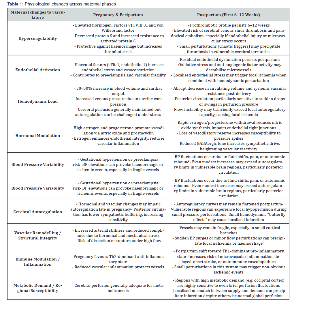
The resulting autoregulatory compromise reduces cerebral resilience and leaves the brain more susceptible to fluctuations in vascular load and endothelial stress [9]. Concepts from chaos theory offer a useful metaphor: in systems balanced at the edge of stability, even small perturbations can cascade into dramatic, disproportionate outcomes. In such a state, transient hypertensive spikes—often clinically unremarkable—can serve as the tipping point that initiates vascular injury and perfusion deficits. Although haemorrhagic strokes are more common in this population, ischaemic events—particularly those affecting the occipital lobes—present unique diagnostic challenges. The posterior circulation is disproportionately susceptible to these perturbations. This may be due to its reduced sympathetic innervation and narrower autoregulatory margins compared to the anterior circulation, possibly reflecting evolutionary trade-offs that favour high metabolic function (e.g., visual processing) at the cost of reduced vascular resilience.
The puerperium, then, is a phase of heightened vascular vulnerability. In this context, previously tolerable physiological shifts may surpass thresholds for vascular stability. The postpartum state reveals latent fragilities in the cerebrovascular system, particularly in metabolically active regions like the occipital cortex, which are exquisitely sensitive to momentary drops in perfusion. In the case described, an ischaemic episode selectively affected the occipital lobe, manifesting as an isolated homonymous hemianopia. The patient had no antecedent history of migraine, epilepsy, or prodromal features of PRES or fulminant pre-eclampsia. Her visual loss was objectively corroborated with field testing, distinguishing it from the more subjective and variable deficits seen in migraine aura or postictal states. It is likely that a convergence of transient hypertension, postpartum endothelial fragility, and metabolic vulnerability within the occipital region culminated in this focal insult, while sparing other cerebrovascular territories.
Homonymous hemianopia (HH)
Homonymous hemianopia is defined as the loss of the same side of the visual field in both eyes—either left or right. This type of loss reflects an interruption in neural pathways responsible for transmitting information from the contralateral visual field to the occipital cortex. In this case, HH arose as a complication of an occipital lobe infarct. More commonly, however, it is associated with retro-chiasmal damage involving the optic tract, lateral geniculate nucleus, optic radiations, or occipital cortex. It is also a recognised feature of posterior reversible encephalopathy syndrome (PRES), which classically presents with headache, seizures, encephalopathy, and visual disturbances in the context of acute hypertension. HH may also occur in eclampsia but is generally accompanied by seizures, proteinuria, or other systemic signs of end-organ involvement. Additional differential diagnoses include migraine with aura and seizure-related visual disturbances, though these tend to be more transient, subjective, and variable.
Patients with HH often develop subtle compensatory behaviours, such as head turning, line-skipping while reading, or manifestations of visual-spatial neglect. Diagnosis requires a high index of suspicion and detailed clinical evaluation, with confrontation visual field testing serving as a crucial bedside tool. MRI is typically used to confirm the diagnosis and to localise the cortical insult. Prognosis is highly variable. Some patients experience spontaneous improvement over several months, driven by neuroplasticity and partial sparing of the occipital pole. However, persistent visual field loss is not uncommon and may significantly impact daily function—particularly reading, orientation, and driving. Rehabilitation is therefore essential to optimise outcomes. Effective strategies include saccadic eye movement training, scanning exercises, and the use of optical devices such as prism lenses to enhance visual awareness. Visionspecific occupational therapy plays a vital role in equipping patients with compensatory techniques and guiding environmental modifications to improve safety and independence. Early referral to neuro-ophthalmology and rehabilitation services should be considered standard practice—especially in cases where visual deficits compromise mobility or functional autonomy.
Postpartum Stroke: Clinical Variability and Diagnostic Vigilance
Several illustrative cases from the literature highlight the clinical and temporal heterogeneity of postpartum neurovascular presentations. Ubaid et al. described a 35-year-old woman who, ten days postpartum, developed visual disturbances and gait instability secondary to a right parietal infarct. In another report, a 26-year-old woman presented ten weeks after delivery with visual symptoms and was diagnosed with a left thalamic infarction. Wildman and Rimawi contributed a further example, detailing a case of pontine infarction during pregnancy— demonstrating that ischaemic events may involve not only cortical but also brainstem structures. Together, these cases underscore the wide range of anatomical territories and timelines in which maternal stroke may manifest. Consequently, clinical vigilance should not be confined to the immediate postpartum period. Events may arise days to weeks after delivery and may present without overt or classical neurological signs. This variability calls for a low threshold for imaging and a broad differential diagnosis encompassing the entire neuroaxis—particularly when symptoms are subtle, ambiguous, or out of keeping with the presumed risk profile. These examples reinforce the necessity for obstetric and neurological teams to adopt a proactive, rather than reactive, approach to postpartum neurological symptoms. Even mild or transient complaints—especially those affecting vision, balance, or cognition—should prompt early neuroimaging to exclude potentially serious pathology.
Acute Management Considerations
The acute management of ischaemic stroke in the immediate postpartum period presents a distinct clinical dilemma. While intravenous thrombolysis with alteplase remains the cornerstone of treatment for acute ischaemic stroke in the general population, its use in postpartum women is complicated by concerns regarding haemorrhagic transformation, uterine bleeding, and impaired surgical wound healing (19). Emerging case-based evidence suggests that thrombolysis may still be considered safe and effective when the risk of maternal disability clearly outweighs the risk of bleeding—particularly if delivery occurred more than 48 hours prior and there is no active haemorrhage, uterine atony, or evidence of coagulopathy (19). Such decisions require careful risk-benefit analysis and should be guided by stroke severity, imaging findings, and the patient’s haemodynamic stability.
Similarly, mechanical thrombectomy—now widely accepted as standard of care for large-vessel occlusions—may offer a viable alternative or adjunct in postpartum patients [20]. Early intervention is particularly warranted when vessel patency and limited infarct core are confirmed on imaging. However, the presence of recent caesarean section, ongoing uterine or vaginal bleeding, or concerns regarding anaesthetic risk must inform collaborative decision-making involving neurology, obstetrics, and anaesthesia teams. Where thrombolysis or thrombectomy is not indicated or feasible, secondary prevention becomes the focus. In patients without evidence of cardioembolic source or thrombophilic disorder, antiplatelet monotherapy—typically aspirin—is considered appropriate. Conversely, in cases where deep vein thrombosis or intracardiac thrombus is suspected, anticoagulation with low molecular weight heparin (e.g., clexane) may be warranted, with careful balancing of thrombotic versus haemorrhagic risk in the postpartum context. This complex risk calculus necessitates a multidisciplinary, individualised approach that honours both maternal safety and the imperative to prevent long-term neurological disability.
Public Health and Long-Term Implications
Postpartum stroke carries significant long-term and public health implications, particularly in terms of maternal morbidity and potential years of life lost. These challenges are especially acute in rural and regional health settings, where limited access to neuroimaging and acute stroke services constrains the window for timely intervention and effective early care. The impact of postpartum stroke reaches well beyond the acute episode. Survivors frequently endure persistent functional, visual, and psychosocial sequelae, which can disrupt maternal–infant bonding, destabilise family dynamics, and impede a return to independent living. A seamless transition from tertiary care to communitybased follow-up is essential to mitigate the risk of recurrence and to support recovery. This includes personalised strategies for risk factor modification, access to psychological support, and functional rehabilitation programs attuned to the unique needs of young mothers. Crucially, these cases underscore that obstetric medicine must extend its vigilance beyond the delivery suite. The postpartum period should be reframed not as a clinical endpoint, but as a critical phase of ongoing risk management—a window for proactive surveillance, targeted education, and timely intervention that can decisively shape long-term maternal outcomes.
Conclusion
Stroke in young mothers may present subtly, with symptoms easily mistaken for benign postpartum complaints. This case highlights the imperative to include cerebrovascular events in the differential diagnosis of visual disturbances during the postpartum period—even in the absence of overt systemic illness, significant hypertension, or hallmark neurological signs such as thunderclap headache or collapse.
Published case reports detailing infarcts in parietal, thalamic, pontine, and occipital regions reinforce the anatomical and temporal heterogeneity of peripartum stroke. While some events occur against a backdrop of recognised risk factors, many arise without warning, in patients with no clear antecedent pathology. In our case, gestational hypertension emerged as the most plausible contributor. Though clinically mild, it may have assumed disproportionate significance within the shifting haemodynamic and autoregulatory landscape of the postpartum brain—a milieu rendered more fragile by the physiological transitions following birth.
The consequences of postpartum stroke are profound, extending beyond maternal morbidity to affect infant development, family dynamics, and long-term psychosocial wellbeing. This case underscores the evolving responsibility of modern maternity care to remain vigilant for neurological symptoms that fall outside traditional risk thresholds. Even mild or isolated complaints— especially those involving visual function—should prompt timely neurological evaluation and imaging to exclude cerebral ischaemia or haemorrhage.
Through this report, we advocate for a broader index of suspicion, reframing the postpartum period as a phase of ongoing cerebrovascular vulnerability where early recognition and intervention may profoundly alter outcomes for mothers and their families.
References
- Sheth H (2024) Postpartum stroke incidence, risk factors, and short-term outcomes in a national cohort study. Neurology.
- Karmarkar P, Camargo L (2023) Isolated visual symptoms following delivery: Diagnostic utility of neuroimaging and the challenge of low suspicion. Journal of Clinical Neuroscience pp. 105.
- Gao F (2022) Posterior reversible encephalopathy syndrome (PRES) in the postpartum period: A systematic review and meta-analysis of clinical features and prognosis. BMC Neurology 22(1).
- Cipolla MJ, Vitullo L (2020) Cerebral autoregulation in pregnancy and preeclampsia: A comprehensive review of human and animal studies. Experimental Biology and Medicine 245(15).
- Maina I (2021) The global burden of stroke in women of reproductive age: A systematic analysis of trends and risk factors. International Journal of Stroke 16(8).
- Bushnell C (2020) Stroke in women: Incidence, risk factors, and outcomes. Stroke 51(3).
- Leffert LR (2021) Anesthetic and obstetric management of peripartum stroke: A narrative review. Anesthesia & Analgesia 133(1).
- Camargo L, Singhal AB (2023) Cerebrovascular events in the peripartum period: A peak vulnerability window. Journal of Thrombosis and Haemostasis 21(9).
- Luo B (2023) Endothelial dysfunction and its role in cerebral vascular stability during pregnancy and postpartum: A focused review. Frontiers in Physiology p. 14.
- Arai Y (2020) Reversible cerebral vasoconstriction syndrome (RCVS) in the peripartum period: A critical consideration in differential diagnosis. Neurology India 68(1).
- D'Amato JE (2022). Pregnancy and postpartum-associated stroke: Risk factor profile, incidence, and management. Clinical Neurology and Neurosurgery pp. 219.
- Méndez TL (2024) Isolated posterior circulation symptoms in young women: A call for early imaging. Journal of Stroke and Cerebrovascular Diseases 33(3).
- Zimmermann M (2021) Homonymous hemianopia as an isolated symptom of occipital infarction in the young adult population: A case series and review. Neuro-Ophthalmology 45(6).
- Fugate JE (2020) Clinical spectrum of posterior reversible encephalopathy syndrome (PRES): Implications for diagnosis and treatment. The Lancet Neurology 19(2).
- Kamel H (2021) Cerebral venous sinus thrombosis in pregnancy and postpartum. Stroke 52(4).
- Brazdil M (2022) Varicella-zoster vasculopathy presenting as a posterior circulation stroke in the postpartum period. Case Reports in Neurology 14(1).
- Singh A (2023) Cerebrovascular instability in normotensive postpartum women: A case for microvascular insult. European Journal of Neurology 30(2).
- Miller BA, Bushnell C (2022) Prevention and recognition of peripartum stroke: A multidisciplinary call for vigilance. Obstetrics & Gynecology Clinics of North America 49(2).
- Abbott LR (2020) Intravenous thrombolysis for acute ischemic stroke in the postpartum period: A systematic review of safety and outcomes. Stroke 51(10).
- Nguyen TN (2023) Mechanical thrombectomy for large vessel occlusion in pregnancy and postpartum: A multi-center experience. Journal of NeuroInterventional Surgery 15(7).

























