Aortic Valve Disease How Do We Treat? Report of the First Case of Ozaki’s Surgery Performed in Angola
Fernando L Bundi*, Guiomar Dos Anjos Correia*, Valdano Manuel and Adriana Bernardo
Department of Cardiovascular and Thoracic surgery, Complexo Hospital Complex for Cardio-Pulmonary Diseases, CDAN, Luanda, Angola
Submission: December 05, 2023; Published: December 13, 2023
*Corresponding author: Guiomar Dos Anjos Correia, Department of cardiovascular and Thoracic surgery, Hospital Complex for Cardio-Pulmonary Diseases CDAN, Luanda, Angola
Ferrnando L Bundi, Department of cardiovascular and Thoracic surgery, Hospital Complex for Cardio-Pulmonary Diseases CDAN, Luanda, Angola
How to cite this article: Fernando L B, Guiomar Dos Anjos C, Valdano M, Adriana B. Aortic Valve Disease How Do We Treat? Report of the First Case of Ozaki’s Surgery Performed in Angola. Ann Rev Resear. 2023; 10(3): 555789. DOI: 10.19080/ARR.2023.10.555789
Abstract
Neocuspidization of the aortic valve with autologous pericardium or Ozaki surgery represents a new and advanced surgical strategy in the management of patients with aortic valve disease and has proven to be effective in the treatment of aortic valve diseases of different causes or entities, representing a versatile alternative to the complex repair of the aortic valve. aortic valve, with better hemodynamics compared to biological aortic valve replacement since it restores the physiological function of the aortic valve and left ventricular remodeling. We present a case of a patient admitted with a significant double aortic lesion with stenotic predominance in NYHA functional class III/IV diagnosed by echocardiography. The patient underwent Ozaki cardiac surgery. without operative complications, and with satisfactory evolution.
Keywords: Aortic Valve; Neocuspidization; Ozaki’s Surgery; Echocardiography; Stenosis gradient
Abbreviations: LVEF: Left Ventricular Ejection Fraction; PTCA: Transpercutaneous Coronary Angiography
Introduction
Aortic valve disease accounts for nearly 50% of all valve disease and affects millions of people worldwide. The gold standard treatment has always been aortic valve replacement, in which the native aortic valve is replaced with a biological or mechanical prosthesis1. Conservative surgical treatment of the aortic valve in patients with aortic valve insufficiency has not had as satisfactory results as that of mitral insufficiency. However, attempts to surgically preserve the aortic valve are, in fact, an expression of the complications that heart valve prostheses present in their evolution, both due to the limitations imposed on patients who receive mechanical prostheses, and the possibility of reoperation to exchange the biological prosthesis that undergoes a degenerative process. Developed in Japan in 2007 by Dr. Ozaki. The Ozaki technique is capable of completely reconstructing an aortic valve with a minimum diameter of 13 mm from the patient’s own pericardium. Therefore, methods used to preserve the aortic valve must be considered, standardized and analyzed in the long term [1,2].
Method
Report of the first case of Ozaki’s Surgery performed in Angola.
Case Report
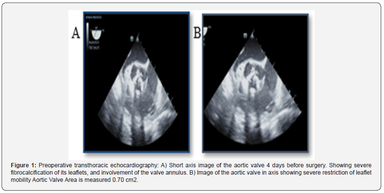
This is the case of a 53-year-old man admitted to the hospital’s emergency department with heart failure associated with aortic valve disease. Past medical history revealed previous hospitalization for Hyperuricemia with crisis and respiratory symptoms, although these have not been well clarified. To determine the etiology, a preoperative transthoracic echocardiogram was performed and showed a left ventricular ejection fraction (LVEF) of 66%, moderate concentric hypertrophy of the left ventricular wall and mild systolic dysfunction. The results of the aortic valve were as follows: Significant fibro calcification of its leaflets, which generates an important limitation in its opening and moderate incompetence in its closure. Aortic valve area 0.70 cm2.
a) Stenosis gradient: maximum pressure gradient 84.8 mmHg, mean pressure gradient 55 mmHg. Double aortic lesion with a predominance of significant aortic stenosis, aortic annulus - 23.5 mm of undefined etiology, due to the presence of significant fibro calcification that compromised the entire valve apparatus. The left ventricular tip diastolic and systolic diameters were 53 mm and 34 mm, respectively. The thicknesses of the interventricular septum and posterior left ventricular wall were 16 mm and 14 mm.
b) Other Tests Performed: Hemoglobin 13 g/dL, hematocrit 38.8%, white blood cells: 5,700 u/L, Neutr: 58.1%. Platelets: 196 u/L, Clucose: 90 mg/dL, Urea: 36 mg/dL, Creatinine: 0.6 mg/dL, Troponin T 0.024 ug/L, Uric Acid 8.0 mg/dL, C-Reactive Protein 0.839 mg/L dL. Transpercutaneous coronary angiography (PTCA) without coronary lesions (Figure 1).

Surgical Technique
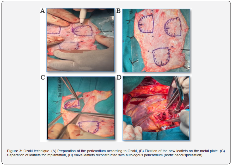
The surgery was performed through a classic median sternotomy, cardiopulmonary bypass was established through cannulation of the artery and double-stage cannulation (superior and inferior vena cava) through the right auricle. Cardiac arrest was achieved by injecting anterograde cardioplegia. A pericardiectomy was performed and fixed on a plastic plate extracted from the Ozaki kit, prepared in glutaraldehyde solution for 5 min to improve stability (OKAZI technique) and rinsed three times for 6 min using saline saline solution. The leaflets were manufactured from autologous pericardium according to Ozaki’s rules. The ascending aorta was incised transversely, and the aortic valve was explored where the echocardiographic alterations were verified, with retraction and calcification of the aortic valve leaflets and the valve leaflet was removed, was reserved for histological study. The valve was reconstructed by implanting the leaflets manufactured and sutured with 5/0 proline to the aortic annulus. Surgery without complications and functional reconstructed aortic valve. Perioperative transesophageal echocardiography was performed (Figure 3A) to confirm the function of the new valve (Figure 2a,b,c,d) and (Figure 3a,b).
Discussion
In this case, aortic valve neocuspidization or Ozaki surgery was performed in a patient with a double aortic lesion of stenotic predominance with significant fibro calcification of its leaflets, surgery performed using cardiopulmonary bypass and myocardial protection, with 145 minutes of by-pass and 94 minutes of cross clamping. The valve was reconstructed using the patient’s autologous pericardium, duly prepared, whose surgical result was satisfactory with good valve functionality. This innovative technique offers multiple advantages: to begin with, it allows us to replace the aortic valve with the body’s own tissue without having to replace to a prosthesis, which will require permanent anticoagulation in the case of a mechanical prosthesis, and which does not follow body growth or the high risk of endocarditis [3], or scheduled reoperation in case of dysfunction over time. Furthermore, in cases where an adequate-sized prosthesis is not available, we can avoid using the pulmonary valve itself, as the Ross operation does, thus preserving the integrity of the rest of the heart’s structures [4,5].

Conclusion
The Ozaki technique is a new procedure used in surgery for aortic valve disease, which preserves the natural dynamics of the aortic root and valve, not intervening on it like the Ross procedure and not fixing it with a rigid artificial ring like aortic prostheses and in our context (Angola) it is important due to the precariousness of primary health services and the availability of regular follow-up in cardiology consultations, thus reducing the costs of purchasing and importing prostheses and reducing the need for permanent anti-coagulation in patients.
References
- Ozaki S, Kawase I, Yamashita H, Shin U, Mikio T, et al. (2018) Midterm outcomes after aortic valve neocuidization with gluterakdehyde-treated autologous pericardium. J Thorac Cardiovasc Surg 155(6): 2379-2387.
- Krane M, Boehm J, Prinzing A, Ziegelmueller J, Holfeld J, et al. (2021) Excellent Hemodynamic Performance After Aortic Valve Neocuspidization Using Autologous Pericardium. Ann Thorac Surg 111(1): 126-133.
- Ivanov Y, Drury NE, Stickley J, Botha P, Khan NE, et al. (2020) Strategies to minimise need for prosthetic aortic valve replacement in congenital aortic stenosis-value of the Ross procedure. Semin Thorac Cardiovasc Surg 32(3): 509-519.
- Riggs KW, Colohan DB, Beacher DR, Alsaied T, Powell S, et al. (2019) Mid-term outcomes of the supported Ross procedure in children, teenagers, and young adults. Semin Thorac Cardiovasc Surg 32(3): 498-504.
- Ross DN (1963) Aortic valve reconstruction surgery. Lancet 1(7281): 571-574.


