Formation and Microscopic Appearance of Fireworks-like Objects Created from Synthetic DNA (E. coli) Crown Cells with Adenosine-DNA (E. coli)
Shoshi Inooka*
Japan Association of Science Specialists, Japan
Submission: May 20, 2023; Published: May 30, 2023
*Corresponding author: Shoshi Inooka, Japan Association of Science Specialists, Japan
How to cite this article: Shoshi I. Formation and Microscopic Appearance of Fireworks-like Objects Created from Synthetic DNA (E. coli) Crown Cells with Adenosine-DNA (E. coli). Ann Rev Resear. 2023; 9(2): 555760. DOI: 10.19080/ARR.2023.09.555760
Abstract
Techniques for producing artificial cells (DNA crown cells), in which the outer membrane surface is covered with DNA were established in 2012. Such cells, hereafter referred to as synthetic DNA crown cells, can be readily synthesized using sphingosine (Sph)-DNA-adenosine mixtures and can proliferate within egg white or in vitro. However, how synthetic DNA crown cells proliferate within egg white or grow in vitro has not yet been clarified in detail. Synthetic DNA (E. coli) crown cells formed assemblies, which were reconstructed either spontaneously or in the presence of salts. The formation of new compounds, such as adenosine-DNA compounds, may occur within the reconstructed assembly. In this study, adenosine-DNA (E. coli) compounds were first prepared then the possible effect on adenosine-DNA (E. coli) by cell formation was investigated. The present experiments showed that fireworks-like objects were formed from synthetic DNA (E. coli) crown cells together with adenosine-DNA (E. coli) compounds. The findings suggested that adenosine-DNA played a role in the new of cell formation mechanism and participated in cell generation.
Keywords: Synthetic DNA crown cell; Adenosine-DNA; Fireworks-like structure; Cell regeneration
Introduction
Approaches for generating artificial cells, which are cells covered with DNA, were first reported in 2012 [1,2]. These artificial cells have been designated as DNA crown cells [3]. DNA crown cells can be synthesized using sphingosine (Sph)-DNA and adenosine-monolaurin (A-M) [4,5], and proliferate within egg white and grow in vitro [6]. However, it is unclear how synthetic DNA crown cells proliferate within egg white or grow in vitro [6]. In a previous report [7,8], the assembly of synthetic DNA crown cells was shown to occur spontaneously or in the presence of inorganic ions, and that synthetic DNA crown cells were modified. Many DNA crown cells were retained within such modified assemblies. Moreover, when monolaurin was added to the modified assemblies, most of the assemblies were transformed to crystal-like substances. The findings showed that original synthetic DNA (E. coli) crown cells were reconstructed twice; when they first formed assemblies and when they formed crystal-like structures. After reconstruction, new cells (regenerated cells) were formed. The findings showed that new cells may have been formed by a new mechanism which differed from that used in the original preparation of synthetic DNA crown cells. Therefore, it was expected that new compounds, such as adenosine-DNA compounds, would be produced. The present study examined whether adenosine-DNA (E. coli) compounds could be prepared. Then, the effect on adenosine-DNA (E. coli) cells due to the formation of generated cells was examined.
Materials and Methods
Materials
The following materials were used in this study. Sph (Sigma, USA and Tokyo Kasei, Japan), DNA (E. coli, B1 strain; Sigma-Aldrich, USA), adenosine (Sigma and Wako, Japan), monolaurin (Tokyo Kasei, Japan), and A-M compound (synthesized from a mixture of a 0.1 M solution of monolaurin and 0.1 M solution of adenosine), which was prepared using distilled water. Ethanol (99.5) (Sigma-Aldrich, USA). In addition, RBC Lysis Solution (Pharmacia Biotech, USA) and Dulbecco’s minimal essential medium (Sigma-Aldrich, USA) containing 10% bovine serum (D-MEM) was prepared.
Methods
Preparation of DNA crown cells: The generation of artificial cells using Sph-DNA-A-M was performed as described previously [4,5]. Briefly, 180 μL of Sph (10 mM) and 90 μL of DNA (1.7μg/ μL) were combined, and the mixture was heated and cooled twice. A-M compound (100 μL) was added to the mixture, which was then incubated at 37ºC for 15 min. Next, 30 μL of monolaurin was added, and the mixture was incubated at 37ºC for another 5 min. The resulting suspension was used as synthetic DNA (E. coli) crown cells.
Preparation of adenosine-DNA (E. coli) compounds: A 100 μL aliquot of adenosine solution was combined with 100 μL of DNA (E. coli) in micro tubes and mixed. Then, 200 μl of ethanol was added to the mixture and mixed. The mixtures were dried at 37ºC for approximately 2~3 days in an open screw-top tube. The precipitates were then resolved with 100 μL of RBC lysis solution. This solution was used as the stock solution for A-D compounds.
Preparation of diluted solution: A 10 μL aliquot of undiluted solution was combined with 100 μL distilled water to give a 101 dilution. Then, 100 μL of distilled water was added to 10 μL of the 101 solutions to give a 102 dilution. This process was repeated to give dilutions of 103, 104,105, 106, 107, 108, 109 and 1010.
Experimental procedure
Experiments using stock solution
First, 0.25 μL of undiluted solution was added to 0.25 μL of synthetic DNA (E. coli) crown cells. Then, 0.25 μL of adenosine solution, RBC lysis solution and distilled water were added to 0.25 μL of synthetic DNA (E. coli) crown cells. After mixing at room temperature, the resulting samples were observed under a light microscope.
Experiments using diluted solution
a) First, 0.25 μL of diluted solution (101 through 102) was added to 0.25 μL of synthetic DNA crown cells and distilled water. After mixing, the mixtures were incubated at 37ºC for 30 min.
Then, 500 μL of D-MEM were added to the mixtures. The mixtures were then incubated at 37ºC for 2 days and the resulting samples were observed under a light microscope.
b) Then, 0.25 μL of diluted solution (106 through 1010) was added to 0.25 μL of synthetic DNA crown cells and distilled water (without cells). After mixing, the mixtures were incubated at 37ºC for 30 min. Then, 500 μL of D-MEM was added to the mixtures and incubated at 37ºC for 3 days, after which the samples were observed under a light microscope.
For the microscopic observations, 10 μL of the sample was placed on a glass slide and covered with a cover slip.
Results and Discussion
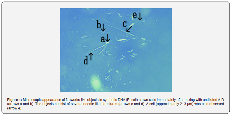
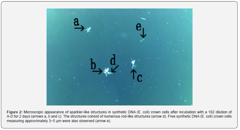
Figure 1 shows the microscopic appearance of fireworkslike objects in a synthetic DNA (E. coli) crown cell mixed with undiluted A-D compounds. However, these objects were not observed consistently. The reason for this is unclear, but, as described previously [9], synthetic DNA (E. coli) crown cells may vary between experiments because preparation with a consistent appearance is difficult. The figure shows several fireworks-like objects (arrows a and b). The objects consist of several needle-like structures (arrows c and d). In addition, a cell (approximately 2~3 μm) was also observed (arrow e). The needle-like objects appeared immediately after the mixtures were prepared and disappeared within approximately 3 hours. Figure 2 shows the microscopic appearance of three sparkler-like structures in synthetic DNA (E. coli) crown cells after incubation with the 102 dilution with the A-D solution for 2 days (arrows a, b and c). The objects consisted of numerous needle-like structures (arrow d). A free synthetic DNA (E. coli) crown cell measuring approximately 3~5 μm was also observed (arrow e). Figure 3 shows fireworks-like objects in synthetic DNA (E. coli) crown cells after incubation with the 106 dilution of the A-D solution for 3 days. Two fireworks-like objects were observed (arrows a and b) after 3 days of cultivation. The objects consisted of numerous needle-like structures. The size of the cell shown in Figure 3 (arrow c) was approximately 2~3 μm.
Figure 4 shows fireworks-like objects in synthetic DNA (E. coli) crown cells after incubation with the 108 dilution of the A-D solution for 3 days. A cluster that appeared to consist of three fireworks-like objects was observed (arrow a). Each object had several needle-like structures (arrow b). The size of the cell shown in Figure 4 (arrow c) was approximately 3~5 μm. Figure 5 shows the magnified view of the structure shown in Figure 4 (arrow a). The object shown consists of assembly-like structures (arrow b) and several rod-like structures (arrows c and d). The structure shown in Figure 4 is described as a cluster. However, it was not a cluster, but may have been derived from an assembly (arrow b), such as that shown in Figure 5 (arrow a). Thus, fireworkslike objects were observed after 2~3 days of cultivation and may disappear. In this study, the effect of adenosine-DNA (A-D) compounds on synthetic DNA (E. coli) crown cells was examined. Here, A-D was prepared using similar methods as those used to prepare A-M compounds [4], and the formation of fireworks-like objects using the mixtures of synthetic DNA crown cells with the compounds was observed.
In previous studies [7,8], it was shown that assembly formation was observed using synthetic DNA (E. coli) crown cells either spontaneously or in the presence of inorganic materials. The assembly contained numerous cells that appeared similar to synthetic DNA crown cells but may differ from the original synthetic DNA crown cells, as described previously [8]. These crown cells are referred to here as modified synthetic DNA crown cells (regenerated cells). Original synthetic DNA crown cells were prepared using Sph-DNA and A-M compounds. However, it is possible that the regenerated cells were formed by another mechanism. The assembly consists of related materials (Sph-DNA and A-M compounds) with the original DNA crown cells. These materials were mixed within the assembly. As a result, it is possible that new chemical reactions based on new combinations of chemical agents (Sph, DNA, adenosine, monolaurin) occur within the assembly and create new compounds, for example, A-D. Based on this assumption, it considered that A-D was formed within the assembly and that it influenced the production of cells. When A-D was added to synthetic DNA (E. coli) crown cells, fireworks-like objects were formed. As is well known, fireworks are enjoyed for their interesting sparkle-like shapes which are formed as a result of gun powder and metals being ignited. Various types of sparkles of various shapes can be made using different combinations of gun powder and metals. In this study, numerous objects that were similar in shape to sparklers were observed. Therefore, these objects are referred to here as cell sparklers as they consist of needle-like objects, threads and a rod. Also, firework sparklers are formed from a ball which contains gun powder and metal, whereas the cell sparklers are formed from an object like assembly which is referred to here as cell bodies. Moreover, the fireworkslike objects that produce sparklers are referred to here as fresh bodies. No fresh bodies were observed during the preparation of the synthetic DNA (E. coli) crown cells before cultures or with RBC lysis solution.

In this study, when undiluted A-D was used, fresh bodies with numerous needle-like objects were formed immediately after the addition of A-D. Fresh bodies with rods were formed when A-D dilutions of 106 and 108 were used. It was clear that the fresh bodies that were formed using undiluted A-D formed due to the effect of the A-D solution, because similar fresh bodies with needle-like structures were not observed in any experiments. On the other hand, objects similar to fresh bodies were observed in the secondary cultures of synthetic DNA (E. coli) crown cells [10]. Therefore, it is unclear whether the fresh bodies that are formed in the presence of diluted A-D solution were formed in response to the addition of A-D. To clarify these issues, it is important to confirm whether the fresh bodies that were formed in the culture experiments were actually formed in response to the presence of materials such as A-D, which was newly produced in the medium. Though it was unclear whether the formation of fresh bodies was attributed to A-D or whether they occurred naturally, the finding that fresh bodies were observed at dilutions above 106 is considered to be important, as described below. Cell sparklers may be produced by the cell body. Cell bodies may consist of materials that were used to prepare the original cells and A-D might act as a catalyst or activating agent. Various types of sparkers, needles, rods, and thread-like objects may be produced as a result of the combination of materials and the A-D concentration.
It has been clearly demonstrated that DNA crown cells are formed by the ring formation of DNA fibers or bunches [8]. Cell sparklers (especially cell needles) may consist of DNA fibers or bunches and it may be possible that new cells are generated from cell sparklers. In the electron microscopy studies on DNA crown cells, the outer membrane of cell was partially degraded and formed linear objects [2]. Moreover, microscopic observations revealed that elongated Sph-DNA objects were observed in the cultures of synthetic DNA (E. coli) crown cells [6]. The elongation of such components was observed to be important in the continuous generation of cells. The findings of the present study showed that A-D elongated components such as Sph-DNA at very low concentrations. The results suggested that elongation of these components occurred without enzymes, such as DNA polymerase, and that cells were generated due to reactions among chemical agents. Thus, A-D may have several functions in the production of DNA crown cells, including cell proliferation [11,12] or cell nest formation [13], and materials such as A-D are believed to exist in nature, especially in egg white. Future research will examine whether A-D-like materials are contained within egg white and whether A-D possesses other functions in addition to cell formation.


References
- Inooka S (2022) Preparation and cultivation of artificial cells. App Cell Biol 25: 13-18.
- Inooka S (2016) Preparation of Artificial Cells Using Eggs with Sphingosine-DNA. J Chem Eng Process Technol l7: 277.
- Inooka S (2016) Aggregation of sphingosine-DNA and cell construction using components from egg white. Integrative Molecular Medicine 3(6): 1-5.
- Inooka S (2017) Biotechnical and Systematic Preparation of Artificial Cells. The Global Journal of Reseraches in Engineering 17(1).
- Inooka S (2017) Systematic Preparation of Artificial Cells (DNA Crown Cells) J Chem. Eng Process Technol pp. 327.
- Inooka S (2022) Preparation of a DNA ( coli) Crown Cell line in Vitro-Microscopic Appearance of Cells. Annals of Reviews and Research 8(1).
- Inooka S (2022) Assemblies Formation of Synthetic DNA ( coli) Crown Cells with Salt. “Reconstruction and Regeneration of Synthetic DNA Crown Cells” American Journal of Biomedical Science & Research 16(1).
- Inooka S (2022) The assembly in synthetic DNA crown cells with inorganic salts and the transformation of DNA crown cells to crystal like substance in the presence of monolaurin Novel Research in Science 11(2): NRS.000759.
- Inooka S (2021) Microscopic observation of assemblies formed in mixtures of DNA (Human placenta) crown cells with Bacillus subtilis cells an archive of organic and inorganic chemical sciences 5(2): 658.
- Inooka S (2022) Microscopic Appearance of Synthetic DNA ( coli) Crown Cells in Secondary Cultures. Novel Research in Science 12(3): NRS.000791.
- Inooka S (2022) Cell Proliferation from the Assembly of Synthetic DNA ( Coli) Crown Cells with Stimulation by Monolaurin Twice. Current Trends on Biotechnology & Microbiology 2(5).
- Inooka S (2023) Cell Proliferation in Assemblies of Synthetic DNA ( coli) Crown Cells in the Presence of MgCl2]] Current Trends on Biotechnology & Microbiology 3(3).
- Inooka S (2023) Formation of Cell “Nest” in Cultured Synthetic DNA (Streptomyces) Crown Cells-Microscopic Appearance of Cells Within “Nests”. Current Trends on Biotechnology & Microbiology 3(4).


