




 ARGH Home




Effect of mammalian target of rapamycin on the activation and migration of hepatic stellate cells induced by transforming growth factor-β
Ji Zhou1, Lili Liu2, Tiancheng Luo2 and Lei Li1*
1Department of Gastroenterology and Hepatology, Shanghai Geriatric Medical Center; Shanghai Institute of Liver Diseases, Shanghai 201104, China
2Department of Gastroenterology and Hepatology, Zhongshan Hospital, Fudan University, Shanghai Institute of Liver Diseases, Shanghai 200032, China
Submission:October 15, 2026;Published:October 29, 2026
*Corresponding author: Shyam S. Meena, Professor & In charge Pediatric Gastroenterology, Hepatology& Nutrition Division, Dept. of Pediatrics, VMMC & Safdarjung Hospital, India
How to cite this article: Shyam S. M, Sailaja G. Post-Viral Gastroparesis: When the Virus Lingers Beyond the Infection. Adv Res Gastroentero Hepatol, 2026; 22(5): 556100. DOI: 10.19080/ARGH.2026.22.556100.
Abstract
Objective: We aimed to explore the effect of the mammalian target of rapamycin (mTOR) on the activation and migration of hepatic stellate cells (HSCs) induced by transforming growth factor-β (TGF-β). Methods: Rat primary hepatic astrocytes (RHStec, HSCs) were induced by TGF-β for 30 min. HSCs were performed intervention with mTOR inhibitor (rapamycin or resveratrol) for 72 h. The migration ability of HSCs was detected by scratch wound healing assay. Western blot and quantitative real-time polymerase chain reaction (qRT-PCR) were used to detect the protein and mRNA expression levels of Rictor, Raptor, α-smooth-muscle actin (α-SMA), collagen (Col) I, Col III, peroxisome proliferator-activated receptor (PPAR)-γ, RhoA and Rho-associated kinase (ROCK) in HSCs, respectively. Results: TGF-β significantly up-regulated the phosphorylation level of mTOR and the protein levels of Raptor and Rictor compared with the control group (p<0.05). Rapamycin intervention for 24 h or resveratrol intervention for 48 h significantly attenuated TGF-β-induced migration of HSCs (p<0.05). Rapamycin or resveratrol attenuated the up-regulation of TGF-β on the mRNA and protein levels of α-SMA, Col-I, Col-III, PPAR-γ, RhoA, and Rock in HSCs (p<0.05). Conclusion: TGF-β can activate the RhoA/Rho signaling pathway through mTOR, thereby regulating HSC activation and migration.
Keywords: TGF-β; mTOR; Activation; Migration; Hepatic stellate cell
Abbreviations: mTOR: Mammalian Target of Rapamycin; HSC: Hepatic Stellate Cells; qRT-PCR: Quantitative Real-Time Polymerase Chain Reaction; SMA: Smooth-Muscle Actin; PPAR: Peroxisome Proliferator-Activated Receptor
Introduction
Hepatic fibrosis is a pathological lesion caused by excessive deposition of extracellular matrix components and abnormal proliferation of fibrous connective tissue in the liver after long-term inflammation caused by various causes [1]. Hepatic stellate cells (HSCs) are the main source of extracellular matrix, and the activation of HSC is the central link in the development of chronic liver disease into hepatic fibrosis [2]. When the liver is stimulated by chronic inflammation, HSC is activated and then secretes a large amount of extracellular matrix, such as type I, III, and IV collagen, promoting the expression of α-smooth muscle actin (α-SMA), cell migration, and microfilament skeleton rearrangement, thereby increasing portal pressure and promoting intrahepatic structural reconstruction, leading to liver fibrosis [2,3]. Hepatic fibrosis is a reversible disease. If not treated in time, fibrotic liver tissue will further develop into cirrhosis and even liver cancer [4]. Therefore, it is particularly important to control and reverse the occurrence of hepatic fibrosis.
Transforming growth factor β (TGF-β) is an important activator of liver fibrosis, which can promote the epithelial/mesenchymal trans differentiation of HSCs by accelerating the activation of HSCs. It affects the collagen expression and extracellular matrix metabolism, and then dominates the initiation and formation of hepatic fibrosis [5]. It has been reported that TGF-β can regulate the actin expression and microfilament cytoskeleton rearrangement of HSCs through the RhoA / Rho signaling pathway, thereby further affecting the cell migration ability [6,7]. The mammalian target of rapamycin (mTOR) is an important regulator of cell growth and proliferation [8]
. mTOR can recruit other proteins and active molecules to form two mTOR complexes, mTORC1 and mTORC2 in the cells, and play a biological function in the form of complexes. mTOR plays an important role in regulating cell migration. It has been reported that mTOR may have the ability to regulate RhoA activity, and TGF-β is reported to simultaneously activate RhoA and mTOR activity in colorectal cancer cells [10]. Therefore, we speculate that TGF-β can regulate cell migration and cytoskeleton reorganization of HSCs through mTOR. In this study, we aimed to explore the effect of mTOR on the activation and migration of HSCs induced by TGF-β.
Material and Methods
Culture and intervention of rat primary hepatic astrocytes (HSCs)
HSCs were purchased from the ScienCell Research Laboratories, Inc. (Carlsbad, CA, USA). HSCs were cultured in high glucose Dulbecco’s modified Eagle’s medium (DMEM, Invitrogen, Carlsbad, CA) medium containing 20% fetal bovine serum (FBS, Sigma, St. Louis, MO) supplemented with 100 U/ml penicillin, and 100 mg/ml streptomycin (Sigma, St. Louis, MO) at 37 ℃ in an incubator with 5% CO2. HSCs were seeded in cell culture plates at a density of 1 × 10⁶ cells/mL and cultured in DMEM without FBS for 24 h. TGF-β (25 ng/mL, Sigma, St. Louis, MO) was added to HSCs for 30 min to induce the activation. After treatment with TGF-β, the cell culture medium was replaced, and mTOR inhibitor rapamycin (10 μmol/L, Rapa, Sigma, St. Louis, MO) or resveratrol (50 μmol/L, Rsv, Sigma, St. Louis, MO) was added in HSCs for 72 h.
Scratch wound healing assay
The migration ability of HSCs was detected by scratch wound healing assay. Cells were seeded in 96-well plates with a density of 1 × 10⁶ cells/well. After the corresponding intervention treatment, the cell medium was replaced with DMEM without FBS. The scratch instrument was used to gently push up the central part of the lower end of the 96-well plate to form a scratch. The 96-well plates were gently washed 2-3 times with serum-free culture medium, and cultured in DMEM with low-concentration FBS at 37 °C and 5% CO2 incubator. Wound closure was photographed at 0, 24, and 48 h using an inverted microscope (Olympus, Tokyo, Japan). The cell migration rate of each group was calculated.
Western blot
After intervention treatment, cells in each group were collected. The cell culture medium was discarded and phosphate buffered solution (PBS) was added to wash 2-3 times. After PBS was fully discarded, the radioimmunoprecipitation assay lysis buffer (Beyotime Biotechnology, Shanghai, China) containing a protease inhibitor cocktail (Roche, USA), and phosphatase inhibitors (Merk, USA) was added, and lysed cells on ice for 30 min. Pyrolysis products were collected and centrifuged at 12000 rpm/min at 4 °C for 20 min. The supernatant was discarded, and the protein was quantified by bicinchoninic acid (BCA) protein assay (Beyotime Biotechnology) according to the manufacturer’s instructions. Equal quantities of protein were separated by SDS-PAGE and transferred onto the PVDF membrane (Amersham Biosciences). The membrane was blocked with TBST solution containing 5 % skim milk at room temperature for 1 h. The primary antibodies including anti-Rictor (Abcam, Cambridge, UK), anti-Raptor (Abcam, Cambridge, UK), anti-α-SMA (Abcam, Cambridge, UK), anti-Col-I (Abcam, Cambridge, UK), anti-Col-III (Abcam, Cambridge, UK), anti-peroxisome proliferator-activated receptor (PPAR)-γ (Abcam, Cambridge, UK), anti- glyceraldehyde-3-phosphate dehydrogenase (GAPDH, Santa Cruz Biotechnology, CA, USA) and anti-RhoA (Abcam, Cambridge, UK) and anti-Rho-associated kinase (ROCK, Abcam, Cambridge, UK) were added and incubated overnight at 4 °C. The membrane was then incubated with horseradish peroxidase (HRP)-conjugated goat anti-mouse or goat anti-rabbit IgG (Santa Cruz Biotechnology, CA, USA) at room temperature for 1 h. Target proteins were detected by an enhanced chemiluminescence (ECL) detection system (Amersham, Little Chalfont, UK).
Quantitative real-time polymerase chain reaction (qRT-PCR)
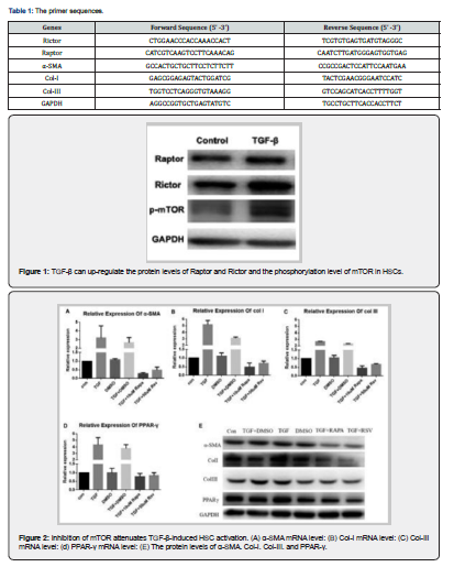
After intervention treatment, cells in each group were collected. The cell culture medium was discarded and phosphate buffered solution (PBS) was added to wash 2-3 times. After PBS was fully discarded, the total RNA of HSCs was extracted by TRIzol method (Sigma-Aldrich, St. Louis, MO, USA) and reverse transcribed into cDNA by reverse transcription kit (Hiscript QRT supermix for qPCR, enzyme, CN). Using cDNA as a template for qPCR reaction. qRT-PCR amplification conditions were as follows: 95 °C pre-denaturation 30 s; 95 °C 5s, 60 °C 30 s, for 40 amplifications; 55-95 °C melting for 30s. GAPDH was used as the internal reference, and the expression of mRNA was calculated by 2-△△Ct. The primer sequences are shown in Table 1.
Statistical analysis
All data and plotting were performed using GraphPad Prism version 7.0 (GraphPad, La Jolla, CA, USA). The data are expressed as mean ± standard deviation (SD). Student's t-test was used for comparison between the two groups. Analysis of variance was used to perform the comparison among groups, and the Bonferroni test was used for pairwise comparison within the group. p < 0.05 was considered statistically significant.
Results
TGF-β activated mTOR signaling pathway in HSCs
We treated HSCs with TGF-β for 30 min, and the results showed that TGF-β significantly up-regulated the phosphorylation level of mTOR protein, the protein levels of Raptor and Rictor compared with the control group, suggesting that TGF-β can activate the mTOR signaling pathway of HSCs (p < 0.05, Figure 1).
TGF-β induced HSC activation through mTOR
The mTOR inhibitor rapamycin (Rapa) or resveratrol (Rsv) was administered to TGF-β-induced HSCs to investigate whether TGF-β can induce HSC activation through the mTOR signaling pathway. The results showed that TGF-β could significantly up-regulate the mRNA and protein expression of α-SMA, Col-I, Col-III and PPAR-γ compared with the control group (p < 0.05), while rapamycin or resveratrol could significantly inhibit the induction of TGF-β on the above-mentioned HSC activation-related genes at protein and mRNA levels (p < 0.05, Figure 2), suggesting that TGF-β could be mediated by mTOR signaling pathway.
TGF-β induced HSC migration through mTOR signaling pathway
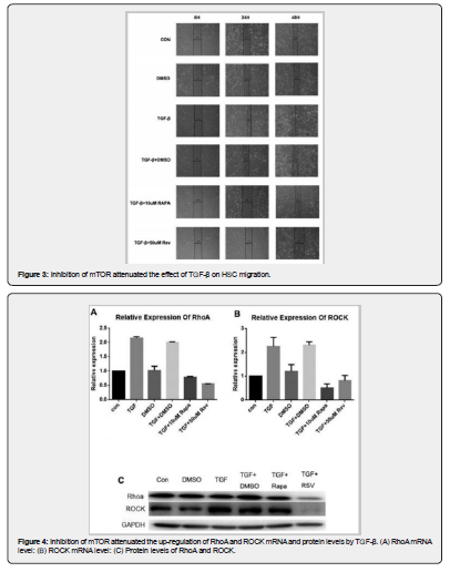
Through scratch wound healing assay, we intend to explore the effect of the mTOR signaling pathway on TGF-β-induced HSC migration. The results showed that compared with the control group, TGF-β intervention for 24 h and 48 h could significantly shorten the cell scratch spacing (p < 0.05), while rapamycin intervention for 24 h could significantly increase the scratch spacing, and resveratrol intervention for 48 h could significantly increase the scratch spacing and weaken the effect of TGF-β on HSC migration (p < 0.05, Figure 3). Those results suggested that TGF-β can induce HSC migration through the mTOR signaling pathway.
TGF-β activated RhoA / Rho signaling pathway through mTOR
The RhoA / Rho signaling pathway plays an important role in TGF-β-induced HSC migration. We intend to further explore whether TGF-β can activate RhoA / Rho signaling pathway through mTOR. Compared with the control group, TGF-β could significantly up-regulate the mRNA and protein levels of RhoA and Rock, while rapamycin or resveratrol could attenuate the effect of TGF-β (p < 0.05, Figure 4). It is suggested that TGF-β can be activated by mTOR.
Discussion
TGF-β-induced HSC activation and migration play an important role in hepatic fibrosis, but the mechanism has not been fully elucidated. In this study, we found that TGF-β can activate the mTOR signaling pathway. The mTOR inhibitor significantly attenuated the up-regulation of TGF-β on HSC activation markers, α-SMA, Col-I, Col-III, and PPAR-γ, the migration of HSC, and the activation of RhoA / Rho signaling pathway. It is suggested that TGF-β can activate the RhoA / Rho signaling pathway through mTOR, thereby promoting HSC activation and migration. The core mechanism of hepatic fibrosis includes excessive deposition of extracellular matrix components and abnormal proliferation of fibrous connective tissue in the liver, and the extracellular matrix is mainly synthesized by HSC in the Disse space of the hepatic sinus [11]. The cytokines involved in the regulation of HSC activation include transforming growth factor (TGF-β), platelet-derived growth factor (PDGF), tumor necrosis factor (TNF-α), connective tissue growth factor (CTGF), and so on [12-14]. Among them, TGF-β is the most important hepatic fibrosis-promoting factor found so far, and its pathway dominates the initiation and formation of tissue fibrosis. The specific regulation methods include accelerating the activation of resting HSC cells, promoting the EMT process of HSC cells, stimulating cytokine secretion, and affecting collagen expression and extracellular matrix metabolism [5]. The Rho protein family is a class of small molecule protein switches that can bind to guanylate GTP and mediate a variety of downstream target proteins and signaling pathways to exert physiological activities. The most important physiological functions are the regulation of cell contractility, intracellular actin expression, and cytoskeletal reorganization [15, 16]. Our previous study found that TGF-β regulates HSC actin expression and microfilament cytoskeleton rearrangement through RhoA, thereby further affecting cell migration.
In addition, TGF-β-regulated cell migration is also related to another signaling protein mTOR. Rik Derynck et al. first reported that mTORC1 can be regulated by TGF-β, forming a TGF-β / PI3K / Akt / mTORC1 signaling pathway to regulate cell movement [17]. In addition, mTORC2 plays an important role in the regulation of TGF-β on the EMT process in tumor tissues [18, 19]. Consistent with this report, in this study, we found that TGF-β can activate the mTOR signaling pathway in HSCs and promote HSC activation and migration through mTOR. It has been reported that mTOR may regulate RhoA activity, and TGF-β signaling can simultaneously activate RhoA and mTOR in the process of regulating EMT [10, 20]. In this study, we found that mTOR inhibitors significantly attenuated the activation of RhoA by TGF-β in TGF-β-induced activated HSCs, suggesting that TGF-β can activate RhoA through mTOR, thereby regulating HSC cytoskeleton rearrangement and affecting cell migration. This study also has some limitations. First of all, this study is an in vitro experiment, and the above results still need to be verified in animal models of hepatic fibrosis. Secondly, mTOR can form two complexes (mTORC1 and mTORC2), and there are some differences in function. In this study, we did not study these two complexes separately. The roles of mTORC1 and mTORC2 in TGF-β-induced HSC activation and migration need to be further clarified.
Conclusion
In conclusion, we found that TGF-β can activate the RhoA / Rho signaling pathway through mTOR, thereby regulating HSC activation and migration.
References
- Friedman SL, Pinzani M (2022) Hepatic fibrosis 2022: Unmet needs and a blueprint for the future[J]. Hepatology 75(2): 473-488.
- Tsuchida T, Friedman SL (2017) Mechanisms of hepatic stellate cell activation [J]. Nat Rev Gastroenterol Hepatol 14(7): 397-411.
- Seki E, Schwabe RF (2015) Hepatic inflammation and fibrosis: functional links and key pathways [J]. Hepatology (Baltimore, Md.) 61(3): 1066 -1079.
- Ellis EL, Mann DA (2012) Clinical evidence for the regression of liver fibrosis [J]. J Hepatol 56(5): 1171-1180.
- Dooley S, Ten Dijke P (2012) TGF-β in progression of liver disease [J]. Cell Tissue Res 347(1): 245-256.
- Li L, Wang JY, Yang CQ, Jiang W (2012) Effect of RhoA on transforming growth factor β1-induced rat hepatic stellate cell migration [J]. Liver Int 32(7): 1093-1102.
- L L, J L, JY W (2010) Role of RhoA in platelet-derived growth factor-BB-induced migration of rat hepatic stellate cells[J]. Chinese medical journal 123(18): 2502-2509.
- Liu GY, Sabatini DM (2020) Mtor at the nexus of nutrition, growth, ageing and disease [J]. Nature reviews. Molecular cell biology 21(4): 183-203.
- Szwed A, Kim E, Jacinto E (2021) Regulation and metabolic functions of mTORC1 and mTORC2 [J]. Physiol Rev 101(3): 1371-1426.
- Gulhati P, Bowen KA, Liu J (2011) mTORC1 and mTORC2 regulate EMT, motility, and metastasis of colorectal cancer via RhoA and Rac1 signaling pathways 71(9): 3246-3256.
- Zhang DY, Friedman SL (2012) Fibrosis-dependent mechanisms of hepatocarcinogenesis [J]. Hepatology 56(2): 769-775.
- Hellerbrand C, Stefanovic B, Giordano F, Burchardt ER, Brenner DA (1999) The role of TGFbeta1 in initiating hepatic stellate cell activation in vivo[J]. Journal of hepatology 30(1): 77 -87.
- Pinzani M (2002) PDGF and signal transduction in hepatic stellate cells[J]. Frontiers in bioscience: a journal and virtual library 7: d1720 -d1726.
- Huang G, Brigstock DR (2012) Regulation of hepatic stellate cells by connective tissue growth factor[J]. Frontiers in bioscience (Landmark edition) 17(7): 2495 -2507.
- Legg K (2011) Cell migration: An ABC of the RHO subfamily [J]. Nature reviews. Molecular cell biology 12(7): 403.
- Petrie RJ, Doyle AD, Yamada KM (2009) Random versus directionally persistent cell migration [J]. Nat Rev Mol Cell Biol 10(8): 538-549.
- Lamouille S, Derynck R (2007) Cell size and invasion in TGF-beta-induced epithelial to mesenchymal transition is regulated by activation of them TOR pathway [J]. The Journal of cell biology 178(3): 437-451.
- Lamouille S, Connolly E, Smyth JW, Akhurst RJ, Derynck R (2012) TGF- β-induced activation of mTOR complex 2 drives epithelial-mesenchymal transition and cell invasion [J]. Journal of cell science 125: 1259-1273.
- Lamouille S, Derynck R (2011) Emergence of the phosphoinositide 3-kinase-Akt-mammalian target of rapamycin axis in transforming growth factor-β-induced epithelial-mesenchymal transition [J]. Cells, tissues, organs 193: 8 -22.
- Jacinto E, Loewith R, Schmidt A (2004) Mammalian TOR complex 2 controls the actin cytoskeleton and is rapamycin insensitive[J]. Nature cell biology 6(11): 1122-1128.