A Rare Case Report of Granular Cell Tumor Involving the Cecum
Lohitha Dhulipalla1, Praneeth Bandaru1, Geethapriya Rajasekaran Rathnakumar2, Andrew Ofosu3, Febin John3, Ying Xian Liu4, Madhavi Reddy3, Elizebeth Guevara2 and Angela Zhu5
1Department of Internal Medicine, The Brooklyn Hospital Center clinical affiliate of Mount Sinai Hospital, USA
2Department of Hematology and Oncology, The Brooklyn Hospital Center clinical affiliate of Mount Sinai Hospital
3Department of Gastroenterology, The Brooklyn Hospital Center clinical affiliate of Mount Sinai Hospital, USA
4Department of Pathology, The Brooklyn Hospital Center clinical affiliate of Mount Sinai Hospital, USA
5SCI2 foundation institute, USA
Submission:May 24, 2021; Published:June 04, 2021
*Corresponding author:Lohitha Dhulipalla, Department of Internal Medicine, The Brooklyn Hospital Center clinical affiliate of Mount Sinai Hospital, USA
How to cite this article:Lohitha D, Praneeth B, Geethapriya R R, Andrew O, Febin J, et al. A Rare Case Report of Granular Cell Tumor Involving the Cecum. Adv Res Gastroentero Hepatol, 2021; 17(2): 555957. DOI: 10.19080/ARGH.2021.17.555957.
Abstract
Granular cell tumor arises from Schwann cells. It is common in females than males. It affects the age group from 10 to 50 years of age. It can arise anywhere in the body but commonly involves the oral cavity, skin, and subcutaneous tissue. Granular cell tumor involving the cecum is very rare. Here, we report a case of Granular cell tumor involving the cecum in a 58-year-old male..
Keywords: Granular cell tumor; Cecal tumor; Benign tumor
Abbreviations: GCT: Granular Cell Tumor; EMR: Endoscopic Mucosal Resection; ESD-Endoscopic Submucosal Dissection
Introduction
Granular cell tumor was first described by Russian pathologist Alexei Ivanovich Abrikossoff and is also known as Abrikossoff’s tumor. It arises from Schwann cells based on immunohistochemical studies. It is usually benign with 1 - 2% of the cases being malignant. The common sites of involvement include skin, subcutaneous tissue, and the oral cavity. Granular cell tumor involving the gastrointestinal tract is uncommon. Here, we describe a case of Granular cell tumor involving the cecum [1].
Case Report
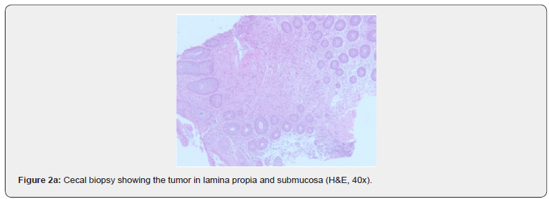
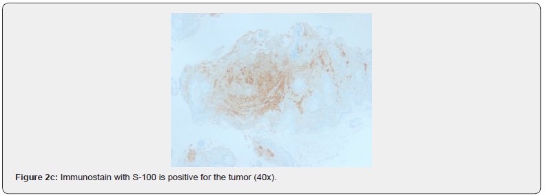
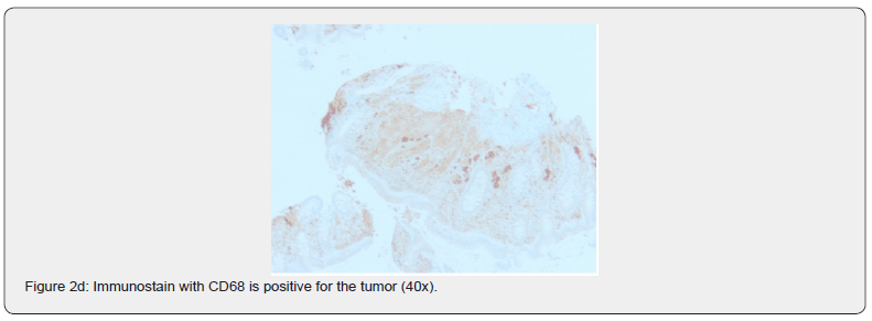
A 58-year-old male with no known past medical history underwent a screening colonoscopy. Colonoscopy showed an 8 mm nodule with yellowish discoloration in the cecum (Figure 1). Biopsy (Figure 2 a & b) was done which revealed a Granular cell tumor. Immunohistochemical stains are positive (Figure 2 c & d) for S-100, Calretinin, CD 68 and negative for AE 1/3, CK7, CK20, CD117, SMA, vimentin, and design. Ki-67 labeling index is <1%. PAS stain is positive for tumor cells [2].


CECT chest abdomen and pelvis were done which showed multiple mesenteric lymph nodes largest of which measures up to 7 mm but are within normal limits in size, with no evidence of metastatic disease in the chest, abdomen, and pelvis. The Tumor within the cecum was not visualized [3].
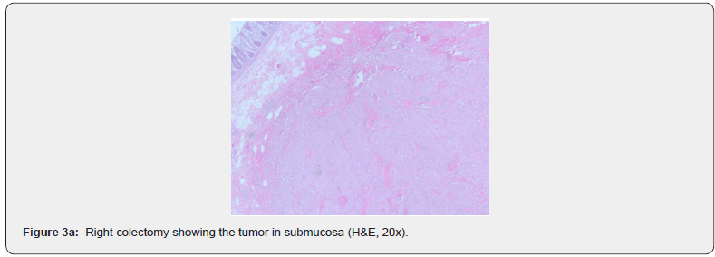
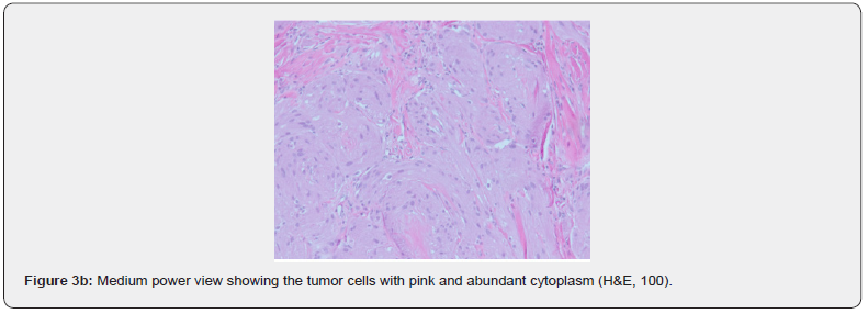
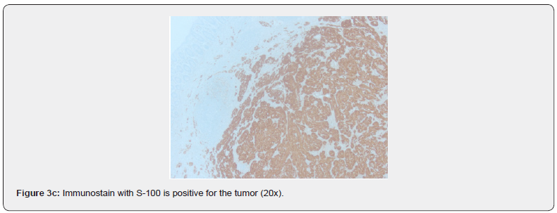
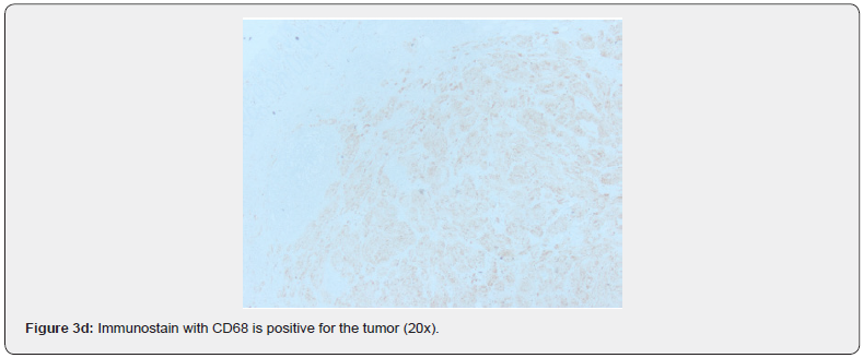
The patient subsequently underwent a right hemicolectomy. Histopathology revealed a Granular cell tumor (0.5x0.3 cm), in the submucosa (Figure 3 a & b) of the cecum. Immunohistochemistry cells positive for CD 68, S-100, negative for calretinin, ki-67 <1%, (Figure 3 c & d) with no muscular propria invasion. Twentyeight reactive lymph nodes were removed and were negative for metastatic carcinoma. Bilateral small bowel, colonic, and soft tissue margins are free of tumor. The patient was discharged without any complications [4].
Discussion
Granular cell tumor can occur anywhere in the body but rarely involves the Gastrointestinal tract. The common site of involvement in the gastrointestinal tract is the esophagus. Most cases of Granular cell tumors involving the Gastrointestinal tract are found incidentally on endoscopy. It presents as a solitary submucosal mass but can be multifocal as well [5].
Colonic GCTs tend to be asymptomatic and usually follow a benign course. Patients who had discomfort may exhibit larger lesions. However, this association is not strong regarding colonic GCTs. Common symptoms are non-specific, including abdominal discomfort, hematochezia and changes in bowel habits [6].
The microscopic criteria as per Fanburg-Smith et al for Granulosa cell tumors include spindling, necrosis, nuclear pleomorphism, high mitotic activity (2 per 10 high power fields at 200 magnification), high nuclear to cytoplasmic ratio, vesicular nuclei with large nucleoli. Tumors that meet 3 or more of the criteria were classified as malignant(histologically), tumors that meet 1 or 2 criteria were classified as atypical, and tumors that don’t fulfill any of the criteria were classified as benign [7].
Malignant tumors are usually larger than 3-4 cm in size, exhibit rapid growth, and ulcerated. The diagnosis of Granular cell tumors is made by endoscopic biopsy, histology, and immunohistochemistry.
Immunohistochemistry stains are positive for S-100, myelin basic protein, and CD 68. Benign granular cell tumors are usually treated with endoscopic resection.
Since there are no established guidelines for the treatment of colorectal GCT, some physicians recommend performing endoscopic mucosal resection (EMR), endoscopic submucosal dissection (ESD), or polypectomy for tumors less than 2 cm. Follow-up colonoscopies seem appropriate after resection if multiple tumors or risk of malignancy exists.
Conclusion
Granular cell tumors involving the cecum are very rare. Most of the tumors involving the gastrointestinal tract are noted incidentally on endoscopy. Although granular cell tumor is predominantly benign, 1-2% of tumors are malignant. Benign granular tumors are treated by endoscopic resection. Here, we report a rare case of Benign Granular cell tumor involving the Cecum.
References
- Meyyappa Devan Rajagopal, Debasis Gochhait, Dasarathan Shanmugan, Adarsh Wamanrao Barwad (2017) Granular Cell Tumor of Cecum: A Common Tumor in a Rare Site with Diagnostic Challenge. Rare Tumors 9(2): 6420.
- Dae-Kyung Sohn, Hyo-Seong Choi, Yeon-Soo Chang, Jin-Myeong Huh, Dae-Hyun Kim, et al. (2004) Granular cell tumor of colon: report of a case and review of literature. World J Gastroenterol 10(16): 2452-2454.
- Kaoutar Znati, Taoufiq Harmouch, Amal Benlemlih, Hinde Elfatemi, Laila Chbani, et al. (2010) Solitary Granular Cell Tumor of Cecum: A Case Report. International Scholarly Research Notices.
- Emad Kandil, Mohamed Abdel Khalek, Obai Abdullah, Salima Haque, Angela Bohlke, et al. (2010) Granular cell tumor arising in the right colon.
- Tagore Sunkara, Savitha V. Nagaraj, Vinaya Gaduputi (2017) Granular Cell Tumor of Rectum: A Very Rare Entity. Case Reports in Gastrointestinal Medicine.
- Devan Rajagopal, Debasis Gochhait, Dasarathan Shanmugan, Adarsh Wamanrao Barwad (2017) Granular Cell Tumor of Cecum: A Common Tumor in a Rare Site with Diagnostic Challenge Meyyappa. Sagepub.
- Yahua Chen, Yangyang Chen, Xiaoqiong Chen, Liang Chen, Wei Liang (2018) Colonic granular cell tumor: Report of 11 cases and management with review of the literature. Oncol Lett 16(2): 1419-1424.


