Gallbladder Cancer: Epidemiology, Diagnosis and Treatment
WM Ali, Maneesh Kumar Bhardwaj*, SAA Rizvi and AZ Rab
Department of General Surgery, Jawaharlal Nehru Medical College and Hospital, India
Submission: August 17, 2018;Published: October 04, 2018
*Corresponding author: Maneesh Kumar Bhardwaj, Junior Resident, Department of General Surgery, Jawaharlal Nehru Medical College and Hospital AMU, India, Tel: ; Email: maneeshkbhardwaj@gmail.com
How to cite this article:WM Ali, Maneesh K B, SAA Rizvi, AZ Rab. Gallbladder Cancer: Epidemiology, Diagnosis and Treatment. Adv Res Gastroentero Hepatol. 2018; 11(1): 555802. DOI:10.19080/ARGH.2018.11.555802.
Abstract
Gallbladder cancer is the most common malignancy of biliary tract. Gallbladder cancer incidence increases with advancing age. It occurs more commonly in females. The incidence rates are very high in Latin America and Asia. Its major risk factors include gallstone, chronic inflammation, primary sclerosing cholangitis, infections, gallbladder polyp etc. it usually presents with symptoms like yellowish discoloration of body, right upper abdominal pain, vomiting, lump in right upper abdomen etc. Investigation profiles include blood investigations and radiological tests like ultrasonography, computed tomography etc. ERCP (Endoscopic Retrograde Cholangiopancreatography) can be used as diagnostic and treatment modality. Treatments of gallbladder cancer include surgery, chemo-radiotherapy and palliative care. Forty-three patients of confirmed gallbladder cancer are included in this study. The main concern of our data analysis is to study epidemiology, diagnosis and treatment measures for gallbladder cancer.
Keywords: Gallbladder cancer; Chemo-radiotherapy; Surgery; Epidemiology; Diagnosis
Introduction
World is facing a lot of problems in now a days. Among these, carcinomas are one of them. An estimated 169.3 million years of healthy life were lost globally because of cancer in 2008.1 About 1,688,780 new cases of cancer are expected to diagnose in year 2017 in US.2 Worldwide there will be 23.6 million new cases of cancer each year by 2030 (estimated) [1]. In 2012, an estimated 8.2 million people died from cancer worldwide [1].
More than half of cancer deaths worldwide occurred in undeveloped countries [2]. The risk of developing cancer in a person depends on factors, including age, genetics, lifestyle and exposure to risk factors [3]. Alcohol, smoking, less physical activity, diet, infections, overweight and obesity are the important risk factors [4]. Prevalence of different risk factors varies by region and country; this is partly why overall cancer incidence rates, and the most common types of cancer, also vary by region and country [1].
Among malignancies of the biliary tract, Gallbladder cancer is the most common accounting for about 80-95% of biliary tract cancers worldwide [3,5,6]. Gallbladder cancer ranks fifth among gastrointestinal cancers [5]. The global rates for gallbladder cancer show differences, reaching epidemic levels for some regions and ethnicities [3,7]. Gallbladder cancer has a particularly high incidence in Chile, Japan, and northern India [3,5]. In the United States, gallbladder cancer accounts for only 0.5% of all gastrointestinal malignancies; less than 5,000 cases occur yearly (1–2.5 per 100,000) [8]. Among Chilean women, gallbladder cancer is the leading cause of cancer death, exceeding breast, lung, and cervical cancers [9,10]. Intermediate frequencies of 3.7–9.1 per 100,000 occur elsewhere in South Americans of Indian descent [6]. Other high-risk regions include Eastern Europe (14/100,000 in Poland), northern India (as high as 21.5/100,000 for women from Delhi), south Pakistan (11.3/100,000), Israel (5/100,000), and Japan (7/100,000) [11]. The incidence is rising in China and has doubled over the past 20 years in Shanghai [12]. In these areas, gallbladder cancer is the most frequent gastrointestinal malignancy and a significant cause of death.
Every year in India there are about 800,000 new cases and 550,000 deaths per annum [13]. Gallbladder cancer is the most common abdominal malignancy in the northern India [14]. The Indian Council of Medical Research Cancer Registry has reported incidence rate of 4.5% in males and 10.1% in females per 100,000 populations in northern India [15].
Material and Method
This is a retrospective study done in Jawaharlal Nehru medical college. The patients included in study were those admitted during the 2016 to 2017. The size of study was the group of 43 patients. Patients were admitted to hospital. Following admission, their, clinical presentation, investigations and management were studied.
It was seen that majority of patients were of age 41-60 years. It was found that 32 patients were female (female 74.5% and male 25.5%). Clinical presentations of patients were not very broad. Symptoms of patients were usually yellowish discoloration of body (present in 13 cases), right upper abdominal pain (present in 34 cases), vomiting (present in 7 cases), lump in right upper abdomen (present in 5 cases), itching all over body (present in 1 case), anorexia (present in 5 cases). There were other symptoms like backache, blood in urine, neck swelling which may be associated with the metastasis of the tumor. Other nonspecific symptoms seen were fever and nausea. Among these symptoms most common was pain abdomen (seen in about 79% of cases) followed by yellowish discoloration of eyes and body (seen in about 30% of cases), vomiting (seen in about 16% of cases) lump in right upper abdomen (seen in about 12% of cases).
On examination, most of patients were vitally stable and one of the most important signs seen in these patients was icterus, which was seen in 13 patients (30.2% of cases). Lymphadenopathy was rarely seen (Table 1).

The patients were evaluated with blood investigations, radiological investigations and pathological investigations. Blood investigations were including haemogram, renal function test, liver function test, prothrombin time, INR. Haemogram and renal functions were usually normal and not reveal any specific correlation. But decreased hemoglobin was seen in 17 cases. Prothrombin time was prolonged in most of cases and INR was also increased. Liver function test include total bilirubin, direct bilirubin, alkaline phosphatase, alanine aminotransferase, aspartate aminotransferase.
In 20 patients (48.5% cases), total bilirubin level was raised and in most of these cases, direct bilirubin was raised up to the more than 50%. Total bilirubin was raised up to 24 mg/dl. In most of cases, total bilirubin was raised in between the range of 5 to 10 mg/dl. Among the all three enzymes included in liver function tests, mostly alkaline phosphatase enzyme was associated with raised level and was raised in 32 patients (74% cases). Alanine aminotransferase enzyme was raised in 13 patients (30.2% cases). Aspartate aminotransferase was seen raised in 12 patients (27.9% cases).
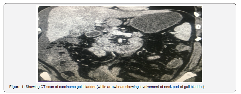
Radiological investigations done in each patient were ultrasonography, chest X-ray and computed tomography. Initially each patient has undergone ultrasonography, which usually suggests a hypoechoic lesion in gall bladder with thickening of gallbladder wall. Cholilithiasis was present in 20 patients (46.5% cases), which suggests an important correlation between the cholilithiasis and gall bladder cancer. Computed tomography scan helps in knowing the involvement of fundus and body of gallbladder. Lesion was having indistinct fat plane with adjacent liver in about 43.7 % of cases. The most involved liver segment was segment 5 (in 38.7%) followed by segment 4b (in 18.2%). Periportal lymphadenopathy was seen in about 17.4% of cases (Figure 1).

Pathological diagnosis was made by the FNAC only in inoperable cases or where the diagnosis was suspected. FNAC was not done in confirmed cases because of risk of spillage of tumor cells along the track. Most of FNAC was suggestive of adenocarcinoma.
After making the diagnosis and staging the disease, we managed the patients. Management consisted of extended cholecystectomy, chemotherapy, radiotherapy and palliative treatment. Extended cholecystectomy was done in stage 1a, 1b and 2. In our study we have done extended cholecystectomy in 8 cases and follow-up of these patients done 3 monthly. In remaining cases, other mode of treatment including chemotherapy, radiotherapy and palliative treatment were given.
Sometime ERCP was also used in few patients as a palliative mode of treatment. ERCP is an invasive procedure.
Discussion
Gallbladder cancer is one of the cancers having good prognosis with early diagnosis. It usually occurs in age group of 40-60 years. It usually occurs in females. Gallbladder cancer presents with symptoms like pain abdomen, yellowish discoloration of body and eyes, lump abdomen, fever, vomiting and anuria. Symptoms usually denote the late presentation of disease thereby bad prognosis of disease. So, at the time of presentation, it is inoperable stage. Important sign in these patients is the presence of icterus. Then patient is investigated by liver function test, prothrombin time, INR, ultrasonography, CT scan. In liver function test, elevated total bilirubin with direct bilirubin is seen. Among enzymes, alkaline transferase is most sensitive in these patients and is elevated in high number of cases. Ultrasonography usually shows thickened gallbladder wall with associated cholilithiasis. CT scan helps us to stage the disease and making further plan of management. Extended cholecystectomy is gold standard of treatment in early stages of disease. Other mode of treatment includes chemoradiotherapy, ERCP, pain management etc.
Conclusion
Gallbladder cancer is one of the major cancers among all gastrointestinal cancers. And it tops the rank among the hepatobiliary cancer in terms of incidence. But at present time, incidence of gallbladder cancer has decreased may be because of increased number of laparoscopic cholecystectomy done. Early diagnosis of gallbladder cancer is very important for increasing the survival of patient. Surgical and medical modalities are the mainstay of treatment. Surgical treatment usually done in early stage of disease. So, it is very important to diagnose early and treatment of it to prevent the progression. And patient must educate about it.
References
- (2014) World cancer factsheet. Cancer research UK.
- (2017) Cancer facts & figures 2017. American cancer society.
- Hundal R, Shaffer EA (2014) Gallbladder cancer: epidemiology and outcome. Clin Epidemiol 6: 99-109.
- Rani Kanthan, Jenna-Lynn Senger, Shahid Ahmed, Selliah Chandra Kanthan (2015) Gallbladder Cancer in the 21st Century. J Oncol 015: 967472.
- Mislav Rakić, Leonardo Patrlj, Mario Kopljar, Robert Kliček, Marijan Kolovrat, et al. (2014) Gallbladder cancer. Hepatobiliary Surg Nutr 3(5): 221-226.
- Lazcano-Ponce EC, Miquel JF, Muñoz N, Herrero R, Ferrecio C, et al. (2001) Epidemiology and molecular pathology of gallbladder cancer. CA Cancer J Clin 51(6): 349-364.
- Chen Chen, Zhimin Geng, Haoxin Shen, Huwei Song, Yaling Zhao, et al. (2016) Long-Term Outcomes and Prognostic Factors in Advanced Gallbladder Cancer: Focus on the Advanced T Stage. PLOS One 11(11): e0166361.
- Eldon A Shaffer (2008) Gallbladder Cancer. Gastroenterol Hepatol (N Y). 4(10): 737-741.
- Roa I, de Aretxabala X (2015) Gallbladder cancer in Chile: what have we learned? Curr Opin Gastroenterol 31(3): 269-275.
- Andia ME, Hsing AW, Andreotti G, Ferreccio C (2008) Geographic variation of gallbladder cancer mortality and risk factors in Chile: a population-based ecologic study. Int J Cancer 123(6): 1411-1416.
- Randi G, Franceschi S, La Vecchia C (2006) Gallbladder cancer worldwide: geographical distribution and risk factors. Int J Cancer 118(7): 1591-1602.
- Hsing AW, Bai Y, Andreotti G, Rashid A, Deng J, et al. (2007) Family history of gallstones and the risk of biliary tract cancer and gallstones: a population-based study in Shanghai, China. Int J Cancer 121(4): 832- 838.
- Kumar NA (1990) Consolidated Report of the Population Based Cancer Registries 1990-1996. National Cancer Registry Programme. New Delhi, India: Indian Council of Medical Research.
- Singh MK, Chetri K, Pandey UB, Kapoor VK, Mittal B, et al. (2004) Mutational spectrum of K-ras oncogene among Indian patients with gallbladder cancer. J Gastroenterol Hepatol 19(8): 916-921.
- (1993) ICMR Annual Report of Population Based Cancer Registries of the National Programme. New Delhi, India: Indian Council of Medical Research.


