Side Attachment Method: Choice for Easy and Safe Intragastric Balloon Insertion
Hyunghun Kim*
Department of Gastroenterology, Kangnam Peter's Hospital, South Korea
Submission: September 11, 2017; Published: September 25, 2017
*Corresponding author: Hyunghun Kim, Department of Gastroenterology, Kangnam Peter's Hospital, Seoul, Korea Address: Kangnam Peter's hospital, 2633 Nambusunhwan-ro, Gangnam-gu, Seoul, South Korea, Tel: +82-2-1544-7522; Fax: +82-2574-9414; Email: relyonlord@gmail.com
How to cite this article: HH Kim, Side Attachment Method: Choice for Easy and Safe Intragastric Balloon Insertion. Adv Res Gastroentero Hepatol 2017; 7(2): 555709. DOI:10.19080/ARGH.2017.07.555709
Abstract
The intragastric balloon is an effective method for weight reduction in obese people. The common problem of intragastric balloon insertion is a blind performance. Practitioners cannot see the insertion from oral cavity to the stomach clearly. To overcome this point, side attachment method was developed. The introduction kit is attached to the shaft of the endoscope side by side, so the introduction kit does not cover the camera of an endoscope. Side attachment method provides clear field of vision during insertion from oral cavity to the stomach, same to diagnostic endoscopy.
Keywords: Intragastric balloon; Endoscopy; Obesity
Abbreviation: IGB, intragastric balloon; SAM, side attachment
Introduction
The intragastric balloon (IGB) technique has become an effective and safe method of achieving significant weight reduction in obese people. IGBs allow patients to feel fullness and finally decrease their food intake. IGBs replace gastric luminal volume and may distend the stomach, potentially inducing neurohormonal effects and changes in motility [-5]. Most IGBs are blindly inserted into the stomach without endoscopic guide, finger technique [6]. End-Ball® (Endalis, Brignais, France) uses endoscope as a guide for passage of the balloon into the stomach; however, the endoscopic view is nearly blind because an introduction kit covers the camera of an endoscope, front attachment method. The common problem of these procedures is a blind performance. If a practitioner can insert an introduction kit with diagnostic visual field, insertion procedure will be easier and safer; blind insertion can damage oral cavity and gastrointestinal tract involuntarily.
Side Attachment Method
Side Attachment Method (SAM) was developed to provide clear visual field during insertion. In SAM, the introduction kit is attached to the shaft of the endoscope side by side, so the introduction kit does not cover the camera of an endoscope. Additionally, SAM with J turn allows a practitioner to notice that an introduction kit is in the stomach. SAM with J turn also enables a practitioner to watch inflation process time to time [8].
Practice of Side Attachment Method
i. A 3.5cm X1cm sized window is made at the distal part of the pouch for releasing a balloon before side attachment to an endoscope.

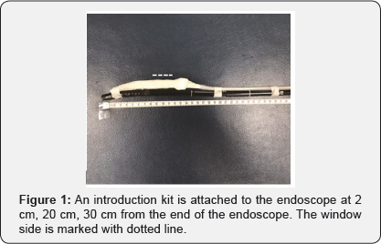
ii.The endoscope was then connected to the introduction kit by side attachment method (Figure 1). The opposite side of the window is attached to the scope by 2.5cm width paper plaster, so the widow is placed at the opposite direction of attached side. The tip of the pouch is attached to the point 2cm distal to the end of the scope, and the catheter is attached to the points 20cm and 30cm distal to the end of the scope
iii. Inserting the endoscope with introduction kit as a diagnostic endoscopy insertion. While passing through the oral cavity, the pyriform sinus, the esophagus, all structures were clearly observed as diagnostic endoscopy.
iv. After inserting an introduction kit into the stomach, J turn is used to confirm whether the introduction kit is in the stomach (Figure 2a).

v. The balloon begins to be filled. When 100 cc air and 150 cc saline were applied, the balloon was pushed out of the pouch; J turn also shows this process (Figure 2b) and filled with saline to its designated volume (600 cc) under endoscopic control.
vi. After then, a feeding catheter was pulled up with an endoscope at the cardia, and the IGB was disconnect from the feeding catheter.
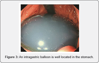
vii. Finishing implantation, follow up endoscopy revealed that the IGB was well placed in the fundus (Figure 3).

Conclusion
SAM is a very useful technique for implanting IGB. Ordinary front visual field during insertion is a pivotal advantage of SAM. Conventional technique caused nearly blind insertion, so practitioners cannot but rely on tactile sensation instead of visual sensation. SAM is a reasonable choice for easy and safe IGB implantation.
References
- Mathus-Vliegen EM (2014) Endoscopic treatment: the past, the present and the future. Best Pract Res Clin Gastroenterol 28(4): 685-702.
- Geliebter A, Westreich S, Gage D (1988) Gastric distention by balloon and test-meal intake in obese and lean subjects. Am J Clin Nutr 48(3): 592-594.
- Geliebter A, Melton PM, McCray RS, Gage D, Heymsfield SB, et al. (1991) Clinical trial of silicone-rubber gastric balloon to treat obesity. Int J Obes 15(4): 259-266.
- Mathus-Vliegen EM, Eichenberger RI (2014) Fasting and meal- suppressed ghrelin levels before and after intragastric balloons and balloon-induced weight loss. Obes Surg 24(1): 85-94.
- Mathus-Vliegen EM, de Groot GH (2013) Fasting and meal-induced CCK and PP secretion following intragastric balloon treatment for obesity. Obes Surg 23(5): 622-633.
- Stimac D, Majanovic SK, Turk T, Kezele B, Licul V, et al. (2011) Intragastric balloon treatment for obesity: results of a large single center prospective study. Obes Surg 21(5): 551-555.
- Buzga M, Kupka T, Siroky M, Narwan H, Machytka E, et al. (2016) Short-term outcomes of the new intragastric balloon End-Ball(R) for treatment of obesity. Wideochir Inne Tech Maloinwazyjne 11(4): 229-235.
- Hyunghun Kim (2017) https://youtu.be/DaCExaz1Ofk


