




 ADOH Home



Abstract
Background: Accurate root canal length determination in biomedical technology is critical to the success of endodontic treatment protocols, as inaccurate measurements may lead to complications such as treatment failure or unnecessary procedures.
Introduction: Conventional medical computed tomography (CCT) technique is widely used due to its effectiveness, simplicity and low cost, providing two-dimensional images, but it is not accurate. In contrast, cone-beam computed tomography (CBCT) technique provides 3D imaging, allowing detailed visualization of the root dimension and structures, which is particularly useful in cases involving complex root curvatures or anatomical variations. However, CBCT has been shown to have some serious limitations, particularly in cases of metal restorations and root fillings, which can cause unwanted image distortions. Moreover, since many hospitals and medical clinics still have only conventional radiology equipment, the authors came up with the idea of finding an empirical relationship, using a practical measurement comparison method that would enable dentists to maximize the use of conventional radiology equipment in implementing the treatment protocol with better accuracy while maintaining patient comfort, low cost and effective clinical applications.
Objective: This research aims to estimate the accuracy of correlation relationship between cone-beam tomography and conventional computed tomography techniques in integration with using Apex Locator, to provide a proposed accurate experimental method for determining root canal length in the diagnosis and treatment in dentistry. In addition to raising the awareness of dental students and increasing the concepts of newly graduated dentists.
Method: Clinical measurement results were obtained using CBCT and CCT techniques for a number of patients and were analyzed using statistical Pearson’s correlation coefficient method.
Conclusion: Analysis of the results showed a new empirical correlation equation between the measurements of both techniques. The empirical relationship concluded may enable the dentists to rely on it in estimating the root canal length for diagnosis and treatment with a high accuracy of up to 98% while ensuring a balance between accuracy and effectiveness in terms of cost, time and patient comfort, which contributes to improving the quality of dental care. Valuable conclusions were also provided for making clinical decisions based on the specific needs of each case.
Keywords:Accuracy in dental treatment; Radiography; Diagnostic; CBCT and CCT techniques; Root canal length measurement
Abbreviations: CCT: Conventional medical computed tomography; CBCT: Cone-beam computed tomography
Introduction
Accurately determining the root length during root canal treatment in biomedical technology is one of the key factors contributing to the success and sustainability of the treatment. Inaccurate root length determination can lead to treatment failure, requiring re-treatment or even tooth loss in some cases. Therefore, modern imaging techniques are vital tools for enhancing diagnostic accuracy and making informed treatment decisions. X-ray techniques play a very important role in diagnosis and then treatment. The most common types of medical X-ray machines used are conventional computed tomography (CCT) radiography and cone-beam computed tomography (CBCT). Both CCT) and CBCT techniques stand out as primary tools in this regard. Conventional CT has been one of the most widely used tools in dental clinics for decades and still providing rapid images at a low cost. However, these images are two-dimensional (2D), making them less capable of providing the detailed information required in complex cases which needs more accuracy. In contrast, CBCT represents a significant advancement in imaging technology, offering three-dimensional images rich in detail, which allows dentists to evaluate teeth and roots more accurately and comprehensively, especially in complicated cases, as well as in the design and manufacture prosthetic organs [1-5]. When CBCT also has many limitations, especially in the cases of metal restorations and root fillings, which may cause unwanted image (produce undesirable) image resolution induced distortions [6- 7]. In addition to imaging techniques, Apex Locator is an essential tool for accurately determining root length. This device relies on measuring the electrical resistance inside the root canal to reliably determine the apical foramen. It is used in all types of root canal treatments to ensure that the correct root length is achieved without damaging the surrounding tissues [8]. However, many hospitals and clinics still rely on conventional CT scanning to differentiate the diagnosis of cancers of the larynx, laryngeal cartilage, soft tissues and lung cancer [9-11]. This, of course, promotes the continued use of conventional CCT scanning and encourages its improvement in terms of both health safety and economic benefits.
Consequently, given the widespread availability of conventional CT techniques in a number of clinics and hospitals, there are also some significant limitations to the use of CBCT techniques. The important question remains: How can we improve the accuracy of conventional radiography measurements, in addition to being a simple, easy and economical tool for imaging procedures, besides being comfortable for patients? A large number of studies have been conducted that focused on the operation, accuracy and comparison of dental X-ray techniques [12-27]. Several previous studies have found no specific consistency in the results of evaluating the accuracy of linear measurements using CT in root canal length determination in Endodontic [28-29]. While correlation between the reference standard of CBCT technique has been presented in a little number of studies in the last ten years [30-35]. While there is no single study that provides a method to deduce an accurate experimental relationship between measurements using CCT and CBCT to maximize the benefit of traditional devices that are still operating efficiently in government clinics and hospitals.
Hence, this research is concerned with trying to find an appropriate answer to the above mentioned question, by presenting an innovative scientific method to improve the accuracy of medical CCT measurements. Thus, there is a need to determine the relationship between the two techniques in terms of accuracy of measurements. In a more professional sense, if a CBCT device is not available when performing diagnostic operations here the conventional device can be used with the use of the proposed method that is the subject of this study. Therefore, this research aims to present a new comprehensive empirical formula based on comparing the practical results between CBCT and CCT radiography in determining root length using Apex Locator in order to improve the accuracy of diagnosis and treatment. In addition, important technical aspects such as radiation dose, cost, time and patient comfort will be compared, with the aim of providing new recommendations regarding the optimal imaging technique for clinical dental practices.
Common Types of X-Ray radiography Techniques in Dentistry
The idea of X-ray computed tomography first appeared in the early 20th century, when Italian radiologist Alessandro Vallibona invented CT using a radiographic film to view a single slice of the body [26-38]. In the early 1930s, Dr. Bernard George Zedsis Des Plantes developed a practical method for applying this technique, known as focal plane computed tomography [39], which relies on the mechanical movement of the X-ray beam source and the capture film in unison to ensure that the desired plane remains in focus while blurring objects outside the plane being examined. In October 1963, William H. Oldendorf received a U.S. patent for his invention of a radiant energy device for investigating selected areas of internal objects obscured by dense materials [40]. The advent of computers led to scientific advances in the late 1960s and early 1970s that led to the development of the first practical CT scanners. The first clinical CT scan was performed in a London hospital in 1971 using a scanner invented by Mr. Godfrey Hounsfield [41]. Then, the first commercial CT technique was installed at Mayo Clinic, USA in 1973.
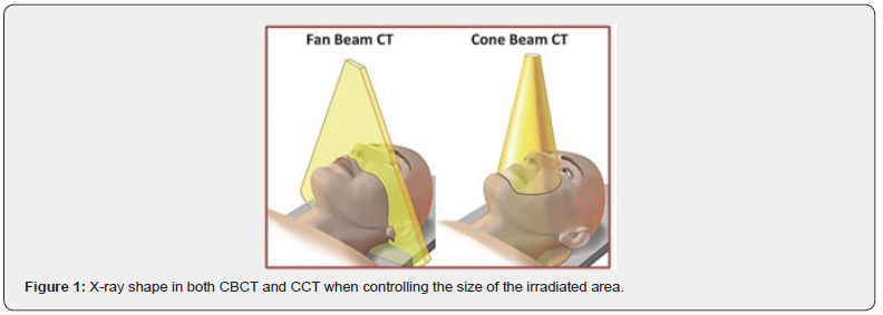
As is known, during the diagnostic process and before the treatment process, CBCT can be used to provide a threedimensional image that clearly reveals the details of the root canals. If CBCT is not available, CCT is used to estimate the approximate length of the root canal. During the treatment process: Apex Locator is used to accurately measure the effective length using the electrical resistance inside the canal. After the treatment process: CBCT or CCT is used to ensure the success of the filling process. If the size of the area of interest to be irradiated is controlled by CCT, the radiation dose can be reduced, see Figure 1. In such control, it can be said that the radiation dose of the CBCT technique will be higher than that of the CCT in dentistry [15]. While without control, the radiation dose of the CCT technique is the largest [16]. Therefore, it will review the theory of work and use of each of them as follows:
CCT Technique
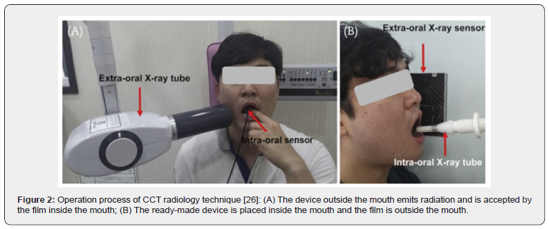
Since 1971, conventional medical computed tomography radiography technique has been one of the primary tools dentists rely on to detect problems that may not be visible during a conventional clinical examination [17]. Through this technique, internal structures of the mouth, such as teeth and jaws, can be imaged, contributing to appropriate diagnosis. Radiography utilizes electromagnetic radiation to capture detailed images that clearly show the teeth and bones. The differences between radiographic techniques lie in the materials and methods used: conventional radiography employs film to capture images, while digital radiography relies on digital sensors connected to computers, significantly reducing radiation exposure. Digital radiography is safer and more effective compared to conventional radiography. Among modern techniques, CBCT is one of the prominent methods that provide highly accurate three-dimensional images, improving diagnostic precision. This technique will be discussed in more detail in the following section. Conventional radiography is widely used in diagnosing interproximal caries and detecting bone loss in the jaw. It also helps determine the need for specific treatments, such as dental implants or orthodontics. During the imaging process, a lead apron is placed to protect the patient from excess radiation, in addition to a thyroid shield. The film is then placed in the patient’s mouth, and the dentist presses the button to release the radiation. It is essential for the patient to remain still during the imaging to obtain an accurate picture. Radiography in dentistry plays a fundamental role in diagnosing and treating various oral health issues, enhancing the dentist’s ability to provide the appropriate treatment to the patient [8]. Conventional X-rays are divided according to the position of the film on which they will be taken, as shown in Table 1. Figure 2 shows how the imaging process is performed.

CBCT technique
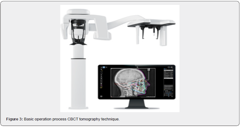
CBCT was initially developed for angiography in 1982, subsequently applied to maxillofacial imaging. Only since late 1990s that it has been possible to produce clinical systems that is both inexpensive and small enough to be used in dental clinics. Emergence of cone beam Computed Tomography has expanded the field of oral and maxillofacial radiology [15]. CBCT is an advanced imaging technique used in situations where conventional dental or facial X-rays do not provide sufficient diagnostic information. This technology is distinguished by its ability to provide detailed 3D images of dental structures, soft tissues, nerve pathways, and bones in the craniofacial region in a single scan. Despite its high effectiveness, it is not used routinely due to the higher radiation exposure compared to conventional X-rays [19]. The absence of clinical reasons or symptoms that require this type of imaging is a justification for not resorting to it, as the decision to use it must be based on a careful evaluation between the desired benefits and possible risks [20]. This technique works by rotating a coneshaped X-ray beam around the patient, generating a series of images that are then combined to create high-quality 3D images. It is more cost-effective and efficient than traditional methods, offering detailed images of bones and skeletal structures, which assist in the evaluation of jaw diseases, dental conditions, nasal cavity structures, and sinuses. However, it does not provide comprehensive information on soft tissues such as muscles and lymph nodes. Despite these limitations, its advantage lies in lower radiation exposure compared to traditional CT scans [21- 30]. The CBCT device has been designed to be more compact and cost-effective, making it easier to implement in outpatient clinics. With these features, it has become an essential tool in the fields of dentistry and maxillofacial surgery, contributing to more precise treatment planning, see Figure 3 for the CBCT device [19].
Working mechanism and technical features of CBCT: The CBCT device operates by rotating a cone-shaped X-ray beam around the patient, capturing sequential images called “projections. These images are processed using advanced techniques to create a 3-D image that reveals fine details of the skeletal structures, teeth, nerves, and soft tissues in the oral and facial regions.
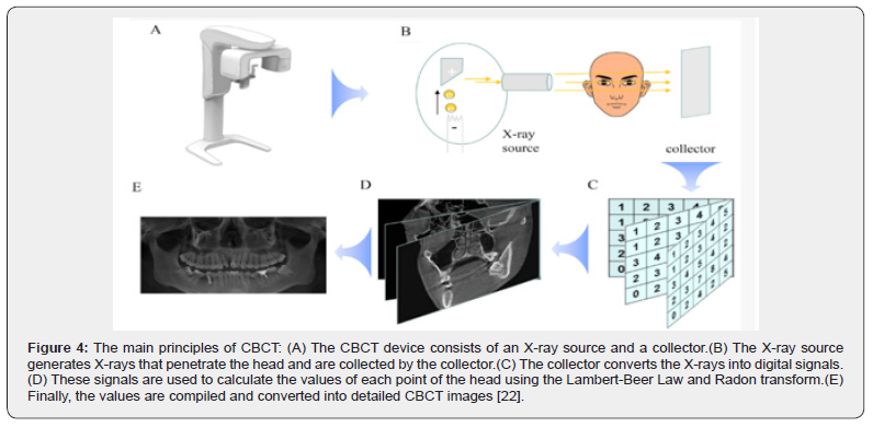
This technique is painless as it projects rays around the area to be imaged in the mouth and these rays are absorbed differently depending on the density of the tissues, resulting in a contrasting image. It only takes about 20-40 seconds to visualize the entire mouth through this technique [42]. This system allows the clinician to assess the health of the teeth and jaw comprehensively in a single scan, with reduced radiation exposure, thereby enhancing the ability of healthcare providers to make precise and informed treatment decisions. As shown in Figure 4, the main principles of CBCT are illustrated, demonstrating the sequence of technical processes that include generating X-rays, collecting the data, and converting it into detailed digital images [21].
Clinical applications and diagnostic benefits: CBCT is used in a wide range of clinical applications, including the evaluation and diagnosis of dental and jaw diseases, analysis of facial skeletal structures, and assessment of the nasal cavity and sinuses. CBCT is preferred for obtaining detailed, precise images of bones, nerves, blood vessels, and soft tissues such as glands and lymph nodes, which aids in more accurate treatment planning [21]. One of the key clinical applications of CBCT is its ability to precisely determine the Apex Location, a feature in which CBCT excels over traditional radiography. By providing detailed three-dimensional images, CBCT allows for the accurate determination of the root apex location, greatly aiding in the assessment of root-related issues such as infections or abnormalities. This also improves diagnostic accuracy in cases requiring surgical intervention, such as root canal treatments or dental implant procedures, by enabling the clinician to determine the relationship between the roots and the surrounding bone structures for precise and safe treatment planning.
Apex Locator
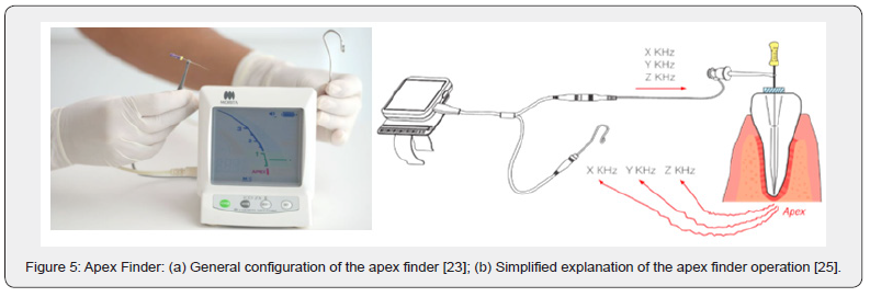
Apex Locator devices are crucial tools in endodontics, used to determine the exact location of the root canal’s apex. These devices measure the distance between the coronal part of the tooth and its apex through an electrical impedance method, a process known as “conductance measurement.” Accurate apex location is vital for successful root canal treatment, as it ensures proper root canal system cleaning, shaping, and filling. Choosing the most suitable device from the many available options is essential to obtain precise and effective results. How does an apex locator work? It works by passing an electrical circuit through the root canal to accurately locate the apex of the tooth. The circuit is completed when the device comes into contact with the gum tissue. Figure 5 shows the general layout and simplified diagram of how an apex locator works.
It is important to understand how the device is used and
operated to ensure correct comparison between imaging
techniques such as CBCT and CCT radiography. This understanding
enhances the accuracy of apex positioning, which helps improve
diagnosis and treatment. It serves as an important reference in the
comparison process, allowing for the evaluation of the accuracy of
apex positioning by Apex Locator, its impact on dental treatment,
and the effectiveness of each device in correctly identifying key
points in treatment. Therefore, the benefits of conductivity
measurement in root canal treatment can be summarized in the
following points:
a) Determine the canal extensions where instruments can
be inserted.
b) Ensure the accuracy of treatment results.
c) Reduce pain and discomfort for the patient.
Modern devices can provide some accuracy, but they may have some limitations, such as the shape of the canal or their ineffectiveness in immature teeth. It is also recommended to avoid their use in patients with pacemakers [23-24].
Advanced and Disadvantage of CBCT and CCT Techniques
For further clarification, during this section we will do a brief scheduled study that includes the advantages and disadvantages of each system as follows in Table1.
Correlation between CBCT and CCT Measurement Results
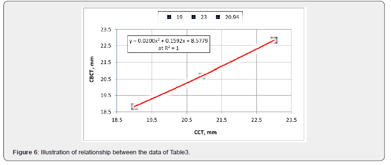
A group of researchers compared the accuracy of two techniques used to determine the actual length of curved root canals: the traditional Apex Locator and CBCT. Measurements were made using these techniques on 18 curved root canals in a group of patients, to examine the compatibility between the two methods in terms of accuracy and quality of measurement. Table 2 shows the results of the comparison [1] after removed the outlier value (number fifteen in the table) according to the statistical basic roles and international standard criteria [45]. Through the current study, the maximum and minimum values were identified and the average value of the measurements was calculated as shown in Table 3. Figure 6 was deduced to illustrate the relationship between the minimum, maximum and average values of the results measured using the two CCT and CBCT techniques in question.


Equation 1 was extracted from the curve of Figure 6 using polynomial regression (second degree), where Y represents the CBCT measurement results and X represents the CCT measurement results. The relationship between the two measurement results using both CCT and CBCT techniques was analyzed using Pearson’s correlation coefficient (R2 = 1) via the CORREL function in Excel (2024 version), indicating a strong positive correlation between the measurements from both techniques as follows:

From table no.3, the accuracy of the measurement results of the conventional CCT technique compared to CBCT can be estimated as follows:
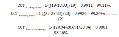
The mean relative error was estimated to be 100% - 98.26% = 1.74% (±0.87%), not more than 10%, which confirms the validity and efficiency of the proposed measurement method. On the other hand, the equation no.3 showed that the error range did not exceed the error limits of CBCT measurements referred to in the previous literatures [46-49]. This it can say that the relative error range that expresses the measurement accuracy of the proposed method was within the limits of ±0.87%, i.e. less than 10% and within the error limits of CBCT measurements which estimate confirms the high degree of confidence in the results and the validity and efficiency of the procedures followed in evaluating the proposed measurement method.
Result Analyses and Evaluation
The results showed that CBCT was more accurate in measuring the working length of root canals compared to conventional radiography (CCT), which provided acceptable but less precise measurements. Table 3 presents the important values of the measurement results, while Figure 6 illustrates the graphical representation, which helped derive the empirical relationship between the measurement results obtained from both techniques. The estimated equation no.1 represents the proposed empirical formula as follows:

From the nonlinear polynomial regression equation 3, the
empirical formulas show that the proposed result has high
sensitivity coefficients of 0.1592 to the measured result of CCT(X)
within low sensitivity coefficients of 0.0200 to the square of the
measured value of CCT(X) with potential measurement result of
8.5779 mm. Although the results showed a significant correlation,
CBCT outperformed conventional radiography by less than 2% in
accuracy, making it the preferred choice for cases requiring precise
measurements. However, despite CBCT’s superiority in accuracy,
CCT remains an economically viable and easy-to-use option due
to the availability of equipment, making it the best choice for
less complex cases and clinics that require fast and cost-effective
techniques. Nevertheless, the nonlinear variation in results may
affect measurement accuracy, making the combined use of both
methods beneficial in certain clinical cases to minimize errors and
enhance precision. Thus, the following can be clearly observed
and evaluated:
1. CBCT and CCT can be considered complementary tools,
where the choice of the most appropriate technique depends on
the complexity of the dental case, ultimately enhancing diagnostic
accuracy and supporting more effective treatment decisions.
Figure 6 presents the comparison analyses and conclusions
reached, along with the advantages of each technique compared
to the other in several aspects.
2. So, it can be said that the derived empirical equation
is novel in its ability to determine the correlation between the
measurements of the two techniques with a high accuracy of
over 98%. This providing a new, easy, and accessible method for
converting the measurements obtained using the CCT technique
into higher-accuracy results comparable to those obtained using
the CBCT technique with high accuracy. This may enable dentists
who do not have CBCT technology and who do have CCT technology
to rely on it to accurately estimate the length of the root canal for
diagnosis and treatment.
Diagnostic and Treatment Procedures
a) During the diagnosis process: CBCT technology is used, if available, to obtain a three-dimensional image that clearly reveals the details of the root canals such as dimensions and curvatures. In the event that CBCT technology is not available, CCT which is characterized by simplicity and speed of use with patient comfort we can be used and with equation 3 can estimate the approximate length of the root canal, accurately.
b) During the treatment process: Apex Locator is used to accurately measure the effective length by determining the electrical resistance inside the canal, which reduces the need for repeated X-rays.
After the treatment process: CBCT or CCT technology is used to ensure the success of the filling process and the absence of gaps. The CBCT device is more accurate than conventional radiography (CCT), especially in difficult cases. The new: in the event that CBCT technology is not available, CCT technology is used to estimate the approximate length of the canal with the possibility of using the proposed method, which is based on use empirical formula (equation no. 2) under study to obtain accuracy comparable to CBCT technology while maintaining simplicity, speed of use and patient comfort. Here, the role of this paper becomes clear in presenting a new empirical formula that enables the dentist who has the traditional CCT technique to obtain highly accurate measurement results.
Conclusions
In this study, a comparative evaluation in biomedical
technology was conducted between clinical measurements using
both CBCT and CCT techniques, along with the use of Apex Locator,
to determine the root canal length in dental treatments. Based on
the comparative analysis using statistical Pearson’s correlation
coefficient method, the following conclusions could be drawn:
i. A new empirical equation (no.3) was deduced to
determine the correlation between the measurements of the two
techniques with an average accuracy exceeding 98%, which may
enable dentists to rely on it to estimate the root canal length for
diagnosis and treatment with great accuracy, if CBCT technology
is not available.
ii. The possibility of using the proposed method, according
to empirical equation (no.3), to improve measured results makes
conventional X-ray CCT technique an effective option in terms of
accuracy, in addition to its low cost, scanning speed, and patient
comfort. It also excels in diagnosing other diseases such as cancers
of the larynx, laryngeal cartilage and soft tissues, especially since
CBCT requires specific and binding conditions, longer time in
scanning operations, doses, and cost as mentioned above.
iii. The relative error range, which represents the accuracy
of the measurement using the proposed method, was within
±0.87%, i.e., less than 10%, and within the error range of conebeam
computed tomography (CBCT) measurements. This
estimate confirms the high degree of confidence in the results and
the validity and efficiency of the procedures used to evaluate the
proposed measurement method.
Eventually, CBCT remains the preferred choice for complex cases requiring high accuracy in root canal measurement, while conventional radiography remains suitable, as the chosen new strategy method (proposed empirical equation no.3) ensures the necessary balance between accuracy, cost-effectiveness, time and patient comfort, contributing to improved quality in dentistry and informed treatment decisions based on the specific nature of each case.
References
- Amal Hassan Mohamed, Amal Hassan Mohamed, Geraldine Mohamed Ahmed, Ghada Borhan Abou Hussein, Hamdy Adly Youssef (2022) Comparative Evaluation of Using Cone Beam Computerized Tomography (CBCT) and Conventional Radiography Confirmed with Apex Locator for Working Length Determination in Curved Root Canals (Clinical Study. Acta Scientific Dental Sciences 6.1 (2022): 121-129.
- Salah HR Ali, Adel Ashry and Naeem Assasa (2017) 3D-Design of Artificial Mandible Using CBCT, CAD and FEA. Curr Trends Biomedical Eng & Biosci 8(2): CTBEB.MS.ID.555732.
- Satyapal Johaley, Freny R Karjodkar, Kaustubh P Sansare; et al. Determination of Three-Dimensional Position of Mandibular Foramen Using CBCT Department of Oral Medicine and Radiology. Forensic Sci & Criminal Inves 12(5).
- Slavena Svetlozarova (2019) C-Shaped Root Canal System– A Review of Literature, Advances in Dentistry & Oral Health, Volume 13 Issue 2 - October 2020 DOI: 10.19080/ADOH.2020.13.555859
- Huang Anderson T and Huang D (2018) The Use of Cone Beam Computed Tomography in Orthodontics: A Review. Advances in Dentistry & Oral Health 8(3).
- Scarfe WC, Farman AG (2008) What is cone-beam CT and how does it work? Dental Clinics of North America 52: 707-730.
- Slavena Svetlozarova and Tsvetelina Borisova-Papancheva. Cone Beam Computed Tomography as a Diagnostic Tool in Endodontics- A Review of the Literature. Adv Dent & Oral Health 11(2).
- (2025) Cleveland Clinic, Dental X-rays, Cleveland Clinic Health Library, Ohio, USA.
- Ali YA, Saleh EM, Mancuso AA (1992) Does conventional tomography still-have a place in glottic cancer evaluation? Clinical Radiology 45(2): 114-119.
- Curtin HD (1989) Imaging of the larynx: current concepts. Radiology 173: 1-11.
- Hyun Su Kim, Kyung Soo Lee, Yoshiharu Ohno, Edwin J.R. van Beek., PET/CT versus MRI for diagnosis, staging, and follow-up of lung cancer. JMRI 42: (2): 247-260.
- Kiljunen T, Kaasalainen T, Suomalainen A, Kortesniemi M (2015) Dental cone beam CT: A review. Phys Med 31(8): 844-860.
- Nikita Kapshe, Madhu Pujar, Satyam Jaiswal (2020) Cone beam computed tomography: A review. International Journal of Oral Health Dentistry 6(2): 71-77.
- Kaasalainen T, Ekholm M, Siiskonen T, Kortesniemi M (2021) Dental cone beam CT: An updated review. Phys Med 88: 193-217.
- Sahithya Kailash (2014) CBCT Cone Beam Computed Tomography. Journal of Academy of Dental Education 1(1): 9-15.
- Abdul Khader, Sulabh Jain, Sarah, Shweta Mishra, Sumera Saleem et al. (2024) Comparing Radiation Doses in CBCT and Medical CT Imaging for Dental Applications, J Pharm Bioallied Sci 16 (Suppl 2): S1795–S1797.
- Raymond A Schulz, Jay A Stein, Norbert J Pelc, How CT happened: the early development of medical computed tomography, J Med Imaging (Bellingham), 29; 8(5):052110, 2021. doi: 10.1117/1.JMI.8.5.052110
- Hassn Mazin Al-alaaf, Mohammed Sabah Jarjees (2024) A Review of Enhancement Techniques for Cone Beam Computed Tomography Images, CC BY 4.0, NTU Journal of Engineering and Technology 3(2).
- (2024) Contributors, Cone beam computed tomography, Oral and maxillofacial radiology, San Francisco, USA.
- American Association of Endodontists, Cone Beam-Computed Tomography in Endodontics, ENDODONTICS: Colleagues for Excellence, Summer 2011, Published by the American Association of Endodontists, 211 E. Chicago Ave., Suite 1100, Chicago, IL 60611-2691, USA.
- (2024) American College of Radiology and Radiological Society of North America, Dental Cone Beam CT, RadiologyInfo.org: For Patients, Illinois, USA.
- Wenjie Fan, Jiaqi Zhang, Nan Wang, Jia Li, Li Hu (2023) The Application of Deep Learning on CBCT in Dentistry, Diagnostics 13(12).
- Dentaltix, How the Apical Locator Improves the Efficiency of Dental Root Canals, Dentaltix Blog, Madrid, Spain, 2025.
- (2025) Contributors, Electronic apex locator, San Francisco, USA, 2023. 25 Minimax, Apex Locator: A New Concept for Precision in Root Canal Depth Measurement, Minimax Implant, Macquarie Park, NSW, Australia.
- (2025) An Intraoral Miniature X-ray Tube Based on Carbon Nanotubes for Dental Radiography - Scientific Figure on Research Gate. Available from: An Intraoral Miniature X-ray Tube Based on Carbon Nanotubes for Dental Radiography - Scientific Figure on Research Gate.
- S. Food and Drug Administration, Dental Cone-beam Computed Tomography, FDA: Radiation-Emitting Products, USA.
- George Fokas, Vida M Vaughn, William C Scarfe, Michael M (2018) Bornstein, Accuracy of linear measurements on CBCT images related to presurgical implant treatment planning: A systematic review, October 2018.
- Adib Al-Haj Husain, Victor Mergen, Silvio Valdec (2025) Comparison of cone-beam computed tomography with photon-counting detector computed tomography for dental implant surgery. Int J Implant Dent.
- Freire-Maia B, Machado VD, Valerio CS (2017) Evaluation of the accuracy of linear measurements on multi-slice and cone beam computed tomography scans to detect the mandibular canal during bilateral sagittal split osteotomy of the mandible. International Journal of Oral and Maxillofacial Surgery 46: 296–302.
- Alkan BA, Aral CA, Aral K (2016) Quantification of circumferential bone level and extraction socket dimensions using different imaging and estimation methods: A comparative study. Oral Radiology 32: 145–153.
- Eachempati P, Vynne OJ, Annishka A (2016) A comparative cross-sectional study of pre-implant site assessment using ridge mapping and orthopantomography (OPG) with cone beam computed tomography (CBCT). Research Journal of Pharmaceutical, Biological and Chemical Sciences 7: 1185-1192.
- Pena de Andrade JG, Valerio CS, de Oliveira Monteiro (2016) Comparison of 64-detector-multislice and cone beam computed tomographies in the evaluation of linear measurements in the alveolar ridge. The International Journal of Prosthodontics 29: 132–134.
- Luangchana P, Pornprasertsuk-Damrongsri S, Kiattavorncharoen S (2016) Accuracy of linear measurements using cone beam computed tomography and panoramic radiography in dental implant treatment planning. International Journal of Oral and Maxillofacial Implants 30: 1287-1294.
- Vasconcelos TV, Neves FS, Moraes LA (2015) Vertical bone measurements from cone beam computed tomography images using different software packages. Brazilian Oral Research.
- (2024) The International Society for Computed Tomography (ISCT), Half A Century In CT: How Computed Tomography Has Evolved.
- Alessandro Vallebona (2024) The Evolution of Medical Imaging Technology. 2019.
- Franco Bistolfi, Alessandro Vallebona 1899-1987. Ricordo di un grande radiologo e del suo contributo allo sviluppo delle scienze radiologiche, in Fisica in Medicina, no. 2, 2005, pp. 115-123. URL consulted on 9 may 2016.
- Valk J Bernard (2004) George Ziedses des Plantes, Radiology, 876, MD 1902-1993.
- Jump up to: a b Oldendorf WH, The quest for an image of brain: a brief historical and technical review of brain imaging techniques, Neurology 28(6): 517.
- Jump up to: a b Richmond, Caroline, Obituary – Sir Godfrey Hounsfield, BMJ 329(7467): 687.
- New Teeth Chicago Dental Implant & Cosmetic Dentistry Center, what is a CBCT Scan and Why Do You Need One?, New Teeth Chicago Dental Implant & Cosmetic Dentistry Center, Retrieved January 17, 2025, Chicago, Illinois, USA.
- Sean Hubar J, Paul J Kapsalos (2025) Clinical Applications of Cone Beam Computed Tomography in Dentistry: An Overview of the Application and Interpretation of 3D Radiographic Imaging by Dental Providers, The Journal of Multidisciplinary Care – Decisions in Dentistry.
- (2025) Comprehensive Dental Center, 3D CBCT & Intraoral Scanner & Intraoral Scanner, Retrieved January 17, 2025, Bing Dental PLLC, Katy, Texas, USA.
- ISO 5726-1: 2023 (en), Accuracy (trueness and precision) of measurement methods and results — Part 1: General principles and definitions, International Standards.
- Anter E, Zayet MK, El-Dessouky SH (2016) Accuracy and precision of cone beam computed tomography in periodontal defects measurement (systematic review). J Indian Soc Periodontol 20(3): 235-243.
- Hekmat Allah Sabry Elsawy, Hoda Saleh, Walaa Ahmed (2023) Accuracy of ultra low dose and standard dose CBCT protocols in volumetric measurements of simulated Bone defects versus real measurements: Experimental study, Volume 69, Issue 1 - Serial Number 3, January (Oral Medicine, X-Ray, Oral Biology & Oral Pathology) pp. 273-283.
- Yusong Zhang, Yixin Liu, Tianqi Liu (2024) Evaluation of CBCT reconstructed tooth models at different thresholds and voxels and their accuracy in fusion with IOS data: an in vitro validation study. BMC Oral Health 24: 1571.
- Ibrahim N, Abouelkheir HM, Elkersh NM (2025) Accuracy of measurements on CBCT-generated digital models using different exposure parameters (in vitro study). Orthod Craniofac Res 28(1): 133-141.

























