Maxillary Tuberosity Fracture During Extraction of Maxillary Posterior Teeth
Ramisz Rahman A1* and Sreejee Gopalakrishnan2
1Associate Consultant, Department of Oral and Maxillofacial Surgery, Hannah Joseph Hospital, Madurai, Tamil Nādu, India
2Lecturer, Department of Oral and Maxillofacial Surgery, A B Shetty Memorial Institute of Dental Sciences, Mangalore, Karnataka, India
Submission: January 09, 2023; Published: January 26, 2023
*Corresponding author: Ramisz Rahman A, Associate Consultant, Department of Oral and Maxillofacial Surgery, Hannah Joseph Hospital, Madurai, Tamil Nādu, India
How to cite this article: Ramisz Rahman A, Sreejee G. Maxillary Tuberosity Fracture During Extraction of Maxillary Posterior Teeth. Adv Dent & Oral Health. 2023; 15(5): 555925. DOI: 10.19080/ADOH.2023.15.555925
Abstract
Objective: Maxillary tuberosity fractures are common in oral surgical practice. The bone density at the maxillary tuberosity is the lowest. In patients with enlarged maxillary sinus, the alveolar process fractures may occur during extraction of the molar teeth. The aim was to assess whether maxillary tuberosity fracture during the extraction of maxillary posterior teeth was a common complication through a systematic review.
Materials and Methods: Three electronic databases (PUBMED, Research Gate, and Google Scholar) were searched. Of the 12805 abstracts reviewed for inclusion. These 12805 references were assessed based on the abstracts and titles; these were reduced to 47 relevant manuscripts of which 21 studies were included in this systematic review.
Results: 47 manuscripts were identified as eligible based on the abstracts and titles searched for full-text and analyzed for inclusion. 21 manuscripts were relevant for inclusion in qualitative analysis.
Conclusion: A proper pre-operative clinical and radiological examination is essential to prevent such a complication. If it is thought that there is a high risk of tuberosity fracture during extraction from the pre-operative evaluation, then surgical removal of the tooth is recommended.
Keywords: Maxillary tuberosity fracture; Third molar extraction complication; Tuberosity fracture during extraction
Introduction
Maxillary tuberosity fractures are common in oral surgical practice. It most commonly occurs during the extraction of maxillary molars. The bone density at the maxillary tuberosity is the lowest and hence the tuberosity bone allows easy luxation of the tooth but is also highly susceptible to fracture even under lower applied forces [1,2]. Fractures of the maxillary alveolar process occur most commonly in the maxillary anterior and premolar regions in young adults. In patients with enlarged maxillary sinus, the alveolar process fractures may occur during extraction of the molar teeth [1]. Maxillary tuberosity is an important structure to maintain the stability of maxillary dentures. In cases with large fractures of the maxillary tuberosity, the fractured bone has to be salvaged by maintaining it in place and aided in healing [3]. A correct preoperative clinical and radiographic evaluation along with sound anatomical knowledge might help to reduce and prevent such complications of extraction [1]. The aim was to assess whether maxillary tuberosity fracture during extraction of maxillary teeth was a common complication through a systematic review. The objectives were to determine which maxillary tooth extraction commonly resulted in maxillary tuberosity fracture and also whether maxillary tuberosity fracture during extraction of maxillary teeth was associated with any other complication.
Methods
This manuscript followed the Preferred Reporting Items for Systematic Reviews and Meta-Analyses Protocols (PRISMA - P) for reporting a systematic review.
The PICO question was:
i. Population: Patients with maxillary tuberosity fracture during extraction of maxillary teeth
ii. Intervention: Extraction or Surgical removal of maxillary teeth
iii. Comparison: Extraction or Surgical removal of maxillary teeth with and without maxillary tuberosity fracture
iv. Outcomes: Maxillary tuberosity fracture with or without complications
Inclusion and Exclusion Criteria
Studies were limited to Case reports, Prospective and Retrospective cohort studies reporting the presence of maxillary tuberosity fracture during extraction of maxillary teeth. The population included patients who presented for the extraction of the maxillary teeth between January 2000 and February 2021. Studies in the English language were only included and studies in languages other than English were excluded. Studies on animals and cadavers were excluded. There were no restrictions on the type of clinical setting. Studies at all levels of healthcare settings (such as primary, secondary, and tertiary healthcare) and those conducted in the community were included for maximum representation.
Search methods for identification of studies
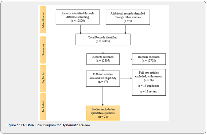
Three electronic databases (PUBMED, Research Gate, and Google Scholar) were searched using the strategies reported in Table 1. The search was performed on March 25th, 2021, with 12805 abstracts reviewed for inclusion, and 21 studies were included in this systematic review (Table 1 & Figure 1).


Data collection and analysis – Selection of studies
Abstracts were initially reviewed to determine if the full-text article should be obtained. If the article fulfilled the criteria or if the authors were unable to decide regarding inclusion, a full-text article was obtained and evaluated (Figure 1).
Results
Results of the search
The initial search strategy yielded 12805 unduplicated references including case reports, prospective and retrospective studies. These 12805 references were assessed based on the abstracts and titles; these were reduced to 47 relevant manuscripts. Of those 12758 references excluded, the main reasons for exclusion were: Teeth extraction with different complications, different etiology for maxillary tuberosity fracture, papers in other languages, abstract/conference proceedings, and editorial/opinion.
Qualitative analysis
All the 47 manuscripts identified as eligible based on the abstracts and titles were searched for full-text and analyzed for inclusion. 21 manuscripts were relevant for inclusion in qualitative analysis.
Characteristics of the participants and interventions
Table 2 provides detailed overview of patient demographics and study characteristics of the included studies. A total of 192 patients in all included studies were comprised in this systematic review.

Results of individual studies
Table 2 summarizes the results reported from the included studies divided by author, place and year, title, type of study, number of cases with incidence rate, tooth extracted, age and gender of the patient, complications and management. The complication encounter in each study was assessed and the management done was tabulated.
Tooth extracted
11 studies reported maxillary tuberosity fracture following maxillary third molar extraction, 5 studies reported following maxillary second molar extraction, 3 studies following maxillary first molar extraction, 1 study reported tuberosity fracture following maxillary premolar extraction and one study did not report the tooth/teeth involved.
Complications and management
Table 2 summarizes the various complications reported in the studies. The incidence rate of maxillary tuberosity in this review ranged between 0.38 - 36.67%. Maxillary tuberosity fracture was the primary outcome and the associated complications reported in the studies included haemorrhage, edema, fractured alveolus, gingival laceration, oro-antral communication, fractured pyramidal process of palatine bone, epistaxis, tachycardia, hypotension, blood loss, maxillary defect, peri-orbital and subconjunctival ecchymosis, diplopia, and facial asymmetry. Each of these complications were managed by varied methods by various authors such as arterial ligation, diathermy for bleeding control etc.
Discussion
The maxillary sinus is the largest paranasal air sinus. It develops during the 3rd and 4th months of intrauterine life [3]. In adults it lies about half inch beneath the level of floor of the nose. Its size varies greatly amongst different individuals [3]. The maxillary sinus enlarges into the alveolar process after the eruption of the permanent maxillary first molar [3]. This extension may weaken the walls of the sinus [3]. Chugh et al [4] found that the bone density at the maxilla was the lowest at the maxillary tuberosity region for both alveolar and basal, cortical and cancellous bones than that at other sites. The presence of least density might be due to the absence of direct mechanical forces in the tuberosity region [4]. The density was found to be 888 Hounsfield unit (HU) in the buccal alveolar cortical bone, 970 HU in the palatal alveolar cortical bone, 362 HU in the alveolar cancellous bone, 933 HU in the buccal basal cortical bone and 411 HU in the basal cancellous bone [4]. The possible etiologic factors for maxillary tuberosity fracture during extraction of maxillary teeth are: a large maxillary sinus extending into the maxillary tuberosity, resorption of the maxillary alveolar process bringing the antral lining close to the oral mucoperiosteum, unerupted maxillary third molar, isolated tooth, tooth with divergent roots, tooth with more roots, tooth with curved roots, tooth with dental anomalies, ankylosed tooth, tooth with hypercementosis, tooth with chronic periapical infection, cyst, multiple extractions [1].
The incidence rate of maxillary tuberosity is varied. The incidence rate of maxillary tuberosity fracture during extraction in cases reported by various authors included in this review ranged between 0.38 - 36.67%. Fractures of maxillary tuberosity pose a serious problem for surgical and prosthetic rehabilitation [5]. The tuberosity fracture can be observed clinical during the extraction and also can be diagnosed radiographically [3,5]. The fracture can be noted by crepitus on palpation from either the buccal or palatal aspect or both or mobility of the posterior maxillary alveolus [3,5]. There may be other clinical signs like hematoma or any soft tissue lacerations near the fracture [5]. There may be an associated oro-antral communication that depends on the size of the tuberosity fracture [5]. In this review, 5 studies reported oroantral communication following tuberosity fracture. Sometimes a tuberosity fracture may be unrecognised until the extracted tooth is examined [3]. In such cases the tuberosity bone may be fused with the extracted tooth and this may have an extension of the maxillary sinus with its mucosal lining [3]. In such cases the extraction site has to be carefully examined for an oro-antral communication and its extent [3]. The tuberosity fracture can be categorised as mild, moderate and severe based on the size of the fracture [6-25].
If the fractured tuberosity is small or if the tooth intended for extraction is infected at the time of fracture, then the fragment should not be left in situ because the fractured complex might not heal due to the persisting infection [1]. But some authors consider immobilization of the fractured fragment using a splint for 3 - 5 weeks and surgical extraction of the tooth at the end of the splinting period [3]. When a large bony fragment is present then one of the following measures can be used: surgical removal of the tooth by root sectioning; detaching the fractured maxillary tuberosity from the roots of the tooth, stabilizing the fractured segment using rigid fixation technique for 4 - 6 weeks and then the tooth can be surgically removed at a later date, the large fragment can be removed and the soft tissue can be sutured back with airtight closure if the fragment is already detached from the maxilla [1]. Bone marrow stromal cells from the maxillary tuberosity can be reliable, accessible and easy-to-harvest intraoral sources of osteoprogenitor cells for bone tissue engineering as an alternative to the iliac crest bone marrow, hence the preservation of maxillary tuberosity during tooth extraction is of great importance [26].
Conclusion
Since the density of the bone at the maxillary tuberosity is relative less and the extension of the maxillary sinus into the alveolar region weakens it further, care should be taken to avoid tuberosity fracture during teeth extraction. Also, the patients must be informed about this potential risk during extraction and its complications prior to extraction of maxillary teeth, especially the molars. A proper pre-operative clinical and radiological examination is essential to prevent such a complication. If it is thought that there is a high risk of tuberosity fracture during extraction from the pre-operative evaluation, then surgical removal of the tooth is recommended.
References
- Chrcanovic BR, Freire-Maia B (2011) Considerations of maxillary tuberosity fractures during extraction of upper molars: a literature review. Dent Traumatol 27(5): 393-398.
- Park HS, Lee YJ, Jeong SH, Kwon TG (2008) Density of the alveolar and basal bones of the maxilla and the mandible. Am J Orthod Dentofacial Orthop 133(1): 30-37.
- Norman JE, Cannon PD (1967) Fracture of the maxillary tuberosity. Oral Surg Oral Med Oral Pathol 24(4): 459-467.
- Chugh T, Ganeshkar SV, Revankar AV, Jain AK (2013) Quantitative assessment of interradicular bone density in the maxilla and mandible: implications in clinical orthodontics. Prog Orthod 14(1): 38.
- Hadziabdic N, Komsic S, Sulejmanagic H (2011) Maxillary tuberosity fracture as a post-operative complication-case study. HealthMed 5(6): 2272-2278.
- Shah N, Bridgman JB (2005) An extraction complicated by lateral and medial pterygoid tethering of a fractured maxillary tuberosity. Br Dent J 198(9): 543-534.
- Polat HB, Ay S, Kara MI (2007) Maxillary tuberosity fracture associated with first molar extraction: a case report. Eur J Dent 1(4): 256-259.
- Altuğ HA, Sahin S, Sencimen M, Dogan N (2009) Extraction of upper first molar resulting in fracture of maxillary tuberosity. Dent Traumatol 25(1): e1-2.
- Bertram AR, Rao AC, Akbiyik KM, Haddad S, Zoud K (2011) Maxillary tuberosity fracture: a life-threatening haemorrhage following simple exodontia. Aust Dent J 56(2): 212-215.
- Venkateshwar GP, Padhye MN, Khosla AR, Kakkar ST (2011) Complications of exodontia: a retrospective study. Indian J Dent Res 22(5): 633-638.
- Evrosimovska B, Velickovski B, Menceva Z (2012) Fracture of the maxillary tuberosity: A case report. Balkan Journal of Stomatology 16(2): 125-128.
- Pourmand PP, Sigron GR, Mache B, Stadlinger B, Locher MC (2014) The most common complications after wisdom-tooth removal: part 2: a retrospective study of 1,562 cases in the maxilla. Swiss Dent J 124(10): 1047-1051.
- Thirumurugan K, Munzanoor RR, Prasad GA, Sankar K (2013) Maxillary tuberosity fracture and subconjunctival hemorrhage following extraction of maxillary third molar. J Nat Sci Biol Med 4(1): 242-245.
- Azenha MR, Kato RB, Bueno RB, Neto PJ, Ribeiro MC (2014) Accidents and complications associated to third molar surgeries performed by dentistry students. Oral Maxillofac Surg 18(4): 459-464.
- Sebastiani AM, Todero SR, Gabardo G, Costa DJ, Rebelatto NL, et al. (2014) Intraoperative accidents associated with surgical removal of third molars. Brazilian Journal of Oral Sciences 13: 2762-80.
- Cilasun Ü, Kan B, Sinanoğlu Ea (2015) Is Maxillary Tuberosity Fracture Possible While Upper First Premolar Extraction? Case Report 1(3): 189-192.
- Baba J, Iwai T, Endo H, Aoki N, Tohnai I (2017) Maxillary tuberosity fracture and ophthalmologic complications following removal of maxillary third molar. Oral Surgery 10(1): 43-47.
- Olborska A, Osica P, Janas-Naze A (2017) Iatrogenic fracture of the maxillary tuberosity–a case report. Journal of Education, Health and Sport 7(12): 155-168.
- Tay ZW, Zakaria SS, Zamhari AK, Lee SW (2018) Dentoalveolar fracture: A complication of extraction of upper left first molar. Clin Case Rep 6(11): 2096-2098.
- Mohan Naik, Vikas Dhupar, Francis Akkara, Praveen Kumar (2021) Fractures of Maxillary Tuberosity During Extraction of Maxillary Molar- A Case Report and Review. Med res chronicles 5(5): 391-377.
- Salik A, Amjad S, Rahman T, Ansari K (2019) Study of complications of surgical removal of maxillary third molar. Journal of Oral Medicine, Oral Surgery, Oral Pathology and Oral Radiology 5(1): 1-3.
- Sayed N, Bakathir A, Pasha M, Al-Sudairy S (2019) Complications of Third Molar Extraction: A retrospective study from a tertiary healthcare centre in Oman. Sultan Qaboos Univ Med J 19(3): e230-e235.
- Shazia Khatoon, Samir Jain (2021) A Retrospective study to assess the complications of surgical removal of maxillary third molar. European Journal of Molecular & Clinical Medicine 7(11): 5455-5459.
- Tsolov R, Gerdzhikov I (2020) Case report: fracture of the maxillary tuberosity during tooth extraction and subsequent treatment. Knowledge International Journal 40(4): 651 - 653.
- Paul JK, Elias AM (2021) Assessment of the complications encountered during and after surgical removal of maxillary third molar: An observational study 7(1): 411-413.
- Cicconetti A, Sacchetti B, Bartoli A, Michienzi S, Corsi A, et al. (2007) Human maxillary tuberosity and jaw periosteum as sources of osteoprogenitor cells for tissue engineering. Oral Surg Oral Med Oral Pathol Oral Radiol Endod 104(5): 618.e1-12.


