Differences of Annual Radiographic Alveolar Bone Loss Rates of Anterior and Posterior Teeth of Individuals Affected with Secondary Occlusal Traumatism between with and without Perioprosthetic Therapy
Guey-Lin Hou*
Former Professor, Graduate Institute of Dental Sciences, Department of Periodontics, and Periodontal Prosthetic Center, School of Dental Medicine, Kaohsiung Medical University, Kaohsiung City, Taiwan
Submission: June 30, 2021; Published: July 08, 2021
*Corresponding author: Guey-Lin Hou, Former Professor and Chairman, Dental Department, Periodontal Prosthetic Center, Chang-Gung Memorial Hospital, Kaohsiung City, Taiwan
How to cite this article: Guey-Lin Hou. Differences of Annual Radiographic Alveolar Bone Loss Rates of Anterior and Posterior Teeth of Individuals Affected with Secondary Occlusal Traumatism between with and without Perioprosthetic Therapy. Adv Dent & Oral Health. 2021; 14(4): 555892. DOI: 10.19080/ADOH.2021.14.555892
Abstract
The purpose of present study was to analysis and investigate the differences of annually radiographic alveolar bone lose (ARABL) rates between the anterior and posterior teeth affected SOT in individuals with and without periodontal therapy, respectively. The study samples consisted of a total 78 individuals, (anterior teeth: 40 and posterior teeth: 38) those present was based on some retrospective analysis from 78 individuals with secondary occlusal traumatism (SOT). In addition, the anterior teeth of 12 treated and 28 untreated individuals were assigned the test group and the control group; The 24 posterior teeth of treated individuals and 14 untreated individuals were assigned as the test group and control group, respectively.
Results showed the mean alveolar bone level gain of the mesial and distal wall in the treated group for anterior teeth with SOT, was significantly higher (p<0.05) as compare to that of the untreated group got a severe alveolar bone loss using two sample t-test. The mean alveolar bone level gain of the mesial and distal wall in the treated group for posterior teeth with SOT was strong significantly higher (p<0.0001) as compared to that of the untreated group using two sample t-test. The mean alveolar bone level gain of the mesial and distal walls in the treated group for posterior teeth with SOT was significantly higher (p<0.05) than that of the untreated group using two sample t-test. The mean RABL gain of the mesial and distal walls in the treated group with SOT was significant higher (p<0.001) than the untreated group using two sample t-test. It is concluded that the RABL gain of the mesial, distal and both mesial and distal walls in the treated group for both anterior and posterior teeth with SOT, was significantly higher as compare to that of the untreated group with a remarkable boss.
Keywords: SAP; ARABL; SOT; Treated NSPT; Untreated NSPT
Abbreviation: ARABL: Annually radiographic alveolar bone lose; SOT: Secondary occlusal traumatism; HP: Healthy periodontium; CP: Chronic periodontitis; AgP: Aggressive peri- odontitis; PI: Plaque index; GI: Gingival index; PPD: Initial probing pocket depths; CAL: Clinical attachment levels; SD: Standard deviation; TPP: Provisional prosthesis
Introduction
The cross-sectional and longitudinal studies employing techniques, relating to measuring the rate of clinical attachment lose in individuals with healthy periodontium (HP), chronic periodontitis (CP), and aggressive peri- odontitis (AgP) with secondary occlusal traumatism (SOT), were documented in the majority of investigators [1]. There is little or no reports regarding studies related to the annually radiographic periodontal bone loss rates among the individuals with periodontal health, chronic periodontitis and aggressive periodontitis with and without SOT from using digital scanning radiographic image analysis [2-6]. The reduction in periodontal bone height with increasing age in healthy, and in those chronic periodontitis and aggressive periodontitis, however, wide variations were noted at the different grades of disease stages, and among different types of periodontitis [1,2,7,8]. Little or limitted data, concerning annual alveolar bone loss rates, in the long-term studies among the teeth of individuals with SOT, was available between the treated and untreated periodontal therapy. The purpose of present study was to analysis and investigate the differences of annually radiographic alveolar bone lose (ARABL) rates between the teeth affected SOT in individuals with and without periodontal therapy, respectively.
Materials and Methods
The study samples consisted of a total 78 individuals, (anterior teeth: 40 and posterior teeth: 38) those present was based on some retrospective analysis from 78 individuals with secondary occlusal traumatism (SOT). In addition, the anterior teeth of 12 treated individuals were assigned the test group, where anterior teeth of 28 untreated individuals were assigned as the control group (Table 1). The 24 posterior teeth of treated individuals were assigned as the test group; where 14 untreated individuals were assigned as the control group (Table 2), respectively. Proper informed consent was obtained from the patients and control individuals. All the samples, which reporting or referred to Periodontal Clinics of Dental School, Kaohsiung Medical University Hospital from 1981 to 2001 were collected in the study. A periodontal charting was performed to record oral examination of both clinical and periodontal parameters evaluation included age, sex, dental history, plaque index (PlI) [9], gingival index (GI) [10], initial probing pocket depths (PPD), clinical attachment levels (CAL). Annual radiographic alveolar bone loss of teeth was also measured mesially and distally.
Measurements of ARABL using DSRIA [11-13]
Proximal RABL was defined as bone defects of at least 2 mm distance between tha CEJ (point A) and the alveolar bone crest (point B). Deeper defects were recorded as the % of the ratio of RABL to root length. The radiographic CEJ (point A), alveolar bone crest (point B) and root apex (point C) were used as three reference points for calculating RABL (Figure 1).
Results
Table 1 indicated the mean and standard deviation (SD) of RABL (mm) in the mesial wall of anterior teeth with secondary occlusal traumatism (SOT) with and without periodontal therapy. Results showed that the mean (SD) on the anterior teeth with SOT of the mesial wall in the treated group (6) was gain (+0.65±1.09 mm), where the untreated group (14) were alveolar bone loss (-1.56±0.72 mm), respectively. The mean alveolar bone level gain, for anterior teeth with SOT of the mesial wall in the treated group, was non-significantly as compare to that of the untreated group got a severe alveolar bone loss (f value = 2.8774, p> 0.05) using two sample t-test. Also, results indicated that the mean (SD) on the anterior teeth with SOT of the distal wall in the treated group(6) was gain (+0.17±0.88 mm), where the untreated group (14) were alveolar bone loss (-1.62±0.57 mm), respectively. The mean alveolar bone level gain, for anterior teeth with SOT of the distal wall in the treated group, was non-significantly as compare to that of the untreated group got a severe alveolar bone loss (f value = 0.1044, p> 0.05) using two sample t-test. Results showed that the mean (SD) of RABL (mm) on the anterior teeth with SOT of the mesial and distal wall in the treated group (12) was gain (+0.41±0.59 mm), where the untreated group (28) were alveolar bone loss (-1.59±0.47 mm), respectively. The mean alveolar bone level gain of the mesial and distal wall in the treated group for anterior teeth with SOT, was significantly higher (f value = 6.0262, p<0.05) as compare to that of the untreated group got a severe alveolar bone loss using two sample t-test (Table 1).
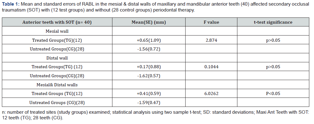
Table 2 demonstrated that the mean and standard deviation (SD) of RABL (mm) in the mesial wall of posterior teeth with SOT with and without periodontal therapy. Results showed that the mean (SD) on the posterior teeth with SOT of the mesial wall in the treated group (12) was gain (+1.21±1.08 mm), where the untreated group (7) was alveolar bone loss (-3.15±1.42 mm), respectively. The mean alveolar bone level gain of the mesial wall in the treated group for anterior teeth with SOT, was significantly higher (f value = 5.9928, p<0.05) as compare to that of the untreated group got a severe alveolar bone loss using two sample t-test. Results indicated that the mean(SD) of RABL (mm) on the posterior teeth with SOT of the distal wall in the treated group (12) was gain (+0.78±0.57 mm), where the untreated group (7) was alveolar bone loss (-3.31±0.75 mm), respectively. The mean alveolar bone level gain of the distal wall in the treated group for posterior teeth with SOT, was significantly higher (f value =18.6869, p<0.001) as compare to that of the untreated group using two sample t-test (Table 2). Table 2 showed that the mean(SD) of RABL (mm) on the posterior teeth with SOT of the mesial and distal wall in the treated group (24) was gain (+1.00±0.46 mm), where the untreated group (7) was alveolar bone loss (-3.23±1.03 mm). The mean alveolar bone level gain of the mesial and distal wall in the treated group for posterior teeth with SOT was strong significantly higher (f value =18.4995, p<0.0001) as compare to that of the untreated group using two sample t-test.
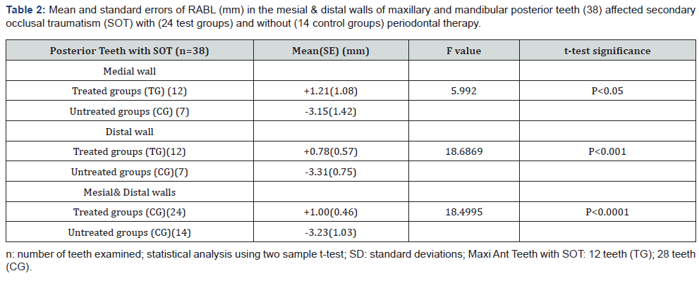
Table 3 illustrated that the mean(SD) of RABL (%) on the anterior teeth with SOT of the mesial wall in the treated group ( 6 ) was gain (+4.43±7.81 %), where the untreated group (14) was alveolar bone loss (-10.74±5.12 %). The mean alveolar bone level gain of the mesial wall in the treated group for anterior teeth with SOT was non-significantly (f value =2.6410, p>0.05) as compare to that of the untreated group using two sample t-test. Results indicated that the mean(SD) of RABL (%) on the anterior teeth with SOT of the distal wall in the treated group (6) was gain (+1.24±6.01 mm), where the untreated group (14 ) was alveolar bone loss (-10.65±0.75 mm), respectively. The mean RABL of the distal wall in the treated group for anterior teeth with SOT, was non-significant (f value =10.5379, p>0.05) as compare to that of the untreated group using two sample t-test (Table 2). Table 3 indicated that the mean(SD) of RABL (%) on the anterior teeth with SOT of the mesial and distal wall in the treated group (12) was gain (+2.84±4.30 %), where the untreated group (28) was alveolar bone loss (-10.69±3.27 %). The mean alveolar bone level gain of the mesial and distal wall in the treated group for posterior teeth with SOT was significantly higher (f value =5.5540, p<0.05) than that of the untreated group using two sample t-test.
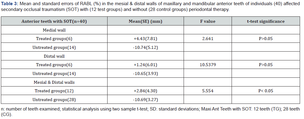
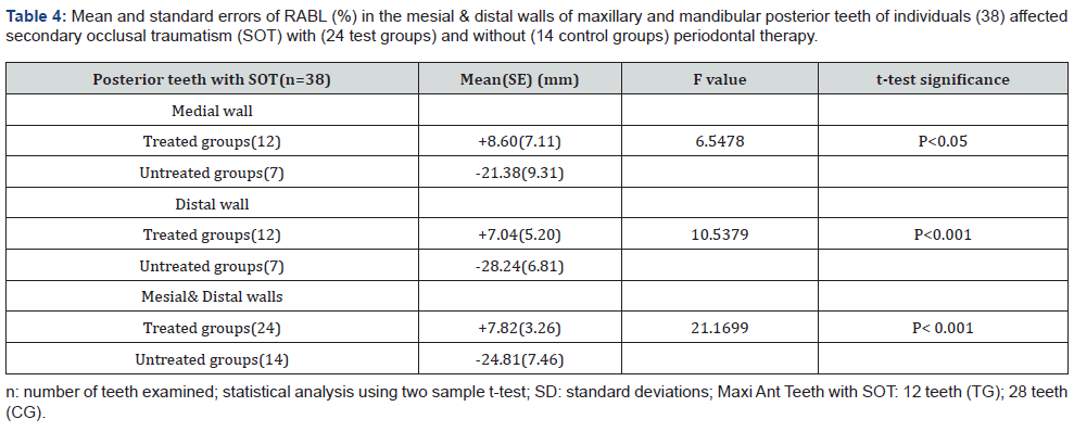
Table 4 showed the mean(SD) of RABL (%) on the posterior teeth with SOT of the mesial wall in the treated group (12) was gain (+8.60±7.11%), where the untreated group (7) was alveolar bone loss (-21.38±9.31 %). The mean RABL gain of the mesial wall in the treated group with SOT was significantly higher (f value =6.5478, p<0.05) than that of the untreated group using two sample t-test. Results indicated that the mean(SD) of RABL (%) on the posterior teeth with SOT of the distal wall in the treated group (12) was gain (+7.04±5.20 %), where the untreated group (7 ) was alveolar bone loss (-28.24±6.81 %). The mean RABL of the distal wall in the treated group with SOT was remarkably significant higher (f value =16.9685, p<0.001) than that of the untreated group using two sample t-test (Table 2). Table 4 illustrated that the mean(SD) of RABL (%) on the posterior teeth with SOT of the mesial and distal wall in the treated group (24) was gain (+7.82±3.26 %), where the untreated group (14) was alveolar bone loss (-24.81±7.46 %). The mean RABL gain of the mesial and distal wall in the treated group with SOT was significant higher (f value =21.1699, p<0.001) than the untreated group using two sample t-test.
Discussion
The pilot study from our earlier report indicated that DSRIA showed in higher values of correlation coefficients for the intra-examiner (r=0.994, p<0.001) and inter examiner (r=0.995, p<0.001) reliability tests to measure alveolar bone loss. Therefore, the present study also uses the DSRIA to assess proximal RABL [11]. Primary trauma from occlusion is possibly first caused by alteration of occlusal forces and /or over loading periodontium to withstand occlusal forces [14]. Polson et al. [15] reported that no change of above reasons probably because the supra-crestal gingivals are not affected and therefore prevent the apical migration of the junction epithelium. Secondary cause of periodontal defects of alveolar bone loss is common reason of SOT because of the burden force increasing on the less remaining tissues and heavy leverage on the weaken periodontium [16-18]. Present study showed that the mean RABL on the anterior teeth with SOT of the mesial and distal wall in the treated group was gain in contrast to the untreated group were alveolar bone loss, respectively. The RABL gain in the treated group for anterior teeth with SOT was significantly higher as compared to that of the untreated group got a severe alveolar bone loss. Therefore, present data revealed that the teeth with SOT in the treated group remarkably bone gain as compared to that of untreated cases with SOT [11-13].
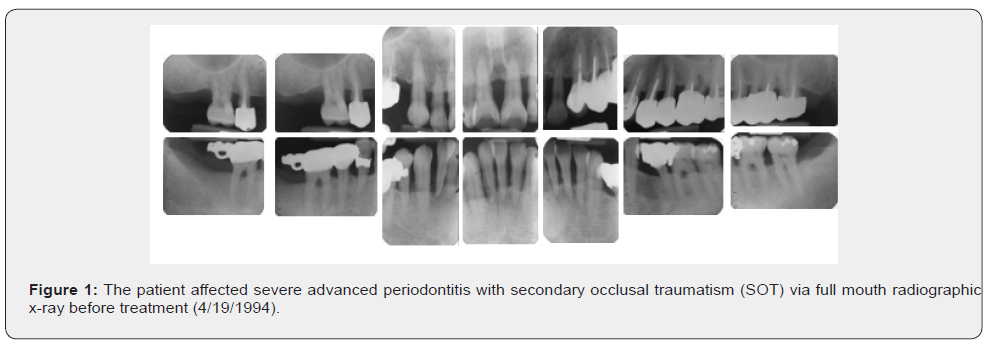
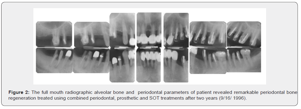
The posterior teeth with SOT of both with and without periodontal therapies revealed remarkable difference of mean RABL among mesial, distal and both walls. Results also showed that the mean of the posterior teeth with SOT for the mesial wall in the treated group was gain, in contrast to that of the untreated group with alveolar bone loss. The gain of RABL of the mesial wall in the treated group for anterior teeth with SOT was significantly higher as compared to that of the untreated group got a severe alveolar bone loss. The mean RABL in the treated group with SOT was remarkable bone gain with strong significant higher (p<0.001) than that of the untreated group using two sample t-test, irrespective of both anterior, posterior, or both groups and mesial, distal wall and both walls ( Figure1 & 2).
Little or limited reports was presented regarding changes of RABL of anterior and posterior teeth affected SOT between the treated and untreated groups. According to the present results, we can concluded that the mean of RABL in either both anterior and posterior teeth with SOT in the treated group revealed bone gain, or where the untreated group revealed alveolar bone loss, respectively. The RABL gain of the mesial, distal and both mesial and distal walls in the treated group for both anterior and posterior teeth with SOT, was significantly higher bone gain as compare to that of the untreated group with a remarkable boss.
Lindhe & Nyman [19] and Rosling [20] reported that the employment of proper periodontal therapy did not diminish the decreased tooth mobility. Teeth affected SOT might still exhibit a progressively increased mobility and splinting of these teeth might necessary using therapeutic provisional prosthesis (TPP) either unilaterally or bilaterally cross-arch design. Our recent two studies also documented and revealed that a more long-term follow-up case series of preservation of similarly compromised teeth affected SAP with SOT using the strategies of non-surgical periodontal therapy, TPP, and crown and sleeve- coping telescopic denture were made to resolve SOT. The conclusions from former and recent studies for stabilizing the teeth affected SAP with SOT seem to be a valuable and better suggestion. The present results were in consistent with therapeutic outcomes of the former studies and showed that remarkable improvement periodontal parameters and radiographic bone fills of angular bony defects (Figure 1 & 2) [21, 22].
References
- Armitage GC (1999) Development of a classification system for periodontal diseases and conditions. Ann Periodontol 4(1): 4-6.
- Boyle WD, Via WF, McFacc WT (1973) Radiographic analysis of alveolar crest height and age. J Periodontol 44(4): 236-242.
- Straham JD (1965) Relation of the muco-gingival junction to alveolar bone margin. Academy Rev 13(1): 23-28.
- Oconner TW, Biggs NL (1965) Interproximal Bony Contours. J Periodontol 35(4): 326-330.
- Selikowitz HS, Sheiham A, Albert D, Willium GM (1981) Retrospective longitudinal study of the rate of alveolar bone loss in humans using bitewing radiograghs. J Clin Periodont 8(5): 4341-438.
- Suomi JD, Plumo J, Barbano JP (1968) A comparative study of radiographs and pocket measurement in periodontal disease evaluation. J Periodontol 39 (6): 311-315.
- Hou GL (2020) Annually radiographic periodontal bone loss rates of teeth affected severe advanced periodontitis with secondary occlusal traumatism. Intern J Dent and Oral Health 6(6): 1-6.
- Manson JD, Nilcholson K (1974) The distribution of bone defect in chronic periodontitis. J Periodontol 45(2): 88-92.
- Silness J, Loe H (1964) Periodontal disease in pregnancy: Correlation between oral hygiene and periodontal condition. Acta Odont Scand 22: 121-135.
- Loe H, Silness J (1963) Periodontal disease in pregnancy: Prevalence and severity. Acta Odont Scand pp. 533-551.
- Hou GL, Hung CC, Yang YS (2003) Rdiographic alveolar bone loss in untreated Taiwan Chinese subjects with adult periodontitis measured by the digital scanning radiographic image analysis method. Dento- maxillofacial Radiology 32: 104-108.
- Hou GL (2020) Digital scanning radiographic image analysis of alveolar bone loss in individuals with untreated adult periodontitis and aggressive periodontitis. A crossectional study. Advances in Dentis- try & Oral Health 13(4): 68-75.
- Hou GL (2020) Annually radiographic periodontal bone loss rates of tooth affected severe advanced periodontitis with secondary occlusal traumatism. Intern J Dent and Oral Health 6(6): 1-5.
- Selikowitz HS, Shelham A, Albert D (1981) Retrospective longitudinal study of the rate of alveolar bone loss in humans using bite-wing radiographs. J Clin Periodont 8(5): 431-438.
- Polson AM, Meitner SW, Zander HA (1976) Trauma and adaptation of marginal periodontitis in aquiiel monkeys. III. Adaptation of inter- proximal alveolar bone to respective injury. J Periodont Res 11(5): 279-289.
- Glickman I, Smulow JB, Moreau L (1966) Effect of alloxen diabetes upon the periodontal response to excessive occlusal forces. J Periodontol 37(2):146-155.
- Rothblatt JM, Waldo CM (1953) Tissue response to tooth movement in normal and abnormal metabolic states. J Dent Res 32: 678.
- Stahl SS, Miller SC, Goldsmith ED (1957) The effect of vertical occlusal trauma on the periodontium of protein derived young adult rats. J Periodontol 28(2): 87-97.
- Lindhe J, Nyman S (1975) The effect of plaque control and surgical pocket elimination on the establishment and maintenance of periodontal health. A longitudinal study of periodontal therapy in cases of advanced diseases. J Clin Periodont 2(2): 67-69.
- Rosling B, Nyman A, Lindhe J (1976) The effect of systematic plaque control on bone regeneration in infra-bony pockets. J Clin Periodont 3(1): 38-53.
- Hou GL, Hou LT, Weisgold A (2016) Survival rate of teeth with periodontally hopeless prognosis after therapies with intentional replantation and perioproshetic procedures - a study of case series for 5-12 years. Clinical and Experiment Dental Research 2(2): 85-95.
- Hou GL, Hou LT (2019) Therapeutic outcomes using the Sandwich’s technique in treating severe advanced periodontitis with secondary occlusal trauma: A long-term study for 5.1-39 years. Intern J Dent and Oral Health 5(7): 76-85.


