Effect of different Cusp Inclinations on the Retention of Cement Retained Implant Supported Prosthesis
Ala’a Abou-obaid*
Lecturer, Department of Prosthetic Dental Sciences, College of Dentistry, King Saud University, Saudi Arabia
Submission: May 18, 2017; Published: May 29, 2018
*Corresponding author:Ala’a Abou-obaid, Lecturer, Department of Prosthetic Dental Sciences, College of Dentistry, King Saud University, Saudi Arabia, Email: aiabuoobaid@ksu.edu.sa
How to cite this article:Ala’a Abou-obaid. Effect of different Cusp Inclinations on the Retention of Cement Retained Implant Supported Prosthesis. Adv Dent & Oral Health. 2018; 9(2): 555756. DOI: 10.19080/ADOH.2018.09.555756
Abstract
Background:Occlusal morphology is an important factor which affects the distribution of occlusal load through implant components.
Objective:The aim was to investigate the effect of different cusp inclinations on the retention of cement retained single implant supported prosthesis after artificial aging.
Materials and Methods:Three groups (n=20/group) of implant supported prosthesis were fabricated with different designs of occlusal morphology. Groups A, B and C had (30°, 10°, combination of 30° and 10°) cuspal inclinations. All crowns were cemented on the implant assemblies using zinc oxide eugenol cement. Each assembly consisted of ITI implant measuring 4.1mm×12mm with corresponding 5.5mm synocta abutment mounted in an epoxy resin-glass fiber composite. The crowns were stabilized in chewing simulator and subjected to 200.000 cycles under 6 kg of load. Then, the crowns were subjected to pull-out test.
Results:The highest mean of tensile strength was noticed in group C (52.99 N) and the lowest was found in group A (38.93 N). Group A significantly differed than the other groups (p<0.05). While, no significant difference was noticed between groups B and C (p>0.05).
Conclusion:The increase in cuspal inclination, reduce the retention of cement retained implant supported prosthesis. The combining of (30° and 10°) cuspal inclinations as antagonists increased the retention of the cemented prosthesis significantly compared to 30° cuspal inclination antagonists.
Keywords: Dental Implant; Mastication; Cusp Inclination; Occlusal Form; Chewing.
Introduction
Long term durability and high success rate of dental implants made them a proper choice to restore esthetic and function of lost teeth [1-3]. However, complications still widely reported in both screw and cement retained prostheses [4,5]. Majority of the complications reported with dental implants were related to biomechanical factors rather than biological aspects. Mechanical stress is a high risk factor for restored implant under function [6]. Masticatory loading is more complex with a combination of vertical and horizontal directions rather than one direction [7]. The part of the chewing cycle where occlusal contacts occur and the pathways taken by the mandible are determined by the morphology of the teeth [8].
Occlusal design is one of the most important factor that should be considered in fabrication of different types of prosthesis. Alterations in occlusal designs were recommended to reduce forces transmitted to the bone and minimize implants failure [9-11]. The choice of occlusal design and occlusal schemes varied between prosthesis. Klineberg et al. [12] conducted a systematic review that identified randomized and other trials (1966-2006). They found that occlusal scheme design and occlusal form had a little scientific evidence to indicate which design is superior [12]. Occlusion of implant supported restorations is based on modification of occlusal concepts used for natural teeth to prevent over loading and biomechanical complications [13].
The general guidelines for occlusal scheme in single implant supported prosthesis are: reduced cuspal inclination, wide grooves and fossa, narrow occlusal table and supporting cusps in central fossa to generate forces along the long axis of the implant [12]. Several finite element analysis studies have been conducted to understand the complexity of occlusal design and the effect of cuspal inclinations on stress distribution. Bedi et al. [14] reported that occlusal design has an important role in load transmission with favorable distribution of stress in D1 (cortical bone density) under 202.23 N loading at central fossa with maximum stress of 15.10, 14.14 and 15.76 Mpa for 0°, 10°, 30° cuspal inclination, respectively [14]. Another study evaluated the stress dissipation underneath maxillary and mandibular dentures with various posterior teeth form. They reported greater magnitude of Von Mises stresses with 33° and 20° cuspal teeth and slightly less stress with 0° teeth [15]. Sornsuwan et al. [16] found a significant effect of the occlusal geometrical factors and the scatter observed with all ceramic crown fracture tests. Occlusal cusps considered more significant factor in determining fracture load [16].
In dentate patients, the differences between natural teeth and osseointegrated implants in occlusal load distribution must be taken under consideration. Moreover, factors affecting the retention of implant supported crowns such as abutment, casting, and luting agents based factors have been widely discussed in the literature [17,18]. However, little information reported on occlusal design factors. To the best of author’s knowledge, no studies evaluated the effect of different occlusal forms on the retention of single implant restorations. Therefore, the aim of this study was to evaluate the effect of cuspal inclinations on the retention of the implant supported cement retained restorations. So, the null hypothesis tested was that cuspal inclination has no effect on the retention of single implant supported restorations.
Materials and Methods
Specimen Preparation
Ten pairs of implant assemblies (n=20/group) consisted of implant fixtures measuring 4.1mm×12mm, standard plus implants (ITI system, Straumann, Waldenburg, Switzerland) with corresponding 5.5 mm synocta screw-retained abutments (048.605 ITI system, Straumann, Waldenburg, Switzerland) were prepared. The implants were mounted in cylinders filled with an epoxy resin-glass fiber composite (NEMA Grade G-10 rod, Piedmont Plastics, Charlotte, NC) (modulus of elasticity app. 20GPa) using a dental surveyor.
Fabrication of the crowns
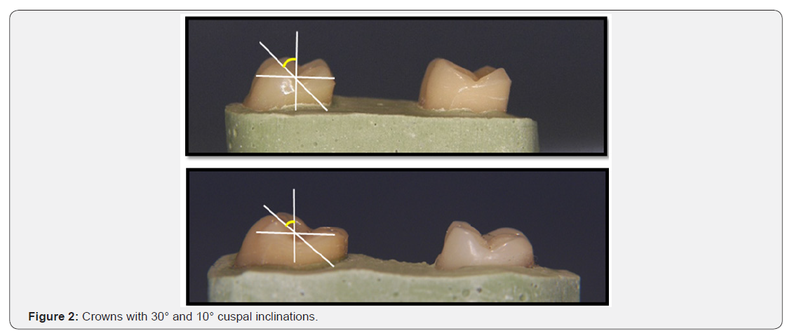
Two moulds were prepared using a clear thermoplastic sheet with 0.020 thickness (Polypropylene Sheets, Buffalo Dental Mfg Co, Inc.) from denture teeth with 30° and 10° cuspal inclinations (Trubyte 30-degree IPN and 10-degree IPN, Dentsply International Inc., York, Pa.) to fabricate the crowns with different designs of occlusal forms (Figure 1). The crowns were fabricated to simulate occlusal forms of maxillary and mandibular first molars. Each plastic coping was placed on the abutment, waxedup, invested and casted with a base metal alloy (Kera NH, Ni 58,40%, Cr 26,91%,Germany) then, veneered with 2 mm thickness of porcelain (IPS InLine, Ivoclar Vivadent AG, Liechtenstein) using the conventional layering technique. Finishing and glazing were done according to the manufacturer’s instructions. The metal ceramic crowns were designed with two wings in the proximal surfaces to secure the restoration into a customized jig during the pull-out test. Fabrication of the crowns was done by one expert technician. Then, specimens were divided into three groups. Group A consisted of crowns with 30° cuspal inclination. Group B consisted of 10° cuspal inclination. While, group C consisted of combination of 30° cuspal inclination opposed by antagonist of 10° inclination (Figure 2).

Cementation of the crowns
Before cementation, each screw retained abutments was tightened to the recommended torque (35 Ncm) and then retightened (to the same torque value) 10 minutes later to minimize embedment relaxation between the mating threads. The abutment screw access opening was covered with vinyl polysiloxane impression material (Virtual, refill light body, regular set wash material, Ivoclar Vivadent, Italy). Twenty-four hours before testing, each crown was uniformly coated with Temp Bond cement (Kerr Co, Italy) and placed on the abutment with finger pressure for 10 seconds and excess cement was removed with dental explorer. Then, each crown was loaded on its long axis with a 2 kg weight. The specimens were then stored in the room temperature (37°C).
Testing Procedure
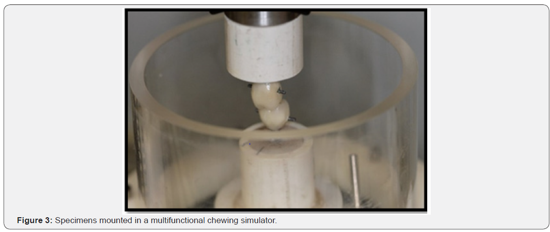
Each specimen was horizontally secured and mounted in a multifunctional chewing simulator (Chewing simulator, CS-4.2, SD Mechatronik, Germany) (Figure 3). Antagonists are coupled via horizontal and vertical traverses and then the specimens subjected to 200.000 cycles under a load of 6 kg to simulate one year of occlusal loading [19]. Artificial saline was used to reduce the coefficient of friction between two opposing surfaces.
Pull-out test
Each specimen was secured in the universal testing machine (Instron, Model 8500 Plus Dynamic Testing System, Instron Corp., England). The crowns and their antagonists were subjected to a pull-out test at a 1mm/min crosshead speed. The load required for dislodgment of the crown was recorded in (N).
Statistical Analysis
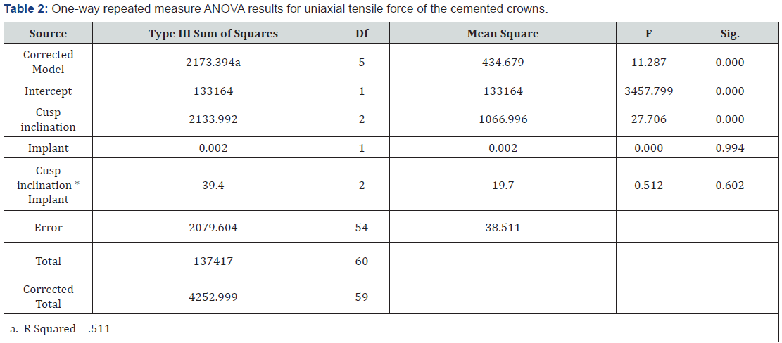
The statistical analysis were performed using the SPSS 16.0 program (SPSS Inc., Chicago, IL, USA). The data was normally distributed according to Shapiro–Wilks test. Statistical analyses were performed using one-way repeated measure analysis of variance (ANOVA). All statistical analysis were set at a significance level of p<0.05.
Results
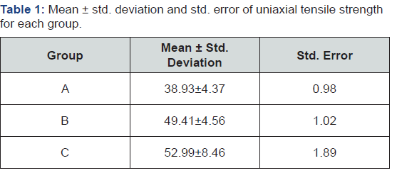
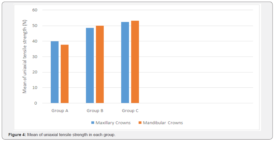
Table 1 showed the mean ± std. deviation of uniaxial tensile strength, and standard error for each group after the pull out test. The highest mean of tensile strength was noticed in group C (52.99 N) and the lowest mean value was found in group A (38.93 N) (Figure 4). One way repeated measure ANOVA (Table 2) showed that different cuspal inclinations had a significant effect on the retention force of the cemented restorations (p<0.05). Tukey Post Hoc test for multiple comparison showed significant differences in the mean tensile strength between group A and the other groups (p<0.05). While, no significant difference was noticed between groups B and C (p>0.05). There was no significant differences (p>0.05) in the retention of maxillary and mandibular crowns in each group (Table 3).

Discussion
Osseointegrated dental implants increased in popularity as an acceptable treatment modality for partially and completely edentulous patients. Biomechanical factors are considered important for long term implant stability [20]. These factors must also considered for the success of the prosthetic restoration. Functional loading is rarely directed along the long axis of implant or tooth and complex bending moments develop as a result [12]. Dental implants are more sensitive to occlusal loading. They differ than natural teeth which have buffering effect to functional loading [21]. Sever stresses and loading directed to the implant may lead to trauma, reduce bone engagement, bone atrophy [22].
The prosthetic design has an impact on the long term survival of implant supported prosthesis [23]. Several biomechanical principles have been suggested to reduce axial and lateral forces such as narrowing occlusal table, reducing cantilever length, centering occlusal contacts and reducing cuspal inclinations [9- 11,24]. Cuspal inclination is the angle between the cuspal incline from the cusp tip to the central groove and the line paralleled to the long axis of the tooth [25]. When cusp inclination increased, the resultant line of force falls away from rotation center of the implant [25-27].
In the present study, the effect of cuspal inclination has been evaluated on cement retained single implant supported prosthesis. Cement retained restorations demonstrated several advantages such as the enhanced esthetic, ease of fabrication, passive fit of casting, reduced cost, lack of accessibility of screw hole and the ease to achieve stable and ideal occlusal contacts [28-30]. However, cemented restorations showed significantly higher rate of technical complications such as abutment loosening compared to screw retained restorations [31]. Retrievability when abutment loosening occurs is one of the common problems in cement retained restoration. Type of luting agent is one of the most important factors controlling the amount of retention attained for cement retained restorations to allow the ease of retrievability without endangering of implant components [29,32,33]. Zinc oxide eugenol used in this study to simulate the clinical condition of temporary cementation.
Screw retained restorations were not included in the study due to presence of screw access hole that occupies more than 50% of the occlusal surface and covered with a restoration that subjected to wear [29,33]. This could affect the size and location of the occlusal contacts and affect the load direction and distribution. Occlusal form of 0 degree has not been evaluated in this study. This based on previous studies that reported several advantages of anatomical compared to non-anatomical denture teeth in providing superior appearance, greater chewing ability and the easiness in cleaning [34-37]. Subjective and objective evaluation of patient’s masticatory performance showed high patient satisfaction with anatomical and semi-anatomical compared to dentures with non-anatomical occlusal forms [36]. Khamis et al. [37] found that mandibular implant supported overdenture with 0° posterior teeth showed lower chewing efficiency compared to 30° teeth or lingualized occlusion with 54.14% of the patients preferred the 30° occlusal form [37]. On the other hand, another study found no significant differences between occlusal forms on the chewing efficiency of edentulous patients due to compressibility of soft tissue and movement of denture bases [38]. According to Wang and Mehta, multi-cuspid teeth increase the masticatory efficiency and distribute the occlusal load effectively compared to flat occlusal surface dentures. Patients with flat posterior teeth showed longer paus and higher biting force in the intercuspal position to compensate for the relative insufficiency of occlusal design [8].
The null hypothesis in this study was rejected since the retention of the cemented crowns significantly affected by the degree of cuspal inclination. Group C demonstrated the highest mean of tensile strength followed by groups B and A, respectively. This was in agreement with Rungsiyakull et al. [7] who conducted a finite element study to evaluate the effect of different cuspal inclinations (0°, 10°, 30°) and different occlusal loading locations (central, 1, 2 mm horizontal offset) on mandibular bone remodeling [7]. They found that 30° of cuspal inclination under a load of 2mm offset resulted in fastest bone remodeling rate and denser cortical bone within 48 months. This explained by the greater magnitude of bending moments associated with higher cuspal inclination that plays a more important role than loading location in resultant changes in bone density [7]. Another study evaluated the correlation between the cuspal inclination and tooth cracked syndrome under a load of 200 N. They found that cracked teeth had steeper cuspal inclinations that generate wedging effect with the antagonist teeth and concentrate tensile stresses on the central groove and cervical region of the molars [39]. According to mathematical analysis by Weinberg & Kruger [40], an increase of 10° in cuspal inclination resulted in 30% increase in bending moment [40]. Antenucci et al. [41] assessed the influence of cusp inclination (10°, 20°, 30°) on stress distribution in implant supported prosthesis under a 200 N of oblique load using a 3 Dimensional finite element analysis (3D FEA) [41]. They found an average of 18% increase in bending moment is observed for every 10° increase in cuspal inclination. Differences between the studies attributed to the asnalysis method used (mathematical and 3D FEA) [40]. Based on the findings reported by Antenucci et al. [41] the amount of bending moments in this study was 54 % in group A and 36% in group B which explained the significantly lower tensile strength in group A compared to other groups. The amount of generated bending moments in group C which has a combination of 30° and 10° could be lower or approximately similar to group B demonstrated by the insignificant differences of the mean of tensile strength between both groups. Behrend et al. [42] evaluated the relationship between tooth displacement and different cuspal inclinations of maxillary canine under vertical impact-type force of 21.5 g. They found that tooth restored with onlay of 65° inclination, generates lateral forces that was 6 times than that with 25° inclination. This resulted in greater crown displacement that is double in length and lower in angulation compared to the 25° onlay [42]. Another study reported maximum implant displacement at 2 mm offset when cusp inclination increased from 0° to 30°. This was explained by the amount of bending moment directed to the implant-abutment fixation. Bending of the abutment cause displacement to the implant [14]. Similarly, the decrease in retention with the increase of cuspal inclination resulted from the increase in bending moment directed to the implant abutment interface and caused displacement of the prosthesis.
There were no significant differences in the retention of the cemented prosthesis and the antagonist in each group. Mankani et al. [15] reported more stresses generated in the mandibular model compared to maxilla where the denture bearing area is reduced [15]. In this study, the insignificant differences in retentive forces were due to similar support of the prosthesis obtained from the use of single implants.
In the current study, group C showed significantly lower tensile strength compared to group A. This could be explained by the decrease in bending moment and lateral forces that resulted in less stresses on the prosthesis. Another possible reason is the type of occlusal contact occur in maximum intercuspal position with the opposing antagonist with reduced cuspal inclination that generate less lateral force component compared to contacts in steeper cusps. This in accordance with Sharry et al. [43] who found that reducing the inclinations will increase the contact areas during the functional movement [43]. Hidaka et al. [44] found that broadened occlusal contact areas would be helpful in mitigating excessive occlusal forces on teeth [44]. Moreover, the differences in the degree of cuspal inclination between the occluding pairs allow a slight freedom during lateral excursions that could play important role in the increase in crown retention. Although, this design has the highest mean of retention. It was insignificant with group B but significantly differed with group A. This allows the use of the combination of cusp inclination in clinical situation where the patient had previous single implant prosthesis as antagonist. This design can easily fabricated by a dental technician either by the recommended technique explained earlier or by the use of computer-aided design and computer-aided manufacturing technology. The possible disadvantage in this design is that the stability of the occlusal contact due to differences in the inclinations is questionable. Comparison with other studies is difficult as no previous mechanical studies conducted in this field. Further studies are recommended to evaluate the type, consistency and size of contacts occurs during occlusal function, the amount of transmitted force to the bone, and the suitability of the design with different types of opposing prosthesis (Antagonists).
The limitations of this study includes the difference between the chewing simulation and the complex motor and sensory masticatory process, the lack of chewing simulation with different food consistency in which the amount and direction of the forces in the cusps may be altered during the chewing cycles and the lack of evaluation of the effect of parafunction.
Conclusion
Based on the findings of this study, the following conclusions can be drawn:
a) The increase in cuspal inclination, reduce the retention of cement retained single implant supported prosthesis.
b) The combining of (30° and 10°) cuspal inclinations increased the retention of the cemented prosthesis significantly compared to 30° cuspal inclination antagonists.
Acknowledgment
The author would like to thank the College of Dentistry Research Center and Deanship of Scientific Research at King Saud University, Saudi Arabia for funding this research project (FR 0282). The author would like to acknowledge the Dental Bio- Materials Research Chair at the College of applied medical science for their assistance in using the chewing simulator machine.
References
- Simonis P, Dufour T, Tenenbaum H (2010) Long-term implant survival and success: a 10-16-year follow-up of non-submerged dental implants. Clin Oral Implants Res 21(7): 772-777.
- Krennmair G, Seemann R, Schmidinger S, Ewers R, Piehslinger E (2010) Clinical outcome of root-shaped dental implants of various diameters: 5-year results. Int J Oral Maxillofac Implant 25(2): 357-366.
- Lambert FE, Weber HP, Susarla SM, Belser UC, Gallucci GO (2009) Descriptive analysis of implant and prosthodontic survival rates with fixed implant-supported rehabilitations in the edentulous maxilla. J Periodontol 80(8): 1220-1230.
- Naert IE, Duyck JAJ, Hosny MMF, Van Steenberghe D (2001) Freestanding and tooth-implant connected prostheses in the treatment of partially edentulous patients. Part I: An up to 15-years clinical evaluation. Clin Oral Implants Res 12(3): 237-244.
- Salinas TJ, Eckert SE (2007) In patients requiring single-tooth replacement, what are the outcomes of implant- as compared to toothsupported restorations? Int J Oral Maxillofac Implants 22: 71-95.
- Misch CE (2006) Consideration of biomechanical stress in treatment with dental implants. Dent Today 25(5): 80-85.
- Rungsiyakull C, Rungsiyakull P, Li Q, Li W, Swain M (2011) Effects of occlusal inclination and loading on mandibular bone remodeling: a finite element study. Int J Oral Maxillofac Implants 26(3): 527-537.
- Wang M, Mehta N (2013) A possible biomechanical role of occlusal cusp fossa contact relationships. J Oral Rehabil 40(1): 69-79.
- Kim Y, Oh TJ, Misch CE, Wang HL (2005) Occlusal considerations in implant therapy: Clinical guidelines with biomechanical rationale. Clin Oral Implants Res 16(1): 26-35.
- Curtis DA, Sharma A, Finzen FC, Kao RT (2000) Occlusal considerations for implant restorations in the partially edentulous patient. J Calif Dent Assoc 28(10): 771-779.
- Duyck J, Van Oosterwyck H, Vander Sloten J, De Cooman M, Puers R, et al. (2000) Magnitude and distribution of occlusal forces on oral implants supporting fixed prostheses: An in vivo study. Clin Oral Implants Res 11(5): 465-475.
- Klineberg I, Kingston D, Murray G (2007) The bases for using a particular occlusal design in tooth and implant borne reconstructions and complete dentures. Clin Oral Implants Res 18(Suppl 3): 151-167.
- Hsu YT, Fu JH, Al-Hezaimi K, Wang HL (2012) Biomechanical implant treatment complications: a systematic review of clinical studies of implants with at least 1 year of functional loading. Int J Oral Maxillofac Implants 27(4): 894-904.
- Bedi S, Thomas R, Shah R, Mehta DS (2015) The effect of cuspal inclination on stress distribution and implant displacement in different bone qualities for a single tooth implant: A finite element study. Int J Oral Health Sci 5(2): 80-86.
- Mankani N, Chowdhary R, Mahoorkar S (2013) Comparison of stress dissipation pattern underneath complete denture with various posterior teeth form: An in vitro study. J Indian Prosthodont Soc 13(3): 212-219.
- Sornsuwan T, Ellakwa A, Swain MV (2011) Occlusal geometrical considerations in all-ceramic pre-molar crown failure testing. Dent Mater 27(11): 1127-1134.
- Bernal G, Okamura M, Muñoz CA (2003) The Effect of Taper, Length and Cement Type on Resistance to Dislodgement of Cement-Retained Implant-Supported Restorations. J Prosthodont 12(2): 111-115.
- Mohit Kheur G, Parulekar N, Shantanu Jambhekar S (2010) Clinical Considerations for Cementation of Implant Retained Crowns. IJDA 2(2): 180-181.
- Breeding LC, Dixon DL, Nelson EW, Tietge JD (1993) Torque required to loosen single-tooth implant abutment screws before and after simulated function. Int J Prosthodont 6(5): 435-439.
- Rangert BR, Sullivan RM, Jemt TM (1997) Load factor control for implants in the posterior partially edentulous segment. Int J Oral Maxillofac Implants 12(3): 360-370.
- Komiyama O, Lobbezoo F, De Laat A, Iida T, Kitagawa T, et al. (2012) Clinical management of implant prostheses in patients with bruxism. Int J Biomater p. 6.
- Carini F, Longoni S, Pisapia V, Francesconi M, Saggese V, et al. (2014) Immediate loading of implants in the aesthetic zone: Comparison between two placement timings. Ann Stomatol 25(Suppl 2): 15‑26.
- Fu JH, Hsu YT, Wang HL (2012) Identifying occlusal overload and how to deal with it to avoid marginal bone loss around implants. Eur J Oral Implantol 5(Suppl): S91-103.
- Stanford CM (2005) Issues and considerations in dental implant occlusion: What do we know, and what do we need to find out? J Calif Dent Assoc 33(4): 329-336.
- Weinberg LA (1998) Reduction of implant loading with therapeutic biomechanics. Implant Dent 7(4): 277-285.
- Skalak R (1983) Biomechanical considerations in osseointegrated prostheses. J Prosthet Dent 49(6): 843-848.
- Rangert BO, Jemt T, Jorneus L (1989) Force and moments on Branemark implants. Int J Oral Maxillofac Implants 4(3): 241-247.
- Breeding LC, Dixon DL, Bogacki MT, Tietge JD (1992) Use of luting agents with an implant system: part I. J Prosthet Dent 68(5): 737-741.
- Chee W, Felton DA, Johnson PF, Sullivan DY (1999) Cemented versus screw-retained implant prostheses: which is better? Int J Oral Maxillofac Implants 14(1): 137-141.
- Michalakis KX, Hirayama H, Garefis PD (2003) Cement retained versus screw-retained implant restorations: a critical review. Int J Oral Maxillofac Implants 18(5): 719-728.
- Ma S, Fenton A (2015) Screw- versus cement-retained implant prostheses: a systematic review of prosthodonticmaintenance and complications. Int J Prosthodont 28(2): 127-145.
- Taylor TD, Agar JR (2002) Twenty years of progress in implant prosthodontics. J Prosthet Dent 88(1): 89-95.
- Vigolo P, Givani A, Majzoub Z (2004) Cemented versus screw-retained implant-supported single-tooth crowns: a 4-year prospective clinical study. Int J Oral Maxillofac Implants 19(2): 260-265.
- Lang BR, Razzoog ME (1992) Lingualized integration: tooth molds and an occlusal scheme for edentulous implant patients. Implant Dent 1(3): 204-211.
- Sutton AF, Worthington HV, McCord JF (2007) RCT comparing posterior occlusal forms for complete dentures. Journal of Dental Research 87(7): 651-655.
- Sutton AF, McCord JF (2007) A randomized clinical trial comparing anatomic, lingualized, and zero-degree posterior occlusal forms for complete dentures. Journal of Prosthetic Dentistry 97(5): 292-288.
- Khamis MM, Zaki HS, Rudy TE (1998) A comparison of the effect of different occlusal forms in mandibular implant overdentures J Prosthet Dent 79(4): 422-429.
- Lu YL, Lou HD, Rong QG, Dong J, Xu J (2010) Stress area of the mandibular alveolar mucosa under complete denture with linear occlusion at lateral excursion. Chinese Medical Journal 123(7): 917- 921.
- Qian Y, Zhou X, Yang J (2013) Correlation between cuspal inclination and tooth cracked syndrome: a three-dimensional reconstruction measurement and finite element analysis. Dent Traumatol 29(3): 226- 233.
- Weinberg LA, Kruger B (1995) A comparison of implant prosthesis loading with four clinical variables. Int J Prosthodont 8(5): 421-433.
- Falcón-Antenucci RM, Pellizzer EP, de Carvalho PS, Goiato MC, Noritomi PY (2010) Influence of cusp inclination on stress distribution in implant-supported prostheses. A three-dimensional finite element analysis. J Prosthodont 19(5): 381-386.
- Behrend DA (1978) The relationship between tooth displacement and cusp inclination in man. Arch Oral Biol 23(12): 1095-1098.
- Sharry JJ, Askaw HC, Hoyer H (1960) Influence of artificial tooth forms on bone deformation beneath complete dentures. J Dent Res 39: 253- 266.
- Hidaka O, Iwasaki M, Saito M, Morimoto T (1999) Influence of clenching intensity on bite force balance, occlusal contact area, and average bite pressure. J Dent Res 78(7): 1336-1344.


