Ectopic Third Molar in Sigmoid Notch: Report of a Case
JL Del Castillo1*, G DeMaria2 and M Chamorro3
1Department of Oral and Maxillofacial, University Hospital La Paz, Spain
2Department of Oral and Maxillofacial, Hospital La Moraleja-Sanitas, Spain
3Department of Oral and Maxillofacial, University Hospital La Paz, Spain
Submission: December 30, 2017; Published: March 23, 2018
*Corresponding author: JL Del Castillo, Department of Oral and Maxillofacial, University Hospital La Paz, Spain, Tel: +34917277336/+34606409021; Email: delcastillo6@hotmail.com
How to cite this article: JL Del Castillo, G DeMaria, M Chamorro. Ectopic Third Molar in Sigmoid Notch: Report of a Case. Adv Dent & Oral Health. 2018; 8(2): 555731. DOI: 10.19080/ADOH.2018.08.555731
Abstract
The surgical removal of impacted third molars is the most common procedure performed by maxillofacial surgeons, but ectopic placement is quite rare. Only a few cases of ectopic third molars in sigmoid notch have been reported. We present a clinical case of a third ectopic molar located in sigmoid notch and associated with a dentigerous cyst in a 32-year-old patient, which was extracted through intraoral approach. The aetiology of ectopic mandibular third molars has not yet been completely clarified. Treatment should be carefully planned according to the position of the ectopic tooth and the potential for trauma caused by the surgery.
Keywords: Ectopic third molar; Intraoral approach; Sigmoid notch
Introduction
Inclusion of the third molar is a common condition with a frequency of 20-30%. Ectopic mandibular third molars, however are unusual, with their heterotrophic positions reported in the mandibular ramus, in the coronoid process and in the condylar or the sub condylar region [1,2]. Ectopic third molar are those that are impacted in unusual positions, or that have been displaced and are at a distance from their normal anatomic location. Tooth development results from a complicated multi-step interaction between the oral epithelium and the underlying mesenchymal tissue. Ectopic eruption can be associated with developmental disturbances, pathologic processes or iatrogenic activity. Only seven cases of ectopic mandibular third molars in sigmoid notch reported in the literature over the past 40 years [3-9]. Other locations for ectopic third molar are the maxillary sinus and the infratemporal fossa. In these cases, the removal of the ectopic third molar can be difficult, and procedure requires surgical intervention. The aetiology and protocol for extraction are still unclear [10].
In this report we present a clinical case of a third ectopic molar located in sigmoid notch and associated with a dentigerous cyst in a 32-year-old patient, which was extracted through intraoral approach and review the literature reports over the past 40 years.
Case Report
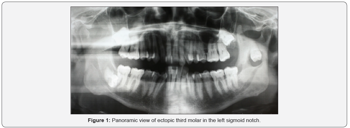
A 31-year-old healthy and asymptomatic Caucasian man was referred to the department of Oral and Maxillofacial Surgery by his Dentist. He reported several episodes of left preauricular pain and swelling of the parotid region that remitted with antibiotic and anti-inflammatory treatment. The patients also experimented limitation of mouth opening. The intraoral examination revealed bulging of the inferior left vestibule that was tender to palpation. Panoramic radiography showed an inverted ectopic third molar in the left sigmoid notch region with an associated cyst (Figure 1). The ectopic third molar was situated with the apex facing the sigmoid notch and the crown facing downward. A small well defined radiolucent area encompassing the crown of the ectopic third molar was observed. A Computed Tomography (CT) scan confirmed these findings.

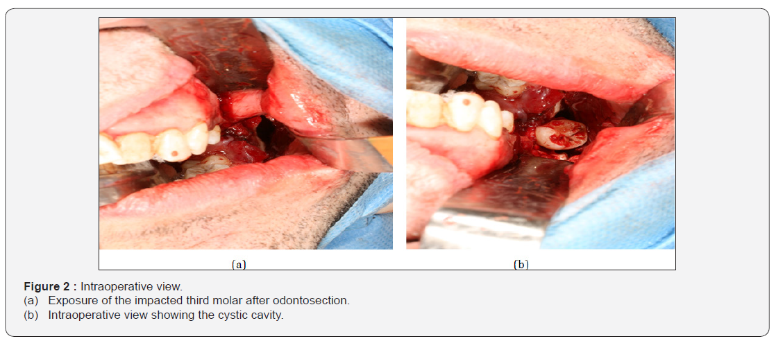
Under naso-tracheal general anaesthesia, an intraoral access was obtained via an incision on the anterior edge of the mandibular ramus and along the external oblique ridge down to the ectopic third molar. After periosteal dissection of the ascending ramus of the mandible, a window in the external cortical bone was created using a 3mm carbide round bur on a straight surgical hand piece and the crown of the ectopic third molar was exposed. A bony window just larger that the crown was created and the tooth was then elevated out of the bony socket by an elevator after odonto section of the ectopic third molar (Figure 2). The associated cyst was removed later and the wound was sutured after irrigation with saline solution.
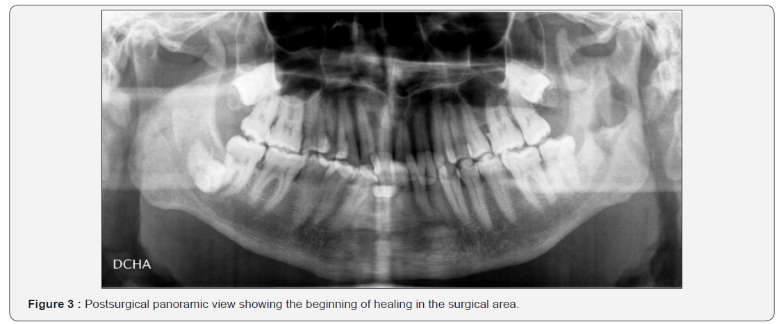
The patient had a satisfactory postoperative recovery. Amoxicillin, 1000mg three times a day was given for a week and Ibuprofen, 600mg three times a day for three days. Mouth opening improved progressively and the function of the inferior alveolar nerve was completely normal one month after surgery (Figure 3). The diagnosis of dentigerous cyst was confirmed by histological examination.


Discussion
The Etiology of ectopic third molars has not yet been completely clarified. Tooth development results from an interaction between the oral epithelium and the underlying mesenchymal tissue. Abnormal tissue interactions during development may potentially result in ectopic tooth development and eruption. Many theories have been put forward including, aberrant eruption, trauma, infection, cyst, tumours and developmental disturbances. In many cases the aetiology cannot be identified. A mandibular third molar may be displaced by a lesion such as a cyst or a tumour. The expansion of a cyst as it develops may result in pressure on the crown of a tooth and displace it in a direction opposite to the path of eruption. The dentigerous cyst is a common lesion [5,8,11]. It is the second most common odontogenic cyst following the radicular cyst and it is more common in males occurring in the second or third decade of life. The radiological image is characterized by a well circumscribed, unilocular and normally symmetric radiolucent image around the crown of an impacted tooth. It is usually found in the region of the third molar, and they are more commonly an isolated finding.
Ectopic eruption of a tooth into a region other than the oral cavity is rare although there have been reports of tooth in the nasal septum, mandibular condyle, maxillary antrum, palate and coronoid process. From 1965 to 2008, only seven reports were made of ectopic third molar in sigmoid notch. Patients are mostly asymptomatic [4,7] and the clinical symptoms caused by these lesions do not differ much from those of other ectopic inclusions or maxillary cysts [5,8,9]. The most common signs and symptoms associated with ectopic third molar in sigmoid notch are:
1. Pain and swelling on the ipsilateral side of the mandible or the preauricular region
2. Limitation of mouth opening, and difficulty in mastication. Granite et al. [4] reported a case of impacted third molar in the sigmoid notch without clinical symptoms and signs.
The diagnosis of this condition can easily be made radiologically with radiological studies (orthopantomogram) and CT scans taken in axial and coronal sections, for confirmation. The precise location of the ectopic third molar, sometimes with high-resolution CT scans, may provide direction in choosing the appropriate surgical method [12].
The usual treatment for a dentigerous cyst associated with an impacted third molar is its enucleation together with the extraction of the tooth. The treatments should be carefully planned according to the location of the ectopic third molar and the possible trauma caused in the surgery. This situation makes it much more difficult to extract the third molar and associated cyst. Treatment of ectopic third molar in the sigmoid notch is recommended to avoid the morbidity caused by the infection of the cyst, risk of fracture, and temporomandibular joint syndrome. In the cases of ectopic third molar in other locations described in the literature several approaches have been used, such as intraoral [13], retromandibular, preauricular and endoscopic. Whenever possible intraoral approaches should be used but logically this will be determined by the location of the ectopic third molar.
The use of endoscopic approach has considerable advantages [11,14], but requires basic endoscopic equipment and special training, and this technique may not be indicated in all locations of an ectopic third molar. If conservative treatment is opted for, a follow-up of the patient will be necessary. The indication for the extraction of an ectopic third molar is in general determined by the presence of symptomatology, or it may be aimed at preventing future complications. In these cases, the removal of the ectopic third molar can be difficult, and procedure requires surgical intervention. The aetiology and protocol for extraction are still unclear.
References
- Bux P, Lisco V (1994) Ectopic third molar associated with a dentigerous cyst in the subcondylar region: Report of a case. J Oral Maxillofac Surg 52(6): 630-632.
- Salmeron JI, del Amo A, Plasencia J, R Pujol, CN Vila (2008) Ectopic third molar in condylar region. Int J Oral Maxillofac Surg 37(4): 398- 400.
- Balan N (1992) Tooth in the sigmoid notch. Oral Surg Oral Med Oral Pathol 73: 767.
- Granite EL, Isaacs M, Kross JF (1985) Asymptomatic impacted mandibular third molar in the subcondylar-sigmoid notch region associated with extensive sclerotic bone. J Oral Med 40(2): 91-92, 97.
- Traiger J, Koral K, Catania AJ, Nathan AS (1965) Impacted third molar and dentigerous cyst of the sigmoid notch of the mandible. Report of a case. Oral Surg Oral Med Oral Pathol 19: 459-461.
- Giardino C, Valletta G (1966) Heterotopia of the lover 3d molar on the level of the sigmoid notch. Clinical case. Arch Stomatol 7(4): 323-327.
- Chongruk C (1991) Asymptomatic ectopic third molar. Oral Surg Oral Med Oral Pathol 71(4): 520.
- Mehta DS, Mehta MJ, Murugesh SB (1986) Impacted mandibular third molar in the sigmoid notch region associated with dentigerous cyst: a case report. J Indian Dent Assoc 58(12): 545-547.
- Nishijima K, Kishi K, Komai M, Maeda K, Wake K (1976) A case of impacted third molar and dentigerous cyst located below the sigmoid notch of the mandible (author`s transl). Nippon Koku Geka Gakkai Zasshi 22(3): 391-395.
- Szerlip L (1978) Displaced third molar with dentigerous cyst – an unusual case. J Oral Surg 36: 551.
- Türner C, Eset AE, Atabek A (2002) Ectopic impacted mandibular third molar in the subcondylar region associated with a dentigerous cyst: a case report. Quintessence Int 33(3): 231-233.
- Bortoluzzi MC, Manfro R (2010) Treatment for ectopic third molar in the subcondylar region planned with cone beam computed tomography: A case report. J Oral Maxillofac Surg 68(4): 870-872.
- Gadre KS, Waknis P (2010) Intra-oral removal of ectopic third molar in the mandibular condyle. Int J Oral Maxillofac Surg 39(3): 294-296.
- Suarez Cunqueiro MM, Schoen R, Schramm A, Gellrich NC, Schmelzeisen R (2003) Endoscopic approach to removal of an ectopic mandibular third molar. Br J Oral Maxillofac Surg 41(5): 340-342.


