Intra-Lesional Steroid Treatment of Central Giant Cell Granuloma of the Mandible
Mohammed H Al-Bodbaij* and Ghada AlQassab
Oral and Maxillofacial Surgery Department, King Fahad Hospital-Hofuf, KSA
Submission: October 24, 2016; Published: November 09, 2016
*Corresponding author: Mohammed H Al-Bodbaij, Oral and Maxillofacial Surgery Department, King Fahad Hospital-Hofuf, Al Salmaniyah South, Al Hofuf 36441, Saudi Arabia, Email: bodbaij@hotmail.com
How to cite this article: Mohammed H Al-B, Ghada Al. Intra-Lesional Steroid Treatment of Central Giant Cell Granuloma of the Mandible. Adv Dent & Oral Health. 2016; 2(4): 555596. DOI: 10.19080/ADOH.2016.02.555596
Keywords:Giant cell granuloma, Steroids, Surgical excision
Introduction
The Central Giant Cell Granuloma (CGCG) is defined by the World Health Organization (WHO) as an intraosseous lesion consisting of cellular fibrous tissue that contains multiple foci of hemorrhage, aggregations of multinucleated giant cells and occasionally trabecule of woven bone [1].
CGCG occurs mainly in children and young adults with more than 60% of all cases occurring before the age of 30 years and female to male ratio of 2:1 (Jaffe 1953). The mandibular / maxillary ratio is from 2:1 to 3:1 [2,3].
CGCG is classified into the following
Nonaggressive: characterized by slow growth that doesn’t cause cortical bone perforation or root resorption. It has low tendency to recur.
Aggressive: characterized by pain, rapid growth, expansion and/or perforation of cortical bone, root resorption and high recurrence tendency [4].
Radiographically, CGCGs present as an expansile radiolucency, either unilocular or multilocular with defined, poorly defined or diffused borders [1].
The treatment of CGCG includes curettage or resection of the lesion [5].
Systemic injections of calcitonin [6] and intralesional injections of corticosteroid were also used [7].
Case Report
In December 2014, a 14 years old Saudi girl presented to the department of Oral and Maxillofacial Surgery at King Fahad Hospital- Hofuf / KSA with a gradually enlarging swelling of the left side of the lower jaw which was diagnosed as central giant cell granuloma in other hospital she had presented to.
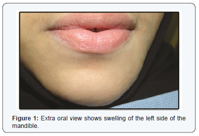
Extra-oral examination, showed non-tender enlarged left side of the mandible. There was no regional lymphadenopathy (Figure 1).
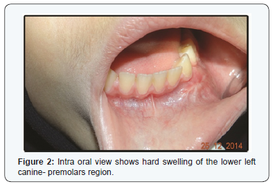
Intra-oral examination showed non-tender hard swelling of canine - premolars region of left mandibular side with linear scar of overlying mucosa. Swelling caused buccal expansion with slight buccal sulcus obliteration,
slight mobility of teeth No. 33 and 34 (Figure 2).
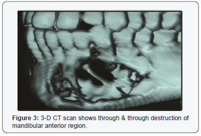
Computerized Tomograph (CT) Figure 3 showed a destruction of the alveolar and basilar bones of anterior region of the mandible extending from tooth 35 to 43.
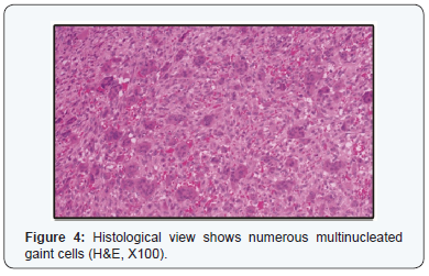
Histological slides were revised which have shown proliferating spindled to plump cells present loosely and interspersed diffusely with numerous multinucleated giant cells; focally collagen matrix and few bony trabeculae were present (Figure 4).
Assays of parathyroid hormone (PTH), calcium, and phosphorus were within normal ranges which ruled out hyperthyroidism.
Intralesional injections of 4 cm’ of a mixture of triamcinolone acetonide 10 mg/ml and lidocaine hydrochloride with adrenaline 1:80,000 in 1:1 ratio was administered on weekly bases for six weeks.
During the last two treatments, slight resistance during injections was found.
One month post-intralesional treatment, there was no obvious changes were noted.
Two months post-intralesional treatment, there was some changes which included slight reduction of teeth mobility 33 & 34 and slight increase of opacification of the lesion on panoramic radiograph.
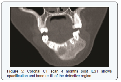
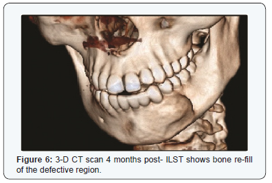
Four months post- ILST, there was no mobility of the teeth No. 33 & 34, harder mass on palpation and CT scan (Figure 5& 6) have shown increased opacity and defect refill.
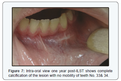
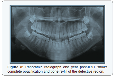
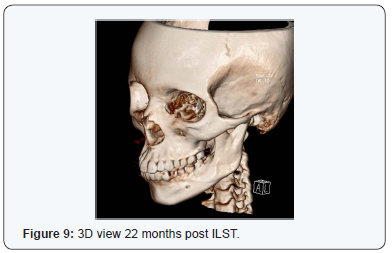
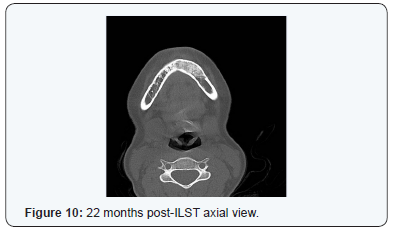
One year later, there was no mobility of the teeth No. 33 & 34, reduced size of bony swelling (Figure 7) and panoramic radiograph has shown complete re-ossification of the region (Figure 8).
Discussion
Central Giant Cell Granuloma (CGCG) is a benign lesion of the jaws, facial bones and the skull of unknown etiology [8].
It commonly affects children and young adults and at least twice as common in females [9].
CGCG is classified into two types nonaggressive and aggressive [4].
The traditional treatment of CGCG of the jaws is surgery which ranges from curettage of the lesion [8] to total en-bloc resection [10] depending on the following factors: aggressive versus nonaggressive behavior, location, size and radiographic appearance. There are no histologic differences between the aggressive and nonaggressive varieties [11].
The commonly considered recurrence rate is between 10% and 20% [3].
Non-surgical approaches have been used, including daily systemic doses of calcitonin [6] with the disadvantage of prolonged time of treatment and intralesional injections with corticosteroids as described by
Terry and J acoway in their protocol [12].
Confirmation of the lesion via biopsy must be performed before the administration of intralesional corticosteroid injections.
Conclusion
This case of CGCG has been treated with six weekly intralesional injections of steroid with very good results. Greater consideration should be given to intralesional steroid as an alternative to surgery in treatment of
CGCGs. The technique is simple, non-expensive and avoids large defects of the jaw post-operatively.
References
- Kramer IR, Pinborg JJ, Shear M (1991) Histologic typing of odontogenic tumors. (2nd edn), Springer-Verlag, Berlin, Germany.
2. Kaffe I, Ardekian L, Taicher S, Littner MM, Buchner A (1996) Radiologic features of central giant cell granuloma of the jaws. Oral Surg Oral Med Oral Pathol Oral Radiol Endod 81(6): 720-726.3. Whitaker SB, Vigneswaran N, Budnick SD (1993) Giant cell lesions of the jaws: evaluation of nucleolar organizer regions in lesions of varying behavior. J Oral Pathol Med 22(9): 402-405.
- Chuong R, Kaban L, Kozakewich H, Perez-Atayde A (1986) Central giant cell lesions of the jaws: a clinicopathologic study. J Oral Maxillofac Surg 44(9): 708-713.
- Bataineb AB, Al-Khateeb T, Rawashdeh MA (2002) The surgical treatment of central giant cell granuloma of the mandible. J Oral Maxillofac Surg 60(7): 756-761.
- Harris M (1993) Central giant cell granulomas of the jaws regress with calcitonin therapy. Br J Oral Maxillofac Surg 31(2): 89-94.
- Carlos R, Sedano O (2002) Intralesional corticosteroids as an alternative treatment for central giant cell granuloma. Oral Surg Oral Med Oral Pathol 93(2): 161-166.
- Jaffe HL (1953) Giant-cell reparative granuloma, traumatic bone cyst and fibrous (fibro-osseous) dysplasia of the jawbones. Oral Surg Oral Med Oral Pathol 6(1): 159-175.
- Whitaker SB, Waldron CA (1993) Central giant cell lesions of the jaws Oral Surg Oral Med Oral Pathol 75(2): 199-208.
- Becelli R, Cerulli G, Gasparini G (1998) Surgical and implantation reconstruction in a patient with giant-cell central reparative granuloma. J Craniofac Surg 9(1): 45-47.
- O Malley M, Pogrel MA, Stewart JC, Silva RG, Regezi JA (1997) Central giant cell granuloma of the jaws: phenotype and proliferation-associated markers. J Oral Pathol 26(4): 159-163.
12.Terry BC, J acoway JR (1994) Management of central giant cell lesions: an alternative to surgical therapy. Oral Maxillofac Surg Clin North Am 6(3): 579-600.


