




 OROAJ Home



Abstract
Introduction: As annual femoral neck fracture (FNF) incidence rises in an aging Indian population, the choice between hemiarthroplasty and total hip arthroplasty (THA) has become increasingly relevant, influenced not only by patient factors of age and activity level but also by the surgeon’s subspecialty special training. Most cases in elderly (55 yrs +) bracket are caused by simple, low-energy falls from standing height at home. Among most a Sub-capital and transcervical fractures are most common. In patients over 80, the location often shifts to the sub-capital region. Elderly Indian patients frequently present with pre-existing conditions, including malnutrition, frailty, diabetes, hypertension, CVDs, COPD, vitamin D deficiencies, and urinary tract infections, that complicate surgical planning. Diagnosis and control of UTI are of utmost importance before operating patients with fracture neck of femur with hemiarthroplasty. In contrast, among the Younge adults up to 70% of femoral neck fractures are caused by high-energy incidents such as road traffic accidents (RTAs) or falls from heights. In 1960’s Treatment focused for young people joint-preserving surgery using Open Reduction and Internal Fixation (ORIF) to avoid long-term complications like avascular necrosis (AVN) as the bone is highly vascular. For the last 3-4 decades traditionally, hemiarthroplasty was favored for older, less active patients, whereas THA is associated with better outcomes for active patients. The THA utilization has increased for FNFs, particularly among adult reconstruction specialists for even older active patient groups up to 70-75 years without much of comorbidities. This shift reflects a growing emphasis on functional outcomes and mobility, and surgeon’s training appears to influence treatment choice beyond traditional age-based guidelines. These findings underscore an evolving approach to FNF management that may create demand pressures on reconstruction and trauma specialists who are managing an expanding case load of complex arthroplasties.
Materials and Methods: This article is based on a few anecdotal cases closely followed by this author over last 60 years, to track the technical developments, practice and outcomes in Femur Neck Fracture cases.
Outcomes: All cases reported had functional restoration of hip joints.
Keywords:Femoral; Avascular Necrosis; Hemiarthroplasty; Arthroplasty; Femoral Neck
Abbreviations:FNF: Femoral Neck Fracture; ORIF: Open Reduction and Internal Fixation; AVN: Avascular Necrosis; FNS: Femoral Neck System; HA: Hemiarthroplasty; THA: Total Hip Arthroplasty; RTAs: Road Traffic Accidents; COPD: Chronic Obstructive Pulmonary Disease; BMC: Baseline Bone Mineral Content; GT: Greater Trochanter; MCH: Medical College hospital
Introduction
Worldwide incidence of hip fractures is estimated to increase by 4.50-6.26 million by 2050 and half of that being in Asia. United States with a population of 332 million reports 300000 hip fractures annually according to CDC data. Hip fractures in the United Kingdom were expected to be around 104,000 in 2025 [1,2]. The incidence of hip fractures in China plateaued from 2012 to 2016 and is expected to decline like in other developed countries. India lacks a centralized registry tracking Femoral Neck Fractures (FNF). Approximate incidence of hip fractures in India is 129 /100,000.
However, epidemiological models indicate that the incidence exceeds 130 cases per 100,000 persons aged >50, translating to roughly 0.2 million hip fractures annually now. Due to increasing life spans, this burden is projected to reach 0.8 million by 2050. Some Studies show crude hip fracture rates of 159 per 100,000 in women and 105 per 100,000 in men over age 50 [3]. Femoral neck fracture trends in India are driven by a rapidly aging population and increasing urbanization. Currently, the data shows a clear split: osteoporotic low-velocity falls dominate among the elderly, while high-velocity trauma, e.g., road traffic accidents, affect young [2,3].
Geographic & Future Considerations Demographic projections suggest that over half of the world’s osteoporotic fractures will be concentrated in Asia by 2050. While the common cause of Femur neck fractures are falls in bathrooms or slipper floors. lower baseline bone mineral content (BMC), changing diets, and increased life spans mean hospitals are actively standardizing geriatric protocols to manage complications such as preoperative urinary tract infections and post-operative mortality [2]. Most cases in elderly (55 yrs +) bracket are caused by simple, lowenergy falls from standing height at home.
Among most a Sub-capital and transcervical fractures are most common. In patients over 80, the location often shifts to the sub-capital region. Elderly Indian patients frequently present with pre-existing conditions, including malnutrition, frailty, vitamin D deficiencies, and urinary tract infections, that complicate surgical planning. In contrast, among the Younge adults up to 70% of femoral neck fractures are caused by high-energy impacts like Road Traffic Accidents (RTAs) and fall from heights. This author is a witness for Indian orthopaedics undergoing a massive transformation, shifting from conservative bed rest to advanced global-standard surgical reconstruction.
Treatment Focusses among young the priority is typically joint-preserving surgery using Open Reduction and Internal Fixation (ORIF) to avoid long-term complications like avascular necrosis (AVN) as the bone is highly vascular. The Femoral Neck System (FNS) & cannulated screws are increasingly preferred, yielding high union rates with minimal femoral shortening. For displaced femoral neck fractures in India, Total Hip Arthroplasty (THA) is generally preferred for active, independent patients due to superior functional outcomes and lower revision rates. Hemiarthroplasty, specifically the bipolar technique, is favoured for sedentary, elderly patients requiring faster, less invasive surgery. In elderly patients over 60 years are subjected to either a unipolar or bipolar Hemiarthroplasty (HA) to mobilize them quickly & reduce the risk of bed-rest complications.
Case Reports
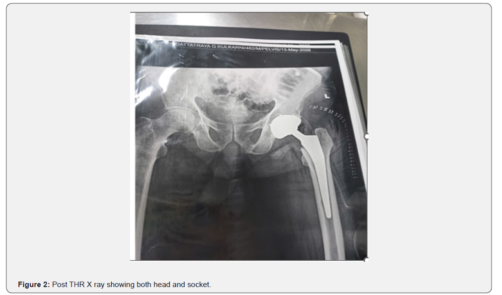
Case 1: Most Recent May 2026 case of THR: Mr. Datta Kulkarni 78 yrs old male patient with an history of self-fall while accessing toilet on 09 May 2026 around 0630 and pain and swelling over left buttock arrived at a private Orthopaedic Clinic in Pune, India. Physical examination showed restricted movements of left hip joint and swelling. An X ray revealed Fracture Neck of Left femur (Figure 1). Initially the resident doctor recommended Open Reduction and Internal Fixation (ORIF) or Hemiarthroplasty keeping cost consideration. Family checked with this author who advised for Left Total Hip Replacement, as he was eligible for THR due to all biochemical parameters, imaging (Chest X Ray, ECG,2D echo, and general health conditions. Left THR was done on 13 May 2026.
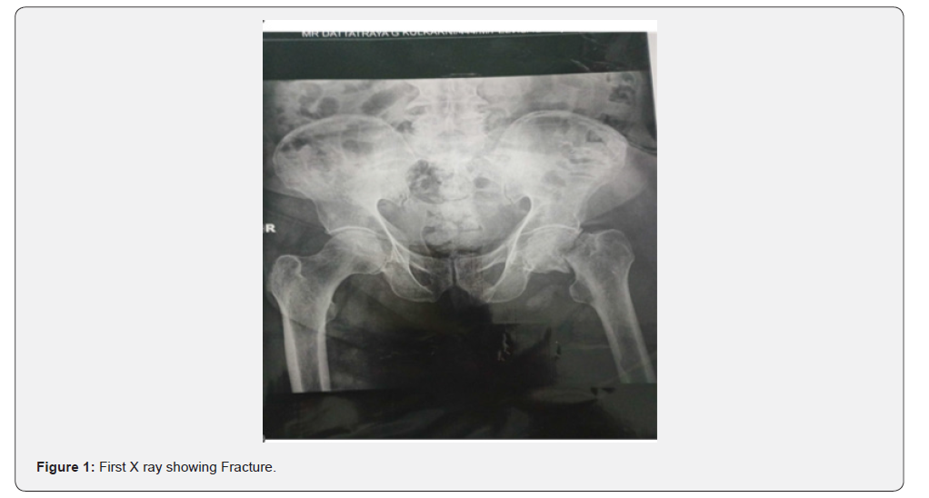
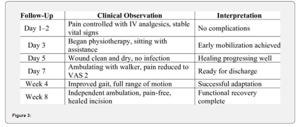
Procedural details: Under Epidural Spinal Anaesthesia and aseptic precautions, patient was put on right lateral position. Incision was taken from tip of greater trochanter (GT) to backwards on Left side. After an incision is made, one finger was placed in sciatic notch & one finger on anterior superior spine; a line is drawn on drapes between these 2 points with Methylene blue. Another line was drawn between sciatic notch & anterior spine on the drapes, and a 2nd orthogonal line was drawn and flexed an additional 10 degrees. Subcutaneous Bursa was incised; External rotators were tied and cut. Capsule was cut in inverted T shape and head of femur extracted. Acetabulum reaming was done from 38 to 52 size. 52 panache cups with liver (DEPUY) put with 2 screws (6.5mm x 25 mm) fixed with good stability Femur side canal entry made. Femur stem trails from 8 to 12 sized. Femur Coralie stem size 12 (DEPUY) put with metal head 36 mm fixed, checked stability. The resulting leg length discrepancy was hardly 0.2 mm. A thorough wash given. Homeostasis achieved; closure done in layers. Aseptic dressing is done. Patients withstood the procedure well. Postoperatively patient was stable and was put on antibiotics and analgesics and other supportive measures for a week. Post-operative recovery was fast with no complications, and he was made to walk with a walker on day 3,4 5 before discharging on 18 May 2026. On a follow-up visit stitches were removed on 21 May 2026 and the wound had healed fully. He is now walking with walker in the home with continued physiotherapy (Figures 1 & 2).
Case 2 Total Hip Arthroplasty (THA) surgery in 2018: A 61-year-old man with untreated diabetes mellitus and hypertension developed pain in the left hip after falling while getting off a bus at around 10:00 p.m. on 25 December 2018. He was first taken to a sub-district hospital, where pelvic radiography confirmed a fracture of the neck of the left femur. Analgesics and traction were if night for pain relief. The following day, he was transferred to a reputed private hospital in Bengaluru, about 300 km away, for further evaluation and management. On the morning of 27 December 2018, the orthopaedic surgeon examined him and advised THA. Considering his age and comorbidities, a cemented femoral stem was selected. The prosthesis included a press-fit acetabular component, neutral polyethylene liner, and ceramicon- polyethylene head/liner construct. The surgeon performed the procedure in the lateral position under fluoroscopic anteroposterior guidance using Moore’s posterior approach. Under epidural spinal anesthesia and aseptic precautions, the patient was placed in the right lateral position. An incision was made from the tip of the greater trochanter posteriorly. After identifying the sciatic notch and anterior superior iliac spine, reference lines were marked on the drapes with methylene blue. The subcutaneous bursa was incised, the external rotators were tied and divided, and the capsule was opened in an inverted T shape. The femoral head was extracted. Acetabular reaming was performed from size 38 to 52, and a size 52 Panache cup with liner (DEPUY) was fixed with two 6.5 mm × 25 mm screws, achieving good stability. The femoral canal was prepared, and stem trials from size 8 to 12 were performed. A size 12 Corail femoral stem (DEPUY) with a 36 mm metal head was implanted and found to be stable. The leg-length discrepancy was approximately 0.3 mm. Thorough lavage was given, haemostasis was achieved, the wound was closed in layers, and an aseptic dressing was applied. The patient tolerated the procedure well and remained stable postoperatively. He received antibiotics, analgesics, and supportive care and was discharged on 2 January 2019 with continued medication. At follow-up on 10 January 2019, ambulation with a walker, quadriceps-strengthening exercises, and medication were advised. Sutures were removed on 15 January 2019, and the wound had healed completely. Physiotherapy continued for another four weeks. He was advised to avoid hip and knee flexion beyond 90 degrees, crossing his legs, lifting the leg to put on socks, and sitting cross-legged on the floor. After six months, he was walking without support and had returned to his pre-injury activities, with a good quality of life.
Case 3 Hemiarthroplasty: This case report is of a 62-yearold male patient who presented to community Health Centre with complaints of right hip pain for two days. The pain worsened with right leg movement and subsided with rest. The onset of pain occurred after the patient fell while walking at home, landing directly on his right hip. For the past decade, the patient had been dependent on walking aid following a right-sided stroke. After the fall, he was unable to stand or walk due to severe pain. An ordinary Xray revealed fracture Neck of Right Femur. He was referred to the nearest Medical College hospital (MCH) for further management. The patient’s medical history revealed hypertension controlled with routine medication, diabetes mellitus treated with metformin three times daily at a dosage of 500 mg. The Orthopaedic surgeon in MCH did hemiarthroplasty which is regarded as the treatment of choice for most elderly patients with displaced femoral neck (Figure 3).
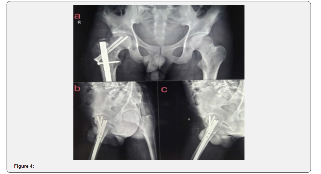
Case 4 Fracture Neck of Femur fixed with screw fixation 20: A 28-year-old male presented to taluka Hospital following a Motorcycle accident with pain in Right hip and swelling in 1985. An X ray revealed the fracture Neck of the Femur. The Orthopaedic surgeon in a private medical college opted for minimally invasive “Miss-a-nail” technique or closed reduction assisted by a traction table under fluoroscopic guidance. The fracture was anatomically reduced and fixed using two to three large partially threaded cannulated screws titanium Screws (can use stainless steel also) driven into the femoral head in an inverted triangle configuration. Post-Operatively strict non-weight-bearing was advised for the first 6 weeks to allow early healing (Figure 4). Rehabilitation began after that with supervised physical therapy and rangeof- motion exercises, with progressive weight-bearing over 3 to 6 months. Good long-term functional recovery after activation without avascular necrosis resulted in 6 months.
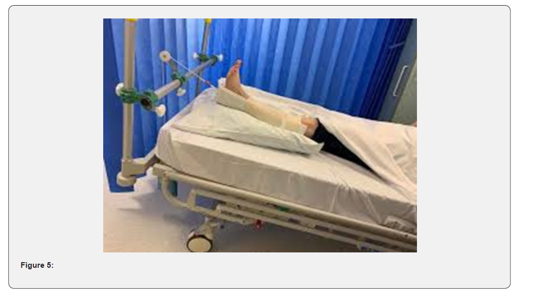
Case Report 5: First ever case of Fracture Head of Femur Author followed in 1969: A female Patient aged 84-year-old with Severe chronic obstructive pulmonary disease (COPD), advanced cardiovascular disease, and type 2 diabetes. She met with a road traffic accident while crossing a road. X’ ray revealed Left femur head fracture, and she was in severe pain. Her physical status classification was too high to tolerate open reduction internal fixation (ORIF); therefore, consensus decision was taken to manage conservatively. A closed reduction was performed under mild sedation. Skeletal traction (using proximal tibial pin) was immediately applied to maintain bone length, overcome muscle spasms, and achieve acceptable alignment. The patient was maintained in continuous skeletal traction for 3 weeks. Once initial soft tissue swelling subsided and early callus formation was visible on radiographs, traction was removed and a long-leg cast was applied (Figure 5). The cast remained in place for an additional 6 weeks, with regular radiographic checks to ensure the fracture did not displace. Because weight-bearing was heavily restricted, conservative physical therapy focused exclusively on non-weight-bearing exercises, maintaining upper body strength, and preventing respiratory complications. Prolonged bed rest led to a grade II pressure ulcer over the sacrum. Strict nursing care, including turning schedules & specialized alternating pressure mattresses, was required. The patient developed minor pneumonia during the third week, successfully treated with intravenous antibiotics and chest physiotherapy. Upon final cast removal, the affected limb showed significant muscle atrophy and reduced joint range of motion, which required a prolonged outpatient physical therapy regimen. Radiographs at 12 weeks confirmed the fracture healed in a functionally acceptable position with minor malunion. The patient was discharged with the ability to ambulate using a walker, though her pre-injury level of independent walking was not completely restored. The primary objective of avoiding the high mortality risks associated with major surgery due to medical fitness failure was managed, and fracture union was accomplished.
Discussion
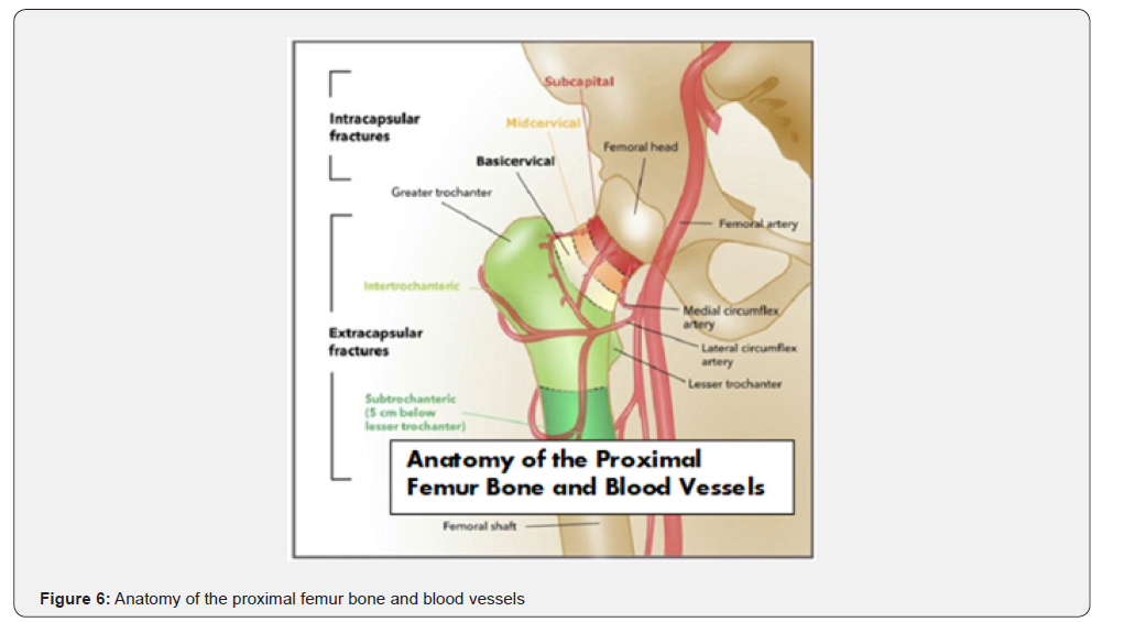
A fracture of the femoral neck (the area connecting the ball of the hip joint to the femur shaft) is an orthopaedic emergency (Figure 6). The standard approach has shifted to early surgical stabilization or joint replacement to prevent severe complications like avascular necrosis [1,2].
Types of Femoral Neck Fractures
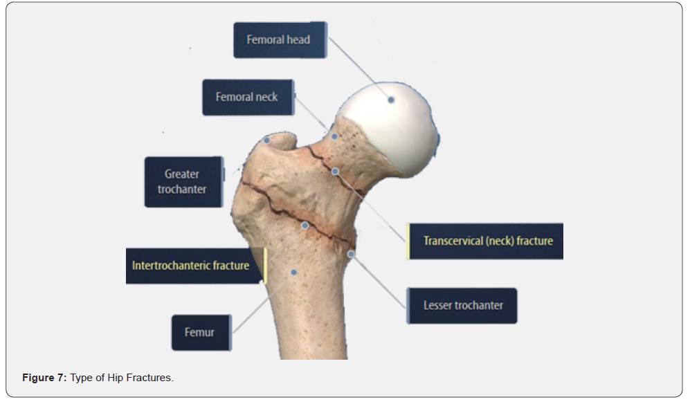
Fractures of the femoral neck are primarily categorized by their anatomical location, the degree of displacement, & the vertical angle of the fracture line [3]. 1.By Displacement (Garden Classification) categorises as Type I: Incomplete, valgus-impacted fracture (stable, non-displaced). Type II: Complete fracture, nondisplaced. Type III: Complete fracture, partially displaced. Type IV: Complete fracture, fully displaced. 2. By Vertical Angulation (Pauwels Classification): The more vertical the fracture line, the higher the shear forces and the greater the risk of the implant failing. Type I: Angle <300. Type II: Angle between 300 and 500. Type III: Angle >500 (highly unstable) [3] (Figure 7).
Types of Hip Fractures
Methods of Fixing the Fracture: Treatment decisions are dictated by the patient’s age, bone quality, and the level of fracture displacement [1,3].
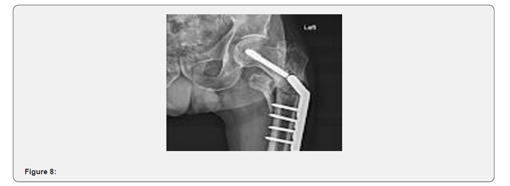
1. Internal Fixation (Osteosynthesis): Usually using three Cannulated crews arranged in an inverted triangle as shown in X ray of case no. 4. This is the gold standard for young, active patients and the elderly with un-displaced (Garden I/II) fractures [4] (Figure 8).
2. Dynamic Hip Screw (DHS): Uses a large lag screw and a side plate, occasionally augmented with a de-rotation screw, to provide compression.
3. Arthroplasty (Joint Replacement):
i. Hemiarthroplasty (HA): Replacing only the femoral head (ball). Most common procedure for frail, elderly patients with displaced fractures, as preserving the joint is unfeasible. In India, cemented modular hemiarthroplasty is being promoted using a cost-effective Indian implant that has confirmed distinct advantage.
ii. Total Hip Arthroplasty (THA): Replacing both the ball and the socket, reserved for active elderly patients to reduce the risk of future revision surgery and improve joint function. India’s Progress Since the 1960s: As this author started studying medicine in 1961-62, he has witnessed Indian orthopaedics undergoing a massive transformation, shifting from conservative bed rest to advanced global-standard surgical reconstruction [2]. 1960s – 1970s (Authors undergraduate days) The Era of Conservative & Early Mechanical Fixation): Managing displaced fractures was largely limited by resources both in terms of equipment and trained orthopaedics. Conservative treatments involved prolonged traction, and spica casts were common but associated with high mortality rates from immobility.
6.3. 1980s – 1990s (Osteosynthesis & Early Arthroplasty): Surgical options began with basic implants like Smith-Petersen nails (often associated with high failure and non-union rates). This era saw the widespread adoption of the Dynamic Hip Screw (DHS) for proximal fractures and the transition to cannulated screws. Indian orthopaedic surgeons also began refining techniques for “neglected” fractures, means those that presented weeks or months post-injury, common in rural India, heavily utilizing McMurray’s osteotomy and muscle pedicle bone grafting to promote healing in younger patients. 2000s – 2026 (Global Standardization & Advanced Arthroplasty): India is now a hub for high-end hip surgery. I) Shift to Arthroplasty: The treatment of displaced fractures in the elderly transitioned aggressively to hemiarthroplasty and Total Hip Arthroplasty (THA) using modern, cemented, and uncemented implants. II)Minimally Invasive Surgery (MIS): Surgeons routinely utilize image intensifiers (C-arms) to perform closed reduction and minimally invasive cannulated screw fixation, allowing for smaller incisions and faster recovery times.
The Indian Council of Medical Research (ICMR) and institutions like the National Institutes of Health (GOI) provide robust clinical guidelines for treating these fractures based on patient demographics. Modern approaches focus strongly on reducing the time from injury to surgery (< 24-48 hours) to prevent complications like avascular necrosis (AVN) [5,6]. There are two major surgical approaches for performing a total hip replacement 1) the posterior approach 2) the anterior approach. The posterior approach to total hip replacement is the most used method and allows the surgeon to have excellent visibility of the joint, more precise placement of implants and is minimally invasive [7] (Figure 9). An Indian study of 80 with 40 patients in each group were recruited to study Hemi-arthroplasty vs THR. The mean age of patients was 73 years in hemiarthroplasty group and 78 years in THR group.
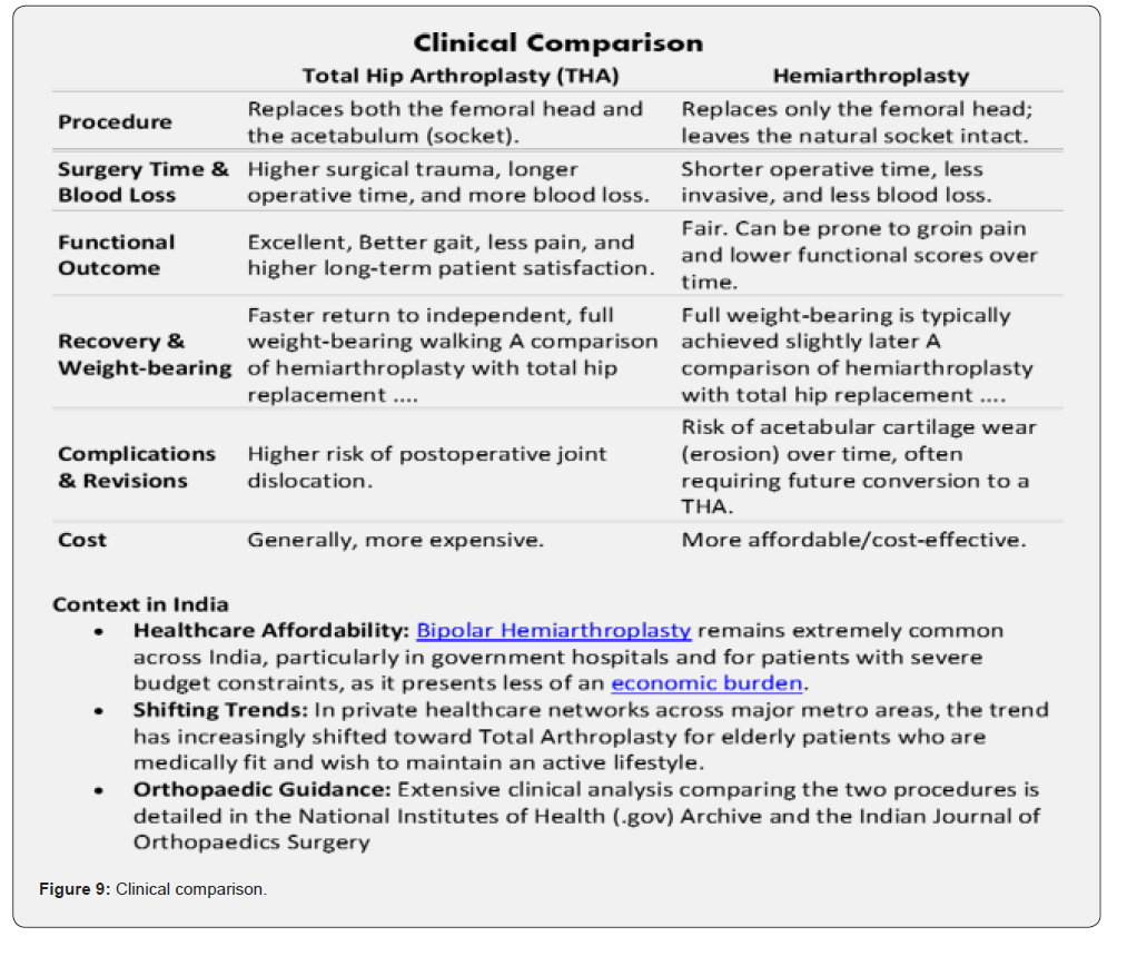
Female to male ratio was 55:45. Mean operative time was 35 minutes in hemiarthroplasty group and 45 minutes in THR. Average intraoperative blood loss was 200cc and 300cc in hemiarthroplasty and THR respectively. The mean hospital stay was 14 days in both the groups. Superficial wound infection was noted in hemiarthroplasty group while in THR group deep wound infection (n=1) and prolonged ICU stay (n=1) were noted. The mean Harris hip score was better in THR group as compared to Hemiarthroplasty group at one year follow-up. The study inferred that the functional outcome at one year was better with THR as compared to hemiarthroplasty in elderly patients with fracture neck femur. Hemiarthroplasty takes less operative time, less blood loss per operation & few complications postoperatively as compared to THR [8].
References
- Hip fractures.
- DK Dhanwal, R Siwach, V Dixit, A Mithal, K Jameson, et. al. (2013) Incidence of hip fracture in Rohtak district, North India. Arch Osteoporos 8(0): 135.
- Neck-femur-fractur (2026) Orthopaedic Surgery.
- N Kaushik, AK Singh, S Durrani, P Kumar, V Bajaj (2025) Miss-a-nail Technique of Femur Neck Fracture with Femur Interlocking Nail in Situ, Case Report. J Orthop Case Rep 15(10): 25-28.
- Senthil Narayanan, UT Vassan, PV Pragasam, Sathyanaryana V (2023) One month and one year mortality of hip fractures in a tertiary care hospital in south India- A retrospective cohort study, Indian Journal of Orthopaedics Surgery 9(4): 243-249.
- Anurag Kedar, Samir Dwidmuthe, Devashis Barick (2018) Is there a need for orthogeriatric unit in the Indian hospital setup for managing hip fractures?. Journal of Orthopaedics and Allied Sciences 6(1).
- V Sharma, B Awasthi, K Kumar, N Kohli, P Katoch (2016) Outcome Analysis of Hemiarthroplasty vs. Total Hip Replacement in Displaced Femoral Neck Fractures in the Elderly. J Clin Diagnostic & Research 10(5): RC11-3.
- Jackson P Tate et. al. (2026) Trends in Hemiarthroplasty Versus Total Hip Arthroplasty for Femoral Neck Fractures Based on Subspecialty Training, Comparative Study, J Arthroplasty 41(3): 770-775.e1.

























