




 OROAJ Home



Abstract
Introduction: The T i b i a l inclination (SLOPE) has been identified as a factor associated with graft failure after anterior cruciate ligament (ACL) reconstruction; However, its association with functional outcomes has been little studied. The purpose of this study is to determine the effect of tibial tilt on functional recovery in patients undergoing ACL reconstruction.
Material and methods: Patients with ACL injury undergoing primary reconstruction from May 2022 to May 2023 were enrolled to measure
tibial angles on MRI and collect pre- and post-surgical Lysholm.
Results: There were 30 patients, of whom 25 had a normal tibia and 13 had an increased tibial inclination. IKDC and Lysholm scores at the end of
follow-up were better in patients with normal tibial tilt. Graft failure was identified only in the group with an increased tibial slope. On the other
hand, a comparative analysis with the control group showed a better functional outcome at the end of follow-up, as assessed by the LYSHOLMS
in the group with normal tibial inclination.
Discussion: In relation to ACL graft failure, the association between increased tibial slope and graft failure was discovered, especially in patients
with >12° tibial slope, as described in various case-control studies [2,11,1,8,14]. It seems to be associated with greater forces experienced by the
ACL graft [14] because of increased tibial tilt, as demonstrated in several in vitro experiments [3, 7,10, 16,17].
Conclusion: Patients undergoing ACL reconstruction and tibial angle augmentation have a functional outcome less than one year of IKDC followup
compared to patients with a normal tibial angle.
Keywords:Anterior cruciate ligament; Osteoarthritis; Magnetic Resonance Imaging; Body mass index; Ligament
Abbreviations:ACL: Anterior Cruciate Ligament; MRI: Magnetic Resonance Imaging; MI: Meniscus Tilt; BMI: Body Mass Index
Case Description
In the knee, the anterior cruciate ligament (ACL) is an important element of the biomechanics of the knee, its main function is to limit the anterior rotation and translation of the tibia during the posture phase of walking. According to the American Academy of Orthopaedic Surgeons, 70% of ACL injuries occur due to non-contact mechanisms [5] during sports activities that involve sudden changes in speed in full extension or axial loading of the knee. Injury to this ligament is one of the main causes of disease and can lead to the loss of sports seasons and school scholarships. From an economic perspective, the cost of treating an ACL injury is significant, with reconstruction and rehabilitation costs exceeding $20,000. ACL reconstruction has become a standard surgical treatment to restore the stability and biomechanics of the knee [1,2,8], and the number of procedures performed increases each year [9].
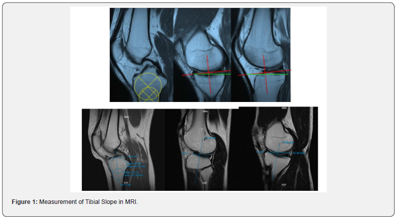
Although ACL reconstruction has shown promising results in addressing common stability issues, such as: Graft failure has been identified, and graft damage is increasing. Risk of cartilage and meniscus injuries [2,5,10] and increased risk of early osteoarthritis [8,6]. We have analyzed and identified various extrinsic factors (development of sports activities, visual impairment, physical contact and interaction with the surface of the shoe) and intrinsic factors (age, sex, body mass index (BMI) [11], reduction of the Q angle [11], neuromuscular control, ligament laxity [9] and knee geometry [1,7]. The latter factor has received considerable attention in recent years as it has been associated with a higher incidence of failure after ACL reconstruction [13]. The tibial slope has a strong linear relationship with the forces experienced by the ACL3 graft between 0 and 30° of flexion, regardless of the position of tunnel [9,14]. The flattening of the tibial slope significantly reduces loads. However, increased tibial tilt increases the strain on the ACL. Graft 3, which leads to failure [1,2,3,13] and poor functional outcome (Figures 1 & 2).
Results
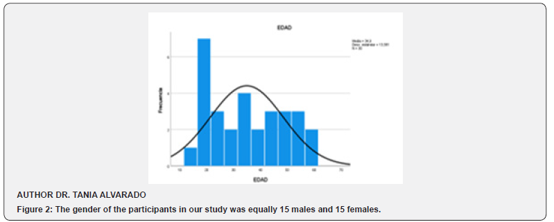
The ages of the participants ranged from 14 to 57 years (mean = 34.9; standard deviation = 13.58). The median age was 36 years, indicating that half of the participants were 36 years old or younger. The 25th and 75th percentiles were 20 and 48 years, respectively, showing most participants in those ages included Figure 2. Histogram of normality of the Age Variable Author: Dr. Tania Alvarado Chavez (Figure 3).
Define the Status and Score of The Tibial Slope in the Study Population
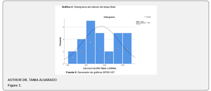
Based on this objective, the slope score was performed numerically from the calculation with MRI and subsequently categorized as normal and abnormal for a better understanding of it. The tibial slope estimates ranged from 6.5 to 12.8 (mean = 9.6; standard deviation = 1.96). The median slope estimate was 9.2, indicating that half of the participants had that score or less. To assess the normality of the variable, the Kolmogorov-Smirnov test was performed. The results of the Kolmogorov-Smirnov test (D = 0.137, p = 0.154) were not significant. Since the p-value (0.154) is greater than the conventional significance level of 0.05, we cannot reject the null hypothesis. This means that there is insufficient evidence to conclude that tibial slope data are not normally distributed. In other words, we can assume that the tibial slope data follows an approximately normal distribution (Figure 4).
Define and Calculate Functional Recovery According to the Lysholm Scale
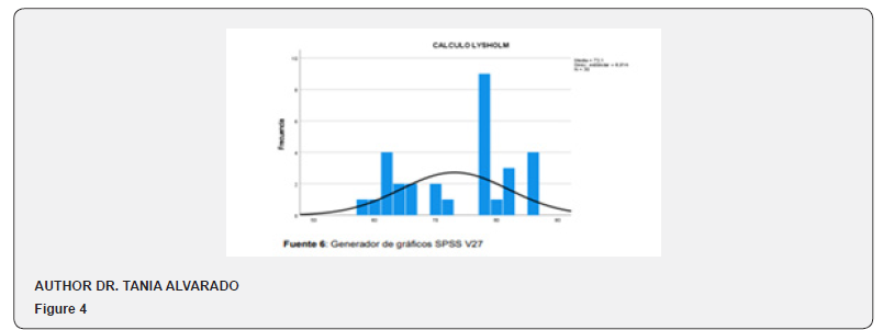
Based on this objective, the Lysholm scale score was performed numerically and later categorized as normal and abnormal for a better understanding of it. The Lysholm calculation ranged from 58 to 86 (mean = 73.10; standard deviation = 8.81). The median of Lysholm’s estimate was 77, indicating that half of the participants had that score or less. To assess the normality of the variable, the Kolmogorov-Smirnov test was performed. The results of the Kolmogorov-Smirnov test (D = 0.238, p = 0.001) were significant. Since the p-value (0.154) is lower than the conventional significance level of 0.05, we can reject the null hypothesis. This means that there is sufficient evidence to conclude that the data from the lysholm calculus are normally distributed. In other words, we can assume that the data follows a non-normal distribution (Figure 5).
Discussion
In our study, we observed that increased tibial slope negatively affected functional outcomes during the first postoperative year in patients undergoing ACL reconstruction. However, some studies have reported no difference in functional outcomes in patients undergoing ACL reconstruction with normal versus increased tibial tilt [2]. In another study, Kiapour et al. [15] found similar results to our study, obtaining better scores on the functional scale patients with tibial slopes within normal parameters after 7 years of follow-up after ACL reconstruction. The natural tibial inclination has been measured between 8 and 10° in magnetic resonance imaging with the method of Hudek et al. [9,12,13].
As the tibial tilt increases beyond 12° degrees, anterior tibial translation increases associated with [3,8] 3- to 10-fold displacements and an increased risk of ACL graft failure [1,13]. In recent years, the use of osteotomy 10 has gained popularity to reduce excessive graft stress after ACL reconstruction. In knees with recurrent failure and excessive tibial slope (>12) [4], anterior distraction osteotomy is indicated to protect the ACL graft from revision surgery for augmented tibial slope [3,14,16]. In most publications, measurements are made by simple x-rays of the knee profile, since only the bone structure is used as a reference [2,11- 15]. More recently, several publications have begun to emphasize the importance of measuring both tibial plateaus independently. Biomechanically, this also seems to be a greater influence of the slope of the external tibial plateau on the cruciate ligament rupture mechanism [15,17].
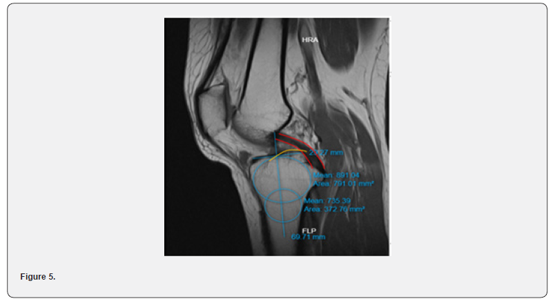
Additionally, it is stated that the measurement error is due to variations in the rotation of the knee at that time. The radiographic throughput rate is relatively high because it is not possible to control the X-ray technique for all measurements. For these reasons, magnetic resonance imaging (MRI) can be a good way to make these measurements [9,16]. An advantage of MRI is that both the tibial plateau and the meniscus tilt (MI) can be measured independently. The measurement of tibial inclination in our study was performed by magnetic resonance imaging. It is a technically simple measurement with the tools of the MRI visualization program and therefore it is considered an accessible study due to its reproducibility and ease of measurement using the techniques described above, because all patients for arthroscopic surgical planning will present their knee n MRI (Figure 1).
Measurement Form
Choose the central sagittal image, define their 3 points which
are:
i. The Tibial Insertion of the PCL
ii. The intercondylar eminence
iii. The anterior and posterior tibial cortices that appear in
a concave shape.
Other studies have evaluated measurements of medial and lateral tibial tilt with plain radiographs and axial computed tomography 9 and have demonstrated a greater association between lateral tibial tilt and graft failure 13. In relation to ACL graft failure, the association between increased tibial slope and graft failure was discovered, especially in patients with tibial slope >12°, as described in various case-control studies [2,11,1,8,14]. It appears to be associated with greater forces experienced by the ACL graft [14] as a result of increased tibial inclination, as demonstrated in several in vitro experiments [3,7,10,16,17].
Having identified patients with changes in the shape of the proximal tibia and anterior cruciate ligament injuries 8, it is necessary to plan the application of individual techniques in addition to ACL reconstruction for greater stability [17] such as proximal tibia osteotomies [10,16]. This reduces the risk of failure [1,18]. These are all good functional outcomes, early return to sport, and prevention of graft failure. This study has limitations because it included follow-up of patients and only covered the first year after surgery. Longer-term follow-up is required to assess the impact of tibial slope on functional outcome after ACL reconstruction.
Conclusion
The main objective of this study was to measure the degree of relationship between the score or measurement of the tibial slope and the score of the Lysholm scale to assess functional recovery after anterior cruciate ligament reconstruction surgery. Based on our general objective, we applied the spearman correlation for this purpose, concluding that there is a negative but strong correlation between these two variables. In practical terms, this result suggests that patients with greater tibial deformity (greater tibial slope) tend to have worse knee function, as assessed by the Lysholm scale. This could indicate that joint deformity is related to a decrease in the health-related quality of life of these patients.
Based on our specific objectives, we determined that the ages did not follow a normality and that the gender was balanced both male and female. The measurements of both the tibial slope and Lysholm scales were calculated separately, allowing an idea of their distribution and normality. Based on the above, we can conclude that the knowledge acquired because of the research is fruitful since it agrees with previous studies. Our recommendation is based on the process methodologically since the most important limitation was the sample size that influenced the calculation of the correlation by non-parametric measures, being possible to make use of Pearson’s correlation. Future research will determine the course of this line of research..
References
- Sauer S, English R, Clatworthy M (2018) The ratio of tibial slope and meniscal bone angle for the prediction of ACL reconstruction failure risk. Surg J (N Y) 4(3): e152-e159.
- Sauer S, Clatworthy M (2018) The effect of medial tibial slope on anterior tibial translation and short-term ACL reconstruction outcome. Surg J (N Y) 4(3): E160-E163.
- Cooper JD, Wang W, Prentice HA, Funahashi TT, Maletis GB (2019) The association between tibial slope and revision anterior cruciate ligament reconstruction in patients ≤21 years old: a matched case-control study including 317 revisions. Am J Sports Med 47(14): 3330-3338.
- Hendrix ST, Barrett AM, Chrea B, Replogle WH, Hydrick JM, et al. (2017) Relationship between posterior-inferior tibial slope and bilateral noncontact ACL injury. Orthopedics 40(1): E136-E140.
- Schuster P, Geßlein M, Schlumberger M, Mayer P, Richter J (2018) The influence of tibial slope on the graft in combined high tibial osteotomy and anterior cruciate ligament reconstruction. Knee 25(4): 682- 691.
- Bates NA, Mejia Jaramillo MC, Vargas M, McPherson AL, Schilaty ND, et al. (2019) External loads associated with anterior cruciate ligament injuries increase the correlation between tibial slope and ligament strain during in vitro simulations of in vivo landings. Clin Biomech (Bristol, Avon) 61: 84-94.
- Grassi A, Signorelli C, Urrizola F, Macchiarola L, Raggi F, et al. (2019) Patients with failed anterior cruciate ligament reconstruction have an increased posterior lateral tibial plateau slope: a casecontrolled study. Arthroscopy 35(4): 1172-1182.
- Bernhardson AS, Aman ZS, Dornan GJ, Kemler BR, Storaci HW, et al. (2019) Tibial slope and its effect on force in anterior cruciate ligament grafts: anterior cruciate ligament force increases linearly as posterior tibial slope increases. Am J Sports Med 47(2): 296-302.
- Dejour D, Pungitore M, Valluy J, Nover L, Saffarini M, et al. (2019) Tibial slope and medial meniscectomy significantly influence shortterm knee laxity following ACL reconstruction. Knee Surg Sports Traumatol Arthrosc 27(11): 3481-3489.
- Wang D, Kent RN 3rd, Amirtharaj MJ, Hardy BM, Nawabi DH, et al. (2019) Tibiofemoral kinematics during compressive loading of the ACL-intact and ACL-sectioned knee: roles of tibial slope, medial eminence volume, and anterior laxity. J Bone Joint Surg Am 101(12): 1085-1092.
- Jaecker V, Drouven S, Naendrup JH, Kanakamedala AC, Pfeiffer T, et al. (2018) Increased medial and lateral tibial posterior slopes are independent risk factors for graft failure following ACL reconstruction. Arch Orthop Trauma Surg (10):1423-1431.
- Bojicic KM, Beaulieu ML, Imaizumi Krieger DY, Ashton-Miller JA, Wojtys EM (2017) Association between lateral posterior tibial slope, body mass index, and ACL injury risk. Orthop J Sports Med 5(2): 2325967116688664.
- Lee CC, Youm YS, Cho SD, Jung SH, Bae MH, et al. (2018) Does posterior tibial slope affect graft rupture following anterior cruciate ligament reconstruction? Arthroscopy 34(7): 2152-2155.
- Samuelsen BT, Aman ZS, Kennedy MI, Dornan GJ, Storaci HW, et al. (2020) Posterior medial meniscus root tears potentiate the effect of increased tibial slope on anterior cruciate ligament graft forces. Am J Sports Med 48(2): 334-340.
- Kiapour AM, Yang DS, Badger GJ, Karamchedu NP, Murray MM, et al. (2019) Anatomic features of the tibial plateau predict outcomes of ACL reconstruction within 7 years after surgery. Am J Sports Med 47(2): 303-311.
- DePhillipo NN, Kennedy MI, Dekker TJ, Aman ZS, Grantham WJ, et al. (2019) Anterior closing wedge proximal tibial osteotomy for slope correction in failed ACL reconstructions. Arthrosc Tech 8(5): E451-E4517.
- Sabzevari S, Rahnemai-Azar AA, Shaikh HS, Arner JW, Irrgang JJ, et al. (2017) Increased lateral tibial posterior slope is related to tibial tunnel widening after primary ACL reconstruction. Knee Surg Sports Traumatol Arthrosc 25(12): 3906-3913.
- Rahnemai-Azar AA, Abebe ES, Johnson P, Labrum J, Fu FH, Irrgang JJ, et al. (2017) Increased lateral tibial slope predicts high-grade rotatory knee laxity pre-operatively in ACL reconstruction. Knee Surg Sports Traumatol Arthrosc 25(4): 1170-1176.

























