Study of the Physeal Trauma in 20 Years
Lázaro Martín Martínez Estupiñan1, Lázaro Martínez Aparicio2, Leonardo Martínez Aparicio3, Sergio Morales Piñéiro4, Roberto Mata Cuevas4 and Claribel Plain Pazos5*
1Doctor in Medical Sciences, II Degree Specialist in Orthopedics and Traumatology, Full Professor, Provincial General University Hospital, Cuba
2I Degree Specialist in Orthopedics and Traumatology, Instructor Professor, Provincial General University Hospital, Cuba
3Resident of 3rd year of Orthopedics and Traumatology, Provincial General University Hospital, Cuba
4I and II Degree Specialist in Orthopedics and Traumatology, Assistant Professor, Provincial General University Hospital, Cuba
5Iand II Degree Specialist in Comprehensive General Medicine, Faculty of Medical Sciences of Sagua la Grande, Cuba
Submission: November 01, 2022; Published: November 15, 2022
*Corresponding author: Claribel Plain Pazos, Faculty of Medical Sciences of Sagua la Grande, Villa Clara, Cuba
How to cite this article: Lázaro M M E, Lázaro M A, Leonardo M A, Sergio M P, Roberto Mata C, et al. Study of the Physeal Trauma in 20 Years. Ortho & Rheum Open Access J. 2022; 20(5): 556047. DOI: 10.19080/OROAJ.2022.20.556047
Abstract
Introduction: Fractures in children present characteristics that, in their evolution and behavior, as well as in the choice of behavior before them, differ from those of adults. Target. Describe the distribution and characteristics of the physeal injuries, according to the injured anatomical region, the causal event and the behavior before the injury.
Method: A prospective and longitudinal study was carried out, taking as a sample the children injured in the musculoskeletal system at the physeal level treated at the Hospital Mártires del 9 de Abril between January 2000 and August 2020. Using scientific research, theoretical and empirical.
Results: Child athletes represent around 11% of children with physeal injuries, the male sex predominates in the sample, with ages between 12 and 14 years, these injuries affect the upper limb with a predominance in the physis of the distal radius, the majority are they are caused by accidents at home and are managed mainly with medical treatment and immobilization.
Conclusion: There are multiple injuries that children can suffer in their development, including physeal trauma; recognizing them, knowing their epidemiology and choosing appropriate behaviors contributes little-studied knowledge to our health system.
Keywords: Children; Injuries; Physeal Trauma
Introduction
Accidents represent an important cause of morbidity and mortality at any age, and the pediatric population is no exception, thus injuries in children continue to be a public health problem throughout the world. The World Health Organization (WHO) estimates that 90% of these are unintentional and are mostly caused by mechanisms such as car accidents, falls and recreational games at home or sports [1,2].
Fractures in children present characteristics that, in their evolution and behavior, as well as in the choice of behavior before them, differ from those of adults. The child is not a small adult, his musculoskeletal system has characteristics that do not occur in adults: there are growth zones (physis), whose injury can cause deformities or shortening, if its management is not adequate, the periosteum is more vascularized, it has a greater cellular component, therefore, greater bone formation capacity, bones have greater elasticity. Children’s bones have a great capacity to absorb trauma and a significant remodeling capacity, determined by the location of the fracture and age [3].
The physeal nucleus is considered a risk factor in children for suffering from SOMA injuries, there is increasing evidence, both clinical, biomechanical, and radiological, where it is referred to as the area most susceptible to injury [4]. Cartilage growth is located in three sites of the immature skeleton: epiphyseal plate, articular surface, and apophyseal attachments of the main muscle-tendon units [5]. There are multiple injuries that children can suffer in their development, including physical traumas, recognizing them, knowing their epidemiology and the choice of behaviors can make a difference in the future. For this reason, the work is carried out on the subject of physeal injuries suffered by the children cared for in our service.
Method
A descriptive, longitudinal study was carried out with the purpose of studying the physeal lesions of the immature skeleton belonging to the Villa Clara province, treated in the orthopedic and traumatology service at the Hospital Provincial General Universitario Martires del 9 de Abril. We worked with an original and intact group, that is to say, already constituted. All children under 18 years of age with a diagnosis of physeal injury (n = 2365) were included in the study. The study period spanned 20 years (September 2000 to August 2020).
The data concerning the general basic characteristics of the children, as well as those related to the lesions presented and their management, were captured in a data collection model and / or extracted from the medical records. To characterize the physeal lesions that made up the sample, the variables were studied: age (under 5 years, between 5 and 8 years, 9-11 years, 12-14 years, 15-17 years); sex; if the child plays sports and is injured in this activity; injured anatomical region, cause of injury (traffic accidents that include accidents on public roads such as pedestrians, bicycles or cars, also accidents that include falls, trauma caused by games or recreational activities, falls from height and sports trauma); type of lesions (according to Salter and Harris classification) [6]; and conduct in the face of injury [7]. The results are expressed in absolute numbers and percentage. This research had the authorization of the administrative bodies of the institution involved and with the approval of the Villa Clara Provincial Scientific Council (Agreement 65/1996, research topic to opt for a scientific degree).
Results
Table 1 shows the distribution of children treated with physeal injuries, grouped into two groups those who practice organized sports activities (child athletes) which represent 11.28 percent (n = 267), the largest number of children are Those who suffer injuries and do not suffer their injuries in organized sports activities, are a great majority, in 20 years of study 2098 children suffered injuries. The age group with the highest number of injuries is found in the group of children between 12 and 14 years old. Male sex predominates with 73.6% of the sample.

Fountain Data collection model.

Fountain. Data collection model.
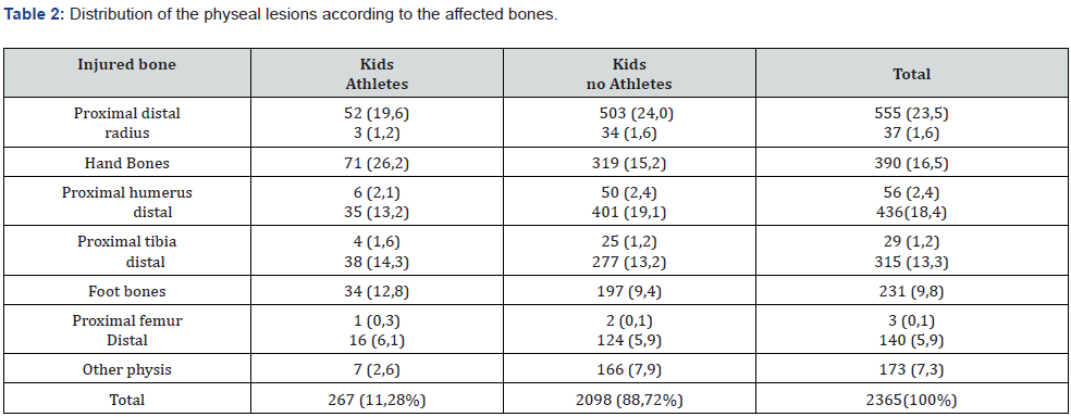
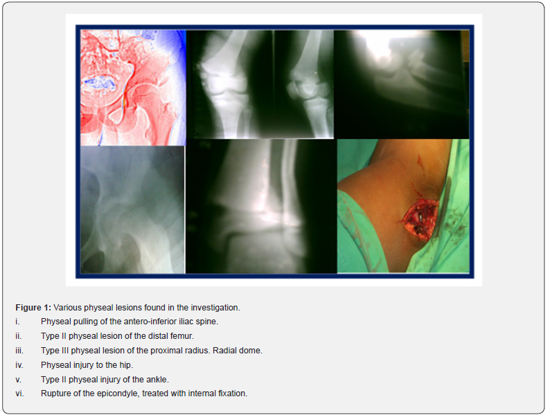
Table 2 shows the distribution of children with acute physeal trauma according to the physis of the injured bone. These children are divided into two groups (athletes and non-athletes). Most injuries occur in the distal radius, followed by injuries to the distal humerus and to the bones of the hand (Figure 1).


Fountain. Data collection model.

Fountain. Data collection model.
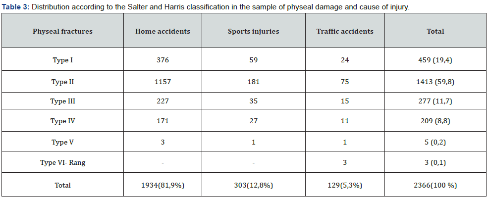
The physeal traumas in the children in the sample are widely distributed in Salter and Harris type II (Table 3), with more than half of the children n = 1413 (59.8%). In only 20 years of study, Rang’s VI injuries have been presented in three of the children, all caused by traffic accidents. Most of the physical traumas are produced by accidents at home n = 1934 (81.9%), followed by sports traumas with 12.8%. Table 4 shows the distribution of behavior according to the Salter and Harris classification, it is appreciable how conservative behaviors are applied in most children with injuries.
Discussion
The effects of trauma on the growth plates are always of interest to orthopedists, considering the impact they may have on the growth process. The incidence of injuries and mechanisms depends on the countries and the social and cultural environment of those affected. In less developed countries they are casual accidents, traffic accidents or civil conflicts and in countries where the cultural and social level is higher, falls from height, traffic accidents, sports dominate. In recent years, musculoskeletal injuries have increased due to the practice of games and sports.
Recent studies speak of an incidence of fractures during childhood of 42% in boys and 27% in girls, increasing linearly from birth to 12 years and later decreasing until 16 years. In the study, it was observed that after five years of age, the incidence of injuries increases with a peak incidence greater than 12 to 14 years, which coincides with other researchers [8,9]. The incidence of injuries and production mechanisms depend on the countries and the social and cultural environment of those affected. In lessdeveloped countries they are casual accidents, traffic accidents or civil conflicts and in countries where the cultural and social level is higher, falls from height, traffic accidents and sports accidents dominate.
The most frequent fractures in children occur in the upper limb with 45.1% in the radius (dominating in its metaphysis and distal physis), 18.4% in the humerus (dominating metaphysis and distal physis), 15,1 % in the tibia, 13.8% in the clavicle and 7.6% in the femur. Physeal fractures represent 21.7% of injuries. The first two represent 75% of epiphysiolysis and are the more benign since the germ plate is not affected. The last four injure the physeal plate and can slow its growth causing epiphysiodesis [5]. Onís González and his collaborators suggest that the incidence of fractures by anatomical regions is as follows: wrist (23.3%), hand (20.1%) , elbow (12%), forearm (6.4%), clavicle (6.4%), leg (6.2%), foot (5.9%) and ankle (4.4%) [8]. Other authors report that 25.5% are sports injuries, they also refer that 15% are fractures, of which 23% occur in the fingers [9].
Osteomyoarticular injuries of traumatic cause are very common in children and mainly those that are due to sports activities, which represent 31%, followed by outdoor activities 25%, domestic accidents 19%, school accidents 13% and accidents in the public roads 12% [10]. In a study in Camagüey province, on proximal humerus injuries, it is described that accidents during children’s play occupied 34.9% of the sample, followed by domestic ones with 25.6% , those of transit 16, 3% and the sports ones with 14% [9].
Type VI Salter-Harris fractures are injuries characterized by ablation of the perichondral ring. Rang was the first to describe this lesion and, later, it was incorporated by Ogden [5] as the sixth type to the classic Salter-Harris classification. Jones, Wolf, and Herman regard them as rare injuries, but closely related to highenergy trauma [11]. Salter and Harris type VI lesions can present in a closed or open manner. Marson, Craxford, and Ollivere [12] evaluated patients with type VI ankle fractures, but these types are rare. Masquijo, Lucas and Allende [13] report similar results, with which this study agrees.
The lesion must be identified by good radiology and proceed to its perfect reduction [3]. For the treatment, it is important to restore the physeal integrity, especially in fractures III and IV, since failure to do so will most likely result in the appearance of a physeal bone bridge. Intra-articular displacement must be reduced to avoid degenerative changes in the future. Regardless of the affected bone, the treatment will always be the same, according to the classification.
Conclusion
Physeal trauma occurs frequently in children between 12 and 14 years old, preferably males, these injuries mostly occur in the upper limb. Salter and Harris type II predominates, and they are resolved with conservative behaviors in their treatment.
Conflict of Interest
The authors of this article declare that they have no conflict of interest with the objectives of the research.
Declaration of the personal contribution of each author to the research
The authors of this article participated in the diagnosis, treatment, study design, and writing of the first version, as well as the final version of the manuscript in equal parts.
References
- Marin Gonzalez AL (2017) Trauma en pediatrí Anestesia en trauma 40(Supl 1): 52-54.
- Bustos CE, Cabrales MR, Ceron RM, Naranjo LM (2014) Epidemiologia de lesiones no intencionales en niños: revisión de estadísticas internacionales y nacionales. Bol Med Hosp Infant Mex 71(2): 68-75.
- Rang M (2010) Rasgos especiales de las fracturas infantiles: los niños no son adultos pequeñ En: De Pablos J, González Herranz P (eds.). Fracturas infantiles. Conceptos y principios. (2ª edn), Barcelona, Ergon p. 31-34.
- Martínez-Estupiñán L (2017) Sports injuries in child athletes (2017) twenty-year study. Medisur 15(6): 819-825.
- Ogden J (2011) Skeletal injury in the child. Springer Verlag, Berlin, Germany.
- Kurup SPS, Padankatti S, Thomas K (2014) Accidental injuries in the Pediatric Emergency Department. International Journal of Emergency Medicine 7(Suppl 1): 9.
- Wang X, Shao J, Yang X (2014) Closed/open reduction and titanium elastic nails for severely displaced proximal humeral fractures in children. Int Orthop 38(1): 107-110.
- Onis Gonzalez E, Varona Perez I, Gil Perez M, Felici C, et al. (2015) Embid Pardo P (2015) Unintentional injuries at school, what are we talking about? Pediatrics Primary Care 17(68): 333-339.
- Rodriguez Rodriguez EI, Tuan Nguyen Pham, Long Nguyen (2016) Tratamiento de las fracturas del extremo proximal del húmero en niños. Rev. Arch Med Camaguey 20(3).
- Patel DR, Yamasaki A, Brown K (2017) Epidemiology of sports-related musculoskeletal injuries in young athletes in United States. Transl Pediatr 6(3): 160-166.
- Jones Ch, Wolf M, Herman M (2017)Acute and Chronic Growth Plate Injuries 38(3): 129-137.
- Marson BA, Craxford S, Ollivere BJ (2019) Management of ankle fractures in children. British Journal of Hospital Medicine 80(4): 201-203.
- Masquijo JJ, Lucas Lanfranchi L, Allende V (2015) Fracturas fisarias Salter-Harris VI de tobillo y pie. Rev Asoc Argent Ortop Traumatol 80(2): 104-113.


