Novel Fixation Method (Tension Band Wiring Over the One-Third Tubular Plate) for Displaced Lateral Malleolar Tip Fracture - Preliminary Report
Mohammad Ayati Firoozabadi* and SMJ Mortazavi
Department of Orthopedic Surgery, Joint Reconstruction Research Center, Tehran University of Medical Science, Tehran, Iran
Submission:July 19, 2022; Published: August 02, 2022
*Corresponding author: Mohammad Ayati Firoozabadi, Joint Reconstruction Research Centre, Tehran University of Medical Sciences, End of Keshavarz Boulevard, Tehran 1419733141, Iran
How to cite this article: Mohammad Ayati Firoozabadi, SMJ Mortazavi. Novel Fixation Method (Tension Band Wiring Over the One-Third Tubular Plate)for Displaced Lateral Malleolar Tip Fracture - Preliminary Report.Ortho & Rheum Open Access J. 2022; 20(2): 556034. DOI: 10.19080/OROAJ.2022.20.556034
Abstract
Introduction: One of the most common fractures around the ankle is a fracture of the lateral malleolus. lateral malleolar tip fracture is one of the types of fractures for which various methods have been suggested to fixation. Surgeons always tend to use a method that, despite the lower cost, has good short-term and long-term results, and also the surgical technique is simple.
Patients and Methods:The present study included assessment functional and radiological outcomes and complication of novel fixation method (tension band wiring over the one-third tubular plate) for lateral malleolar tip fracture. Twenty-one consecutive patients who underwent this novel method were followed retrospectively.
Results: There isn’t significant pain on lateral malleolus was observed in all patients after short term follow up (6 months to 2 years). Radiographs of ankle showed complete union of lateral malleolar tip fracture without any failure or mal union.
Conclusion: Novel fixation (tension band wiring over the one-third tubular plate) is a cheap, simple, safe, and easy surgical procedure for treatment and stabilization of displaced lateral malleolus fracture with type A Denis-Weber.
Introduction
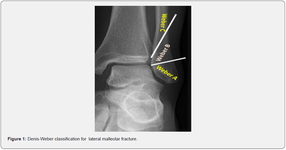
One of the most common fractures around the ankle is a fracture of the lateral malleolus. Denis-Weber classified this type of fracture into three categories based on the position of the fracture line relative to the ankle joint surface (Figure 1). Type A is the infra syndesmotic type and type B is the trans syndesmotic type. In type C, the fracture is supra syndesmotic [1]. In type A fracture, the ankle joint is stable and is mostly treated with plastering, but in cases where the fracture is accompanied by displacement, it requires surgery and fixation [2]. The difficulty of fixing these fractures is in their very small size in the distal part of the fracture. Therefore, to treat this type of fracture, several fixation methods have been proposed (such as installing a longitudinal screw or intramedullary nail, using an anatomical locking plate, tensioning the wiring with a pin, or installing a one-third tubular plate that its one end side is shaped like a hook) [3-5]. In the biomechanical study that my colleagues and I conducted in 2016, we compared the existing methods with our proposed method, which was the wiring tension band fixation on the one-third tubular plate, which was preferable compared to other methods in terms of structure stability (against bending, torsional and compressional forces). In addition, this method is very cheap, and its surgical technique is easy [6]. In this article, we have decided to present a report on the results of the wiring tension band fixation method on the one-third tubular plate in 21 patients who were operated with this method.
Patients and Methods
The present study included assessment functional and radiological outcomes and complication of novel fixation method for displaced lateral malleolus fracture with type A Denis-Weber. Twenty-one consecutive patients who underwent this novel method were followed retrospectively. (Mean age, 40.7 years; age range, 21–70 years; 8 males, 13 females).
Inclusion criteria
• Patients with displaced lateral malleolus fracture (type A Denis-Weber)
• Suitable soft tissue for operation over the lateral malleolus
• Closed fracture
Exclusion Criteria
• Patients with un displaced lateral malleolus fracture (type A Denis-Weber)
• Patients with lateral malleolus fracture (type B or C Denis-Weber)
• Uncontrolled diabetes mellitus
• congenital deformities of the lower extremity,
• Ankle joint infection,
• history of ankle ligament injury or any previous surgery on ankle

Preoperative and Postoperative Ankle Radiographs
Preoperative and postoperative anteroposterior (AP), lateral (Lat.) and mortise view radiographs were obtained in all patients to analyze the displacement of fracture site in lateral malleolus fracture and step amount in healed fracture and degree of ankle osteoarthritis.
Ankle Pain Assessment
Ankle pain was assessed using a Visual Analogue Scale (VAS).
AOFAS Ankle-Hindfoot Score Assessment
American Orthopedic Foot and Ankle Society (AOFAS) Ankle- Hindfoot Rating System is a standardized evaluation of the clinical status of the ankle-hindfoot. It incorporates both subjective and objective information. Patients report their pain, and physicians assess alignment. The patient and physician work together to complete the functional portion. Scores range from 0 to 100, with healthy ankles receiving 100 points. This score usually used to evaluate the ankle, subtalar, talonavicular, and calcaneocuboid joint levels functional outcome and may be useful for fractures, arthroplasty, arthrodesis, and instability procedures.
Surgical Technique
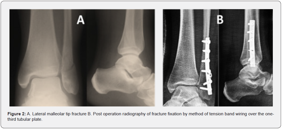
The patients with displaced lateral malleolus fracture with type A Denis-Weber were placed in the supine position under spinal anesthesia. Under adequate pressure of the tourniquet (100 mmHg more than systolic blood pressure) an approximately 7-cm longitudinal incision was made over the lateral skin of the lateral malleolus. Fracture reduction was performed. A 5 or 6 - holes onethird tubular plate was reshaped after bending into the shape of the lateral part of the lateral malleolus. Then the fracture was fixed with the five cortical screws. The second and third screws from the distal side were slightly loosened to allow the tension wiring between the heads of these two screws. After tension wiring band, these screws were completely tightened (Figure 2). The position of the screws and plate was checked with C-Arm to ensure that it was suitable. Finally, after suturing the wound and dressing, for the patient was given a short leg cast for two weeks. Patients were allowed to weight bearing on the affected leg from the same day of surgery as tolerated the pain. After two weeks, patients were advised to wear high top shoes for three months. After removing the cast, the range of motion of the ankle began.

Results
All patients who were followed for a minimum of 6 months. The mean duration of follow-up was 15.8 months (range, 6–24 months). The mean visual analogue scale scores significantly decreased from 7.9 preoperatively on operation day to 0.5 postoperatively on the last follow up. In the ankle radiographies (AP, Lat. And mortise view), which were performed on the last day of treatment follow-up of all patients, complete union was evident. Malunion was not seen. There were no degenerative changes either. In all patients, the range of motion of the ankle was similar to the opposite side. In four patients, they complained about the prominence of the fixation device, and the removal of the device was done for them. The mean AOFAS Ankle-Hindfoot Rating System was 96.7 on the last follow up (range, 90-100).
Discussion
If the ankle is stable and the fracture of the lateral malleolus is not displaced, non-surgical treatment produces excellent results. If the clinical/radiographic findings indicate ankle instability, the appropriate treatment option is surgical fixation, which includes the placement of a plate over the lateral or posterolateral aspect of lateral malleolus or intramedullary fixation. Locking plates and small or minifragment fixation are important and are supplements that the surgeon may consider based on the needs of each patient [1,7,8]. Based on the study conducted by Haroon Rehman and et al. in 2015, they stated that intramedullary nail fixation of distal fibular fractures can perform better than conventional fixation with plates and screws [3].
In a multicenter case series conducted by Vincenzo Giordano and et al. in 2020, intramedullary fixation of lateral malleolus fractures established itself as a suitable and safe option for the treatment of almost all fibula fracture patterns in adults [4].
Magnesium (Mg) bioabsorbable screws are new biomaterials used in fracture fixation. In the current literature, there is only one case of the use of bioabsorbable magnesium screws in ankle fractures, which was reported by Baver and et al. in a patient with a single lateral malleolus fracture, treated with open reduction and intramedullary magnesium screw fixation and then followed up for two years. Fracture union was achieved without any complications such as failure of fixation, loss of reduction, infection, or any other adverse reaction. For this reason, they suggested that absorbable magnesium screws are an alternative method of fracture fixation compared to conventional metal implants because they eliminate the need for implant removal [9]. Locking plates are increasingly used in the treatment of lateral malleolus fractures. Biomechanical studies have shown increased stability with the use of locking versus non-locking plates. However, despite the much higher price of these types of plates compared to previous conventional non-locking plates, the exact advantage of locking plates over non-locking plates in patients with lateral malleolus fractures is unclear. A meta-analysis by Nesar Ahmad Hasami and et al. showed no clear advantage in choosing locking plates over non-locking plates in the treatment of lateral malleolus fractures [10]. In a study conducted by Bankston and et al. on 44 patients with lateral malleolus fractures, it was shown that intramedullary screw fixation of noncomminuted lateral malleolus fractures provides stable fixation with good clinical results. This technique has the advantages of dynamic intramedullary fixation with limited surgical dissection and no subcutaneous hardware [11].
In a retrospective study published by Yenel G¨urkan Bilgetekin in 2018, a total of 62 orthopedic patients who underwent surgery for lateral malleolus fractures were included. In this study, it did not show a significant difference between the use of one-third locking tubular plate and locking anatomical distal fibula plate in the lateral malleolar fixation in terms of clinical and radiological outcomes, the rate of complications and fracture healing time [12]. In a study conducted by Paul and et al. in 2007, the results of lag screw only fixation surgery performed on 25 patients with unstable non-comminuted oblique fractures of the lateral malleolus were retrospectively evaluated. In this study, they demonstrated that lag screw only fixation of the lateral malleolus is a safe and effective method that has a number of advantages over plate osteosynthesis, in particular less soft tissue dissection, less prominent, symptomatic and palpable hardware and a reduced requirement for secondary surgical removal [13]. In a retrospective study conducted by Girgis Latif and et al. on forty-six patients with Weber A and low Weber B displaced lateral malleolus fractures who underwent closed reduction and percutaneous internal fixation with an intramedullary, fully threaded, screw, showed that If reduction of the lateral malleolus fracture was obtained in a closed fixation, fixation may be performed with an axial screw percutaneously. This technique is quick, safe and easy to do with less complication [14]. Qudong Yin et al. also reported the results of fixation of lateral malleolus tip fractures by hook plate with two symmetric sharp teeth hooks in the study they conducted in 2019 [15].
The aim of the biomechanical study conducted by Moghadam et al in 2016 was to compare three common internal fixation techniques and a new fixation technique (tension band wiring over the one-third tubular plate) for the lateral malleolar tip fractures using finite element analysis. In their study, 3D finite element models of fibula and tibia were generated based on computed tomography data that was used for analysis. The model of fixation parts has been added to this model. The simulated results indicated that the most stress was created under the axial bending loads and the stress values decreased with the second technique. However, the results show that the displacement at the fracture under axial bending is more than torsion load. Because of high stresses in the holes of the plate in the first technique, it is recommended to use external fixation to improve this technique [6].
According to the biomechanical results of the Moghadam’s study, we used this new method of fixation (tension band wiring over the one-third tubular plate) in patients with lateral malleolar tip fractures and reported its clinical results.
Conclusion
In summary, our preliminary data demonstrate that novel fixation (tension band wiring over the one-third tubular plate) is a cheap, simple, safe, and easy surgical procedure for treatment and stabilization of lateral malleolar tip fracture. However, several limitations to this study must be noted. First, the follow-up time was relatively short. Second, we didn’t have a control group. Third, the case number was relatively low. Maybe in the near future, this method will become common for displaced lateral malleolus fracture with type A Denis-Weber fixation. According to the biomechanical study that has been done on this method, there is no need to cast in the first two weeks, which will make the patients more satisfied with the treatment.
Declaration of conflicting interests
The authors declare that there is no conflict of interest.
References
- Aiyer AA, Zachwieja EC, Lawrie CM, Kaplan JRM (2019) Management of Isolated Lateral Malleolus Fractures. J Am Acad Orthop Surg 27(2): 50–59.
- Gougoulias N, Sakellariou A (2017) When is a simple fracture of the lateral malleolus not so simple? Bone Jt J 99B(7): 851–855.
- Rehman H, McMillan T, Rehman S, Clement A, Finlayson D (2015) Intramedullary versus extramedullary fixation of lateral malleolus fractures. Int J Surg 22: 54–61.
- Giordano V, Boni G, Godoy-Santos AL, Pires RE, Fukuyama JM, et al. (2021) Nailing the fibula: alternative or standard treatment for lateral malleolar fracture fixation? A broken paradigm. Eur J Trauma Emerg Surg Springer Berlin Heidelberg 47(6): 1911–1920.
- Wissing JC, Laarhoven CJHM van, Werken C van der (1992) The posterior antiglide plate for fixation of fractures of the lateral malleolus. Injury 23(2): 94–96.
- Moghaddam MH, Jalili MM, Ayati Firoozabadi M (2016) A new technique for internal fixation of lateral malleolar tip of fracture. Modares Mechanical Engineering 16(4): 372–382.
- Rydberg EM, Zorko T, Sundfeldt M, Moller M, Wennergren D (2020) Classification and treatment of lateral malleolar fractures - a single-center analysis of 439 ankle fractures using the Swedish Fracture Register. BMC Musculoskelet Disord 21(1): 1–9.
- Sanders DW, Tieszer C, Corbett B, Canadian Orthopedic Trauma Society (2012) Operative versus nonoperative treatment of unstable lateral malleolar fractures: A randomized multicenter trial. J Orthop Trauma 26(3): 129–134.
- Acar B, Unal M, Turan A, Kose O (2018) Isolated Lateral Malleolar Fracture Treated with a Bioabsorbable Magnesium Compression Screw. Cureus 10(4): e2539.
- Hasami NA, Smeeing DPJ, Pull ter Gunne AF, Edwards MJR, Nelen SD (2022) Operative Fixation of Lateral Malleolus Fractures with Locking Plates vs Nonlocking Plates: A Systematic Review and Meta-analysis. Foot Ankle Int 43(2): 280–290.
- Bankston AB, Anderson LD, Nimityongskul P (1994) Intramedullary Screw Fixation of Lateral Malleolus Fractures. Foot Ankle Int 15(11): 599–607.
- Bilgetekin YG, Catma MF, Ozturk A, Unlu S, Ersan O (2019) Comparison of different locking plate fixation methods in lateral malleolus fractures. Foot Ankle Surg 25(3): 366–370.
- McKenna PB, O’Shea K, Burke T (2007) Less is more: Lag screw only fixation of lateral malleolar fractures. Int Orthop 31(4): 497–502.
- Latif G, Al-Saadi H, Zekry M, Hassan MA, Mulla J Al (2013) The Effect of Percutaneous Screw Fixation of Lateral Malleolus on Ankle Fracture Healing and Function. Surg Sci 04(08): 365–370.
- Yin Q, Rui Y, Wu Y, Liu J, Ma Y, et al. (2019) Surgical treatment of avulsion fracture around joints of extremities using hook plate fixation. BMC Musculoskelet Disord BMC Musculoskeletal Disorders 20(1): 1–8.


