Medial Malleolar Stress Fracture caused by Chronic Lateral Ankle Instability - Case Report
Ricardo Monreal*
Medica Vial Orthopedic Clinic, Mexico City, Mexico
Submission:August 13, 2021; Published:August 25, 2021
*Corresponding author: Ricardo Monreal, Medica Vial Orthopedic Clinic, Mexico City, Mexico
How to cite this article:Ricardo M. Medial Malleolar Stress Fracture caused by Chronic Lateral Ankle Instability - Case Report. Ortho & Rheum Open Access J. 2021; 19(1): 556003. DOI: 10.19080/OROAJ.2021.19.556003
Abstract
Background: Stress fractures are most sustained in the lower extremities owing to the repetitive weight-bearing forces. They are overuse injuries that are seen often in athletes, but rare in the general population. his is a rare example of successful treatment of a medial malleolar stress fracture with lateral ankle instability.
Patient concerns: A 30-year-old male patient presented with acute-onset right ankle pain. Four years previously, he had sprained his ankle several times during practice soccer. The symptoms were mild at rest but increased upon walking and training soccer. Plain radiographs of the right ankle showed a vertical fracture line in the medial malleolus and tilting of the talus, demonstrating the lateral instability of the ankle.
Interventions: Surgery was performed under spinal anesthesia using a C-arm image intensifier. The medial malleolar fracture was fixed using two screws and the hemi-Castaing technique was done to repair the complex lateral ligament of the ankle.
Conclusion: According to the bibliographic review, only one previous article published case report (5) describes the relationship between medial malleolar stress fracture and lateral instability of the ankle, supporting the hypothesis that lateral instability of the ankle accelerated the medial malleolar stress fracture. Fatigue fracture of the tibial malleolus associated with lateral ankle instability due to ligament injury should be treated surgically, paying special attention to correcting lateral instability.
Keywords:Ankle fracture; Sprain; Stress fracture; Instability
Introduction
Stress fractures are overuse injuries that are often seen in athletes and are very rare in the general population [1-5]. They most often involve the lower extremities owing to the repetitive weightbearing forces imparted on the bony anatomy, and specific anatomic sites are related to individual sports [4]. The most common site for stress fractures in the lower extremity is the distal third of the tibia; stress fractures of the medial malleolus are rare. Although they are uncommon, it is important that medial malleolar stress fractures be diagnosed and treated early because failure to assess and manage the fracture properly can result in complications such as fracture progression, delayed healing, nonunion, chronic pain, and delayed return to their athletic lives [2,3]. We present the case of a patient with a history of chronic lateral ankle instability who suffered a medial malleolar stress fracture. We proceeded to perform open reduction and osteosynthesis of the tibial malleolus and plasty of the lateral ankle ligament using the hemi Castaing technique.
Case Presentation
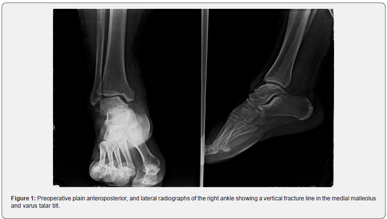
A 30-year-old male patient presented with acute-onset right ankle pain. Four years previously, he had sprained his ankle several times during practice soccer. Subsequently, the ankle pain worsened, and he had tenderness on the medial aspect of his right ankle. The symptoms were mild at rest but increased upon walking and training soccer. Plain radiographs of the right ankle showed a vertical fracture line in the medial malleolus and tilting of the talus, demonstrating the lateral instability of the ankle (Figure 1).
Treatment
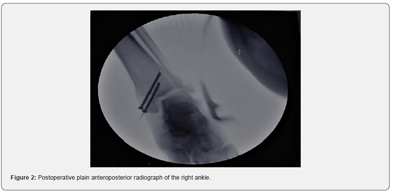
Surgery was performed under spinal anesthesia using a C-arm image intensifier. The medial malleolar fracture was fixed using two screws and the hemi-Castaing technique was done to repair the complex lateral ligament of the ankle [6] (Figure 2). Only the technique of lateral ankle ligament plasty is described below.


Surgical Technique
Description of the hemi-Castaing technique
The principle of most non-anatomical ligamentoplasties is to use the tendon of the peroneus brevis muscle. Due to its position and inherent strength of its distal anchor point, this complete tendon is used to strengthen the lateral ankle joint in the ligamentoplasty initially described by Castaing et al. [6]. The hemi- Castaing procedure is a form of split peroneus tendon tenodesis, which has been infrequently reported in the literature [7,8]. The benefit of a hemi- Castaing is the preservation of the natural ankle stabilizing properties of the peroneus brevis muscle.
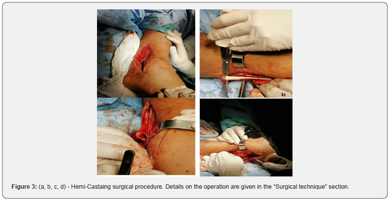
The modification used in this study of this tenodesis, named the hemi- Castaing or Castaing II, makes use of half the peroneus brevis tendon [9]. Hemi-Castaing ligamentoplasty begins with an incision over the lateral ankle, exposing the lateral malleolus and tendon of the peroneus brevis muscle (Figure 3a), approximately 15 cm from its point of insertion.

The tendon is hemi-sected proximally for half of its diameter and then split over the length of the tendon to the level of the most distal point of the fibula, releasing an 8-cm cord of tendon, free proximally, but remaining firmly anchored at its point of insertion at the base of the fifth metatarsal, the so-called hemi-tendon (Figure 3b). A hole is then drilled through the distal part of the lateral malleolus, forming a tunnel running between the directions of the ATFL and CFL (Figure 3c). The hemi tendon is passed through the tunnel from back to front and sutured to itself distally. The foot is maintained in a neutral anatomical position while the sutures are progressively tightened until optimal tensioning is reached (Figures 2 & 3d) [9]. Postoperatively patients are kept in non-weightbearing plaster for two weeks and weight-bearing plaster for six weeks.
Discussion
Rettig et al. [10] reported the first case series of stress fractures of the medial malleolus. They established 3 basic criteria for identifying medial malleolar stress fractures: tenderness over the medial malleolus and joint effusion; pain during activities before an acute episode; and a vertical line from the tibial plafond. Plain radiographs are frequently normal in the early phase because the medial malleolus consists mainly of cancellous bone. Additional imaging using CT, MRI, or nuclear bone scans is recommended [11-13]. In this case, plain radiographs of the right ankle showed a vertical fracture line in the medial malleolus and tilting of the talus, demonstrating the lateral instability of the ankle. Ankle sprain is reported to be among the most common recurrent injuries. About 20% of acute ankle sprain patients develop chronic ankle instability. The failure of functional rehabilitation after acute ankle sprain leads to the development of chronic ankle instability. Unlike acute ankle sprain, chronic ankle instability might require surgical intervention [14].
Lateral ankle sprains are a common consequence of physical activity. If not managed appropriately, a cascade of negative alterations to both the joint structure and a person’s movement patterns continue to stress the injured ligaments. These alterations result in an individual entering a continuum of disability as evidenced by the 30 % of ankle sprains that develop into chronic ankle instability (CAI) and up to 78 % of CAI cases that develop into post-traumatic ankle osteoarthritis (OA) [15]. In this case, the history of repeated ankle sprains and the 3 diagnostic criteria listed above led us to us focus on lateral instability and medial malleolar fracture. Plain radiograph showed a vertical fracture line in the medial malleolus and varus talar tilt [16].
According to the bibliographic review carried out by the author, only one previous article published case report [5] describes the relationship between medial malleolar stress fracture and lateral instability of the ankle, supporting the hypothesis that lateral instability of the ankle accelerated the medial malleolar stress fracture. Therefore, the treatment of fatigue fracture of the medial malleolus associated with lateral ankle instability should be surgical combining the lateral ligament plasty with osteosynthesis of the medial malleolus.
Acknowledgement
The authors declare no conflict of interest in connection with the writing of this article.
References
- Jacobs JM, Cameron KL, Bojescul JA (2014) Lower extremity stress fractures in the military. Clin Sports Med 33(4): 591-613.
- Jowett AJ, Birks CL, Blackney MC (2008) Medial malleolar stress fracture secondary to chronic ankle impingement. Foot Ankle Int 29(7): 716–721.
- Lempainen L, Liimatainen E, Heikkila J, J Alonso, J Sarimo, et al. (2012) Medial malleolar stress fracture in athletes: diagnosis and operative treatment. Scandinavian journal of surgery: SJS: official organ for the Finnish Surgical Society and the Scand Surg Soc 101(4): 261–264.
- Menge TJ, Looney CG (2015) Medial malleolar stress fracture in an adolescent athlete. J Foot Ankle Surg 54: 242–246.
- Hong Seop Lee, Young Koo Lee , Hak Soo Kim, Dhong Won Lee , Sung Hun Won, et al. (2019) Medial malleolar stress fracture resulting from repetitive stress caused by lateral ankle instability A case report. Medicine 98(5): e14311.
- Castaing J, Falaise B, Burdin P (1984) Ligamentoplasty using the peroneus brevis in the treatment of chronic instabilities of the ankle. Long-term review. Rev Chir Orthop Reparatrice Appar Mot 70(8): 653–656.
- Lorenzo G, Calafiore V (2009) Trattamento chirurgico Secondo Castaing nelle instabilita laterali di caviglia. Acta Orthop Ital 34: 11–14.
- Mabit C, Tourne Y, Besse JL, Bonnel F, Toullec E, et al (2010) Chronic lateral ankle instability surgical repairs: the long term prospective. Orthop Traumatol Surg Res 96(4): 417–423.
- Tim Schepers, Lucas M M Vogels, Esther M M Van Lieshout (2011) Hemi-Castaing ligamentoplasty for the treatment of chronic lateral ankle instability: a retrospective assessment of outcome. International Orthopaedics (SICOT) 35(12): 1805–1812.
- Rettig AC, Shelbourne KD, McCarroll JR, M Bisesi, J Watts (1988) The natural history and treatment of delayed union stress fractures of the anterior cortex of the tibia. Am J Sports Med 16(3): 250–255.
- However, diagnosis of a medial malleolar stress fracture may be difficult because of the vague symptoms and insidious onset (11).
- Van den Bekerom MPJ, Kerkhoffs GMMJ, van Dijk CN (2009) Treatment of medial malleolar stress fractures. Oper Tech Sports Med 17: 106–111.
- Jacobs JM, Cameron KL, Bojescul JA (2014) Lower extremity stress fractures in the military. Clin Sports Med 33(4): 591–613.
- Lempainen L, Liimatainen E, Heikkila J, J Alonso, J Sarimo, et al. (2012) Medial malleolar stress fracture in athletes: diagnosis and operative treatment. Scandinavian journal of surgery: SJS: official organ for the Finnish Surgical Society and the Scand Surg Soc 101(4): 261–264.
- Omar A Al-Mohrej, Nader S Al-Kenani (2016) Chronic ankle instability: Current perspectives. Avicenna J Med 6(4): 103–108.
- Erik A Wikstrom, Tricia Hubbard-Turner, Patrick O McKeon (2013) Understanding and treating lateral ankle sprains and their consequences: a constraints-based approach. Sports Med 43(6): 385-393.


