Acute Dislocation of the Elbow - About A case
Luis Crescencio Breton Espinosa1, Claribel Plain Pazos2*, Lazaro Martin Martinez Estupinan3, Sergio Virgilio Morales Pineiro4, Roberto Mata Cuevas4, Leonardo Dominguez Plain5
1I Degree Specialist in Orthopedics and Traumatology, Assistant Professor, Provincial General University Hospital, Cuba
2Specialist of I and II Degree in Comprehensive General Medicine, Faculty of Medical Sciences of Sagua la Grande, Cuba
3Doctor in Medical Sciences, II Degree Specialist in Orthopedics and Traumatology, Full Professor, Provincial General University Hospital, Cuba
4I and II Degree Specialist in Orthopedics and Traumatology, Assistant Professor, Provincial General University Hospital, Cuba
5Resident of 3rd year of Orthopedics and Traumatology, Provincial General University Hospital, Cuba
Submission:April 09, 2021; Published:May 18, 2021
*Corresponding author: Claribel Plain Pazos, Specialist of I and II Degree in Comprehensive General Medicine, Faculty of Medical Sciences of Sagua la Grande, Cuba
How to cite this article:Luis C B E, Claribel P P, Lazaro M M E, Sergio V M P, Roberto M C, et al. Acute Dislocation of the Elbow - About A case. Ortho & Rheum Open Access J. 2021; 18(2): 555985.DOI: 10.19080/OROAJ.2021.18.555985
Abstract
Elbow dislocations occur in any period of life, contrary to supracondylar fractures and other injuries, which occur in specific age periods. The following work aims to present a patient, who was treated in the emergency department of our institution, after an intense trauma to the right elbow. The clinical diagnosis is made and corroborated by X-rays, observing that the proximal portion of the forearm, in addition to being dislocated outward and backward, was fully rotated, therefore the olecranon was in a lateral position. Apparently this injury was produced by a combination of axial compression, internal rotation (pronation) and valgus on the elbow, when the patient fell with the elbow extended. The treatment was closed reduction in the operating room with EV general anesthesia. Radiographs are presented and the subsequent evolution is detailed. Review of the subject is carried out.
Keywords:Elbow; Dislocation; Manual Reduction; Closed or Bloodless
Introduction
Stable and painless mobility of the elbow joint is essential for activities in daily life. The elbow is one of the most common dislocated joints. Acute elbow dislocation has an annual incidence of 6 cases per 100,000 inhabitants. It is more frequent in adults (since in childhood the causal mechanism usually produces supracondylar humerus fracture) and in men [1]. They are the most common injuries to the upper limb after shoulder dislocation. [1-3] Despite this, neurovascular injury associated with acute traumatic elbow dislocation is rare [2]. The elbow represents one of the most stable joints in the body. The close relationship of the humerus-ulnar, capitello-radial, radio-ulnar superior and inferior joints is the result of the high degree of movement that allows flexion and extension, as well as pronation and supination of the forearm. The elbow joint comprises three joints: humerus-ulnar, superior radio-ulnar, and capitello-radial, with normal mobility in flexion-extension from 0 ° to 140 ° and in pronation-supination from 80° to 80° [4]. This joint is made up of static and dynamic stabilizers. Static stability is preserved by the bone and capsuloligamentary structures. Dynamic stability is maintained by the muscles running through the elbow. The mixture of these components achieves a primary and secondary stability against its own movements and deforming forces to which it is subjected. The main stabilizers correspond to: ulnohumeral joint, the medial collateral ligament and the lateral collateral ligament. The secondary restrictors are: radiohumeral joints, common flexor tendons, pronators, and capsule [5].
Case Presentation
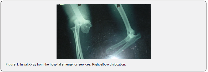
52-year-old male, white-skinned, with a history of left elbow dislocation 4 years ago, which was not treated. He refers to suffering intense trauma to his right elbow days ago when he fell into a ravine; Four days after the trauma, he was evaluated urgently, observing a joint with color changes, greatly increased volume, deformity and total functional impotence. The physical examination revealed the following: pulses present, inability to flex it, and valgus deformity and fixed extension. On palpation, we also noticed the very prominent epitrochlea and the olecranon in lateral or external position. X-rays were indicated where posterolateral dislocation of the right elbow was observed (Figure 1), EKG and emergency blood count that were normal. The elbow, in addition to being dislocated out and back, was fully rotated, therefore the olecranon was in a lateral position. Apparently, this injury was produced by a combination of axial compression, internal rotation (pronation) and valgus on the elbow, when the patient fell with the elbow extended.
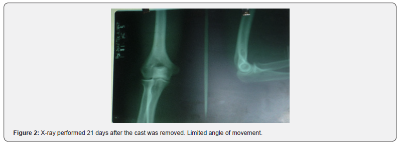
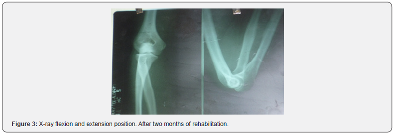
The patient was transferred to the operating room where reduction was carried out under anesthesia in the supine position. Two specialists intervened in the procedure, one of whom fixed the patient’s arm against the examination table while the second specialist performed slow continuous traction. and firm in the direction of the humerus axis by grasping the wrist, keeping the forearm slightly flexed and the wrist slightly supinated. The control X-ray was satisfactory and clinically stable. A posterior brachiopalmar splint was placed for 6 days and a circular brachio-palmar cast for 15 more days, with good recovery of function, which was evidenced in the control X-ray (Figure 2). Nerve paralysis or vascular injury was not observed. The evolution was excellent after two months of rehabilitation (Figure 3).


Discussion
Elbow dislocation is a common injury that accounts for 10% of trauma to the elbow joint, generally caused by sports accidents in young individuals [6,7]. 90% of dislocations occur in a posterior or posterolateral direction and only 1% to 2% move earlier [2]. This injury can be simple (as in the case presented) when it is not accompanied by other injuries, and complex when fractures are present together such as the coronoid process fracture and the radial head fracture, which has been called the terrible triad of the elbow or Hotchkiss triad [6,7]. It is an entity with a high complication rate and requires, in most cases, surgical treatment [1]. The diagnosis of elbow dislocation is clinical, with apparent elbow deformity, functional impotence, and pain clearly present [1]. In the case studied, in addition to the aforementioned symptoms, the diagnosis was corroborated by X-ray of the joint. Simple elbow dislocations are usually benign injuries in the medium and long term, provided that early orthopedic treatment and correct rehabilitation are carried out. Once the reduction is obtained, the evaluation of the elbow stability is imperative. The elbow is considered stable if it remains reduced in a range of motion from 60º of extension to full flexion [7]. In the case studied, the dislocation was simple, as it was not accompanied by a fracture and the reduction was stable.
After reduction, posterior splint immobilization is performed for 1–3 weeks [7]. In the case presented, the reduction was carried out early and afterwards immobilization with a splint and plaster, which affects a good prognosis of evolution. After three weeks of immobilization, rehabilitation of the limb should be performed consisting of controlled flexion and extension with an orthopedic elbow for another 4 weeks. The estimated time of temporary disability is 10 to 12 weeks. [1]. Mobility in this case was recovered after 8 weeks of treatment, with good recovery of limb strength. Complications of traumatic elbow dislocation include neurovascular injury, limited range of motion, and elbow instability [2,3]. Arterial injuries are estimated to occur in approximately 5% to 13% of elbow dislocations. .[two] The incidence of re-dislocation after simple elbow dislocation is low [8]. The aforementioned case did not present neurovascular injury but did have limited mobility ranges and elbow instability; however, its evolution was rapid and satisfactory. Due to the large displacement already described in this case and the severe capsuloligamentous soft tissue injury, ulnar nerve injury was to be expected; In addition, the pressure of the olecranon in external position and maintained against the skin was also to be expected, necrosis of the same, luckily none of these unfavorable events happened. According to the trends of orthopedic or conservative treatment used internationally [1,2], the dislocation was reduced, and immobilization was maintained for 3 weeks so that these highly affected soft tissues had the time necessary for healing; a complete joint function was obtained, with full incorporation to their activities in a short time, without sequelae, after physiotherapy.
References
- De Pablo Márquez B, Bernal PC, Johnson MB, Aparicio NI (2017) Luxación de codo. SEMERGEN-Medicina de Familia 43(8): 574-577.
- Torres Torres V, Cevallos Andrade A, Quispillo Moyota F, Casa Casa G, Peña Toledo J (2020) Luxación abierta postraumática de codo con lesión vascular de la arteria braquial. Revista Ecuatoriana De Ortopedia Y Traumatología 9(Fascículo 1): 49-53.
- Caicedo Cruz N, Ríos Garrido XM (2017) Lesiones óseas y ligamentarias en pacientes con triada terrible del codo Hospital Universitario Mayor 2013-2016. (Doctoral dissertation, Universidad del Rosario) 2017.
- Diaz Carrillo HG, Delgado Rifá E, Álvarez Consuegra W (2017) Actualidades sobre la inestabilidad compleja del codo. Revista Electrónica Dr Zoilo E Marinello Vidaurreta 42(1).
- Alava Muñoz AC (2017) Evaluación funcional postoperatoria del tratamiento quirúrgico de triada terrible de codo en el hospital de las Fuerzas Armadas, periodo 2012 a 2017. (Master's thesis, Quito: UCE) 2017.
- Fonseca García H, Aragón Cervantes I, Chico Gómez M, Sasturaín Miranda ME, Chang Ramírez T (2012) Luxofractura anterior del hombro derecho con luxación posterior del codo: presentación de un caso. AMC 16(1).
- Romero B, Marcos A, Medina JA, Muratore G (2012) Luxación bilateral de codo asociada a lesión de Essex-Lopresti. Rev esp cir ortop traumatol 56 (1): 59-62.
- Vicente Quílez M, Redondo Buil P, López Díaz MDV, Montes García A, Morán Hevia M, Galvez García SG (2018) Fracturas y fracturas-luxaciones del codo en el adulto. Seram.


