




 OROAJ Home



Satisfactory Evolution in Clavicle Fracture with Wrong Predicts
Enrique Arce Morera*
Specialist of I & II degrees in Physical Medicine and Rehabilitation, Master in Bioenergetic Medicine and Nature in A.P.S, Cuba
Submission:April 02, 2021; Published: May 06, 2021
*Corresponding author: Enrique Arce Morera, Specialist of I & II degrees in Physical Medicine and Rehabilitation, Master in Bioenergetic Medicine and Nature in A.P.S, Cuba
How to cite this article: Enrique A M. Satisfactory Evolution in Clavicle Fracture with Wrong Predicts. Ortho & Rheum Open Access J. 2021; 18(2): 555983. DOI: 10.19080/OROAJ.2021.18.555983
Summary
Introduction: The type 2B fracture of the external third of the clavicle (according to the Robinson classification), has a surgical approach or conservative treatment. It was considered this last possibility in the following case.
Objective: to describe the clinical evolution of a patient who presented a clavicle fracture with an unfavorable prognosis.
Presentation of the case: It is a patient with fracture of the external third of the clavicle which had been confirmed by clinical-radiographical examination and failed intent of reduction through Kisner’s wire technique and surgical criterion, which was not developed, the patient received physiotherapeutic treatment at the Agency of Integral Rehabilitation in Dr. Tomás Romay Policlinic.
Conclusion: The patient having clavicle fracture described in the case presentation achieved excellent functional results. She presented a total recovery of her symptoms and reincorporation without limitations to its social activity within 8 weeks and through the appropriate physiotherapeutic treatment. That is why the conservative treatment in this lesion type is supported.
Keywords: Clavicle fracture; Conservative treatment; recovery
Introduction
Clavicle fractures are frequent injuries that cause loss of bone continuity, of mostly traumatic origin, located between the acromioclavicular and sternoclavicular joints. These fractures occur mainly in young men and comprise 4 to 10% of all fractures [1,2]. Powerful muscles are inserted into the body of the clavicle: sternocleid mastoids, clavicular fascicles of the deltoid and pectoralis that act on the movements of the arm [3,4]. When the clavicle is fractured, the fragments already free, directly undergo the contracting action of the indicated muscles that determine the characteristic displacements. The fracture can be: Simple fracture or several fragments: Fracture of the inner end: Fracture of the outer end Fracture of the middle part [5,6]. Among the indications for surgical treatment in these types of fractures are: A prominent fracture on the skin that threatens to perforate the skin, pseudo-osteoarthritis and fracture of the external third with ligamentous injury. The present work reports a patient who presented the characteristics of the latter mentioned whose functional prognosis was unfavorable, given the characteristics of the lesion and was treated with the conservative method for 8 weeks. The objective was to describe the clinical evolution of the patient who presented clavicle fracture with surgical criteria and that through opportune physiotherapeutic actions, a rapid and total recovery was achieved.
Presentation of the case
YRM female patient, 59 years old, with a history of type II diabetes Mellitus, who suffered a fall back and impacted the posterior region of the right shoulder with an irregular surface, presented pain and functional impotence X-ray was performed at the orthopedic service of the shoulder whose image showed a fracture of the external third of the right clavicle type 2B (according to the Robinson classification) [7]. An unsuccessful attempt to reduce with Kirshner wire was made and surgical criteria were suggested, an act that was not carried out due to the patient’s refusal, the shoulder is immobilized with a Velpaut bandage for 2 weeks (see X-ray: solution of contiguity of the external third of the right clavicle with loss of anatomical relationship of the affected surfaces with grade III displacement). Conservative treatment is decided and is referred to the Rehabilitation Service of your health area. Reference to the Comprehensive Rehabilitation Service (S.R.L.) (Figure 1).
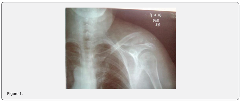
The third week after the injury occurred. First physiatric assessment in the SRI A physical examination is performed: prominence in the super-external region of the shoulder, presence of local ecchymosis, painful shoulder on movements with functional impotence of said joint. The clinical assessment was performed using the score obtained on the Constant A pain scale (5) of 15 points, B: activities of daily living (0) of 20 points, C. range of movement (0) of 40 points, D: abduction force in the scapular plane (0) of 25 points. Total 5 out of 100 points.
Diagnosis
Stiff shoulder due to fracture of the external third, right shoulder, type IIB.
Functional prognosis
Unfavorable, with long-term recovery and possible motor sequelae. As treatment objectives: pain relief, improve joint width of the affected shoulder, train in the activities of the upper right limb. Physiotherapeutic treatment: relaxing massages of the shoulder muscles. Kinesitherapy: pendulum exercises. Magnetotherapy: magnetic field with the equipment Mag 200, trans articular technique, intensity of 50% of the field, frequency 50 Herz, time 20 minutes. 20 treatment sessions. Sixth week after the injury. Second physiatrist evaluation in the SRI. The patient received 5 treatment sessions and continued the exercises at her home. Physical examination is performed: presence of active movements of the shoulder joint, the Constant scale is applied: A: pain (15) of 15 points, B: activities of daily living (8) of 20 points, C: range of motion (24) of 40 points, D: force of abduction in the scapular plane (5) of 25 points. Total 52 out of 100 points. As a physiotherapeutic treatment in her home, the amplitude and strength exercises of said joint are oriented (exercises with dumbbells to strengthen the rotator cuff). Consultation at 2 weeks and evolutionary radiography is indicated. 8 weeks after the injury occurred (see X-ray, the clavicular fracture line is evaluated where signs of bone callus appear because of the osteoblastic process with a pseudarthrosis nucleus) (Figure 2).
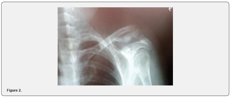
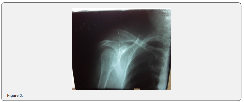
Final evaluation is performed according to the Constant scale: A: pain (15) of 15 points, B activities of daily living (16) of 20 points, C: range of motion (38) of 40 points, D. strength of the abduction in the scapular plane (23) of 25 points. Total 92 out of 100 points. Discharge from the S.R.I is determined without limitations and it is made against a referral to your attending physician with recommendations for your home. Evaluation 2 years after the injury without symptoms or sequelae Rx. exuberant bony callus, distal end of clavicle out of place (Figure 3).
Discussion
Clavicle fractures are usually trivial injuries that only require conservative treatment. Displaced fractures that presumably will not heal properly require surgery. For this, the use of screwretained plates and occasionally needles and cerclage is the norm [10]. Also, as a complication of this, some cases of axillary or subclavian vein thrombosis caused by clavicular fractures have been reported [11]. Surgical treatment presents better functional results than conservative treatment, as expressed by Valero González in his article that considers open reduction and internal fixation of this lesion the method of choice for its treatment [12]. Dr. Moya also considers surgical treatment a good option, given the good functional result and the low incidence of pseudarthrosis that they present [13]. However, according to Garcia CA, the results Functional functions of patients with Type 2B clavicle fractures (according to Robinson’s classification) treated both conservatively and surgically went from good to excellent after 12 weeks of evolution .According to Fernandez Carnero in his analysis of a clinical case with this type of injury states that with intensive physiotherapy treatment, muscle-tendon stability of the entire scapular complex could be obtained after 3 months of treatment [14] Also, when consulting the experience of Dr. Laza Capote and observing the good functional evolution in these cases, He opts for conservative behavior, mainly carrying out scheduled exercises.
Conclusion
The patient with Type 2B clavicle fracture (according to Robinson’s classification) described in this case presentation, achieved excellent functional results, since in just 8 weeks with adequate physiotherapeutic treatment, she presented a full recovery of her symptoms and reincorporation without limitations to her social activity, so conservative treatment is supported in this type of injury.
References
- Moral-Villanueva J, Tamayo-Pacho F, Pineda-Castro OP (2018) Complex fracture of the lateral third of the clavicle. Records Ortop Mex 32(3).
- Checchia CL, Doneux P, Miyazaki AN, Fregoneze M, Luciana A Silva (2018) Treatment of distal clavicle fractures using an arthroscopic technique. J Shoulder Elbow Surg 19(3): 294-298.
- Arches Gutierrez MI, Acnes Segovia JA, Cevallos Mendoza CD, Ollague Armijos R (2020) Conservative treatment in clavicle fracture. Recimundo 3(4): 571-585.
- Lopez JJ (2018) Clavicle fracture, the fractures of Sundays. The bag of the corridor LBDC.
- Joao Roberto Polydoro Rosa, Sacred Caio Checchia, Alberto Naoki Miyazaki (2017) Instabilidade previous traumatic do ombro. Rev Bras 18 Ortop 52(5): 513-520.
- Ávila LJL, Sacred MM, Jacobo EO, Garcia PL, Garcia RC, et al. (2015) Fractures of clavicle distal. 22(1): 49–53.
- Avila Lafuente JL, Moorish Marco S, Jacobo Edo OR, Garcia-Polin Lopez C, Garcia Rodriguez C, et al. Fractures of clavicle distal. Have Spanish of Artroscopia and Surgery to Articulate. 22(1): 49-53.
- Arce ME, Hernandez ET, Armas ML (2016) Acupuncture and kinesioterapia in the treatment of the adhesive capsulitis of the shoulder. R Rev Cuban Med Gene Integr 32(1).
- Chain A (2016) The fracture of the Clavicle in the Sport. Revista Ball World Soccer 26.
- Caballero AJ (2016) Comparison of different technical surgical in unstable fractures of the third clavicle distal. International University of Andalusia. ISBN 978-84-7993-706-5.
- Ubeda PI, Excess-Hidalgo GA (2016) Secondary massive deep veined thrombosis of superior member to fracture of half third of clavicle. Clinical case. it Has Andalusian of Medicine of the Sport 9(3): 99-142
- Valero G, Fernando S, Garcia GA, Aguirre HR (2016) Treatment of the fractures of the clavicle with severe displacement, by means of the open reduction and internal fixation with diverse technical. Rev mex ortop traumatol 8(3): 79-83.
- Moya And, Lamar C (2016) surgical treatment of the fractures diafisarias and conminutas of the clavicle by means of anatomical badge of low profile. 56(2): 127-131.
- Fernandez CJ, Martinez GM (2016) Conservative treatment after clavicle fracture: analysis of a clinical case 26(6): 325-332.

























