




 OROAJ Home



Technical Outcome of Atlantoaxial Trans-articular Screw Fixation without Supplementary Posterior Construct in Rheumatoid Arthritis
Philip M Thomas1, Jack Horan2, Michael Amoo2*, Mohammed Ben Husien2, Derek Cawley1, Jabir Nagaria1 and Ciaran Bolger2
1Acute Services, Department of Neurosurgery, Belfast Health & Social Care Trust, Royal Hospitals, United Kingdom
2National Centre for Neurosurgery, Beaumont Hospital, Dublin 9, Republic of Ireland.
Submission:May 18, 2020; Published: May 26, 2020
*Corresponding author: Michael Amoo, Specialist Trainee, Neurosurgery, National Centre for Neurosurgery, Beaumont Hospital, Dublin 9, Republic of Ireland
How to cite this article: Philip M T, Jack H, Michael A, Mohammed B H, Derek C, et al. Technical Outcome of Atlantoaxial Trans-articular Screw Fixation without Supplementary Posterior Construct in Rheumatoid Arthritis. Ortho & Rheum Open Access J. 2020; 16(2): 555934. DOI: 10.19080/OROAJ.2020.16.555934
Abstract
Objective: To determine the technical outcome of rheumatoid arthritis (RA) patients who underwent atlantoaxial (AA) trans-articular screw (TAS) fixation without supplementary posterior construct.
Methods: To treat AA instability, all 15 RA patients in this study underwent C1-C2 TAS fixation without supplementary posterior construct. The minimum follow-up period was 24 months. Pre- and postoperative sagittal measures of C1-C2, C2-C7, and C1-C7 angles, atlanto-dens interval (ADI), posterior atlanto-dens interval (PADI), and adjacent segment (i.e., C2-C3) anterior disc height (ADH) were retrospectively recorded from lateral X-ray imaging. The presence or absence of superior migration of the odontoid (SMO), cervical sub-axial subluxation, C1-C2 bony fusion, screw pull-out and screw breakage were also noted.
Results: There was little difference between the pre- and post-operative means of all angles measured. Following TAS fixation, mean ADI shortened and mean PADI lengthened. There was no difference in mean measures of C2-C3 ADH. There was no evidence of SMO pre- or post-operatively. Two patients developed anterior subluxation at C5-C6 and of one of these patients also developed anterior subluxation at C2-C3. All patients had evidence of C1-C2 bony fusion. There was no screw pull-out or breakage.
Conclusion: In RA patients who have undergone C1-C2 TAS fixation in the absence of a supplementary posterior construct, the overall technical outcome appears acceptable.
Keywords:Atlantoaxial (AA); C1-C2; Rheumatoid arthritis (RA); Trans-articular screw (TAS) fixation
Introduction
The atlantoaxial (AA) segment is the most mobile region of the spinal column. Approximately 50% of the cervical spine’s rotary movement, and as much as 50 degrees of its rotation, occurs at the C1-C2 joint [1,2]. Instability of this joint can result from various processes, including aplasia or hypoplasia of the odontoid process, laxity of the transverse ligament and assimilation of the atlas. AA instability is associated with various conditions including Down’s syndrome, Klippel-Feil syndrome, osteogenesis imperfecta, neurofibromatosis and rheumatoid arthritis (RA) [3-6].
It is well recognized that RA in the cervical spine causes pathological changes in ligaments, articular cartilage, and bones. In over half of patients with RA, signs of upper cervical spine disease are present as this region is most commonly affected after the hands and feet [7,8]. RA-related neurological impairment is associated with shortened life expectancy [9]. The natural disease process of RA in the cervical spine can result in three principal deformities - AA instability, superior migration of the odontoid (SMO) (also referred to as cranial settling or pseudo-basilar invagination) and/or sub-axial (i.e., below C2) subluxation. These deformities may be seen in isolation or in combination. The most common cervical spine abnormality observed in patients afflicted with RA is AA instability, which accounts for approximately two-thirds of rheumatoid cervical subluxations [10]. It is well recognized that AA instability has the potential to cause progressive cervical pain, neural injury, and vascular compromise if left untreated.
Over the past several decades, various posterior fusion techniques to achieve AA arthrodesis have been reported. Some common posterior block bone-wire/clamp methods that have been used include Gallie fusion (i.e., midline bone graft and sublaminar wiring) [11], ‘Halifax’ bilateral interlaminar clamping of bone graft [12] and Brooks and Jenkins’s technique of bilateral interlaminar bone grafting and sublaminar wiring [13]. However, many spinal surgeons feel the current gold standard technique of posterior AA arthrodesis is trans-articular screw (TAS) fixation [14-16]. C1-C2 TAS fixation was developed by Magerl in 1979 and later fully described by Magerl and Seemann [17]. Classically, three-point fixation of the AA segment can be achieved by the placement of a TAS screw from C2 to the anterior surface of C1 (across the facets, bilaterally) followed by a Gallie-type block bone-wire posterior construct [17].
The biomechanical stability afforded by C1-C2 TAS fixation has been demonstrated in-vitro [18,19]. Given the inherent stability of TAS fixation, patients do not require rigid external bracing following surgery. C1-C2 TAS placement can be a technically demanding procedure with limitations dictated by the extent of C1-C2 disease and/or regional anatomic variability, but despite these challenges, in terms of clinical outcome, results have proven satisfactory with relatively few complications [20-22]. It should also be noted that sublaminar wiring (the third component of a classical ‘three-point’ fixation) is itself not without risk of technical complication (e.g., intra-operative traumatic durotomy and/or injury to spinal cord, late wire failure causing cord injury/ compression etc). There have also been some reports of concern regarding sub-axial instability/subluxation following various posterior C1-C2 arthrodesis procedures [23,24]. With regard specifically to sub-axial subluxation following C1-C2 TAS fixation in patients with RA, it remains unclear the full spectrum of factors associated (causally or otherwise) with sub-axial sagittal alignment change [25-27].
For some time now, we have taken the view that TAS fixation without supplementary posterior construct, even in RA patients, provides sufficient stability to facilitate acceptable clinical results [22]. As far as we are aware, this is the first full report concerning sagittal alignment and other indicators of technical outcome in RA patients who have undergone a modified (two-point) Magerl C1- C2 TAS fixation.
Materials and Methods
We undertook a retrospective, observational review of the radiographic imaging of 15 consecutive patients (13 females, 2 male) who had undergone C1-C2 TAS fixation for the treatment of RA-associated AA instability. All TAS fixations in this series were bilateral, and thus a total of 30 screws were placed. All patients had been operated upon by the same primary surgeon (CB) and these procedures occurred between the years 2000 to 2004 at a single institution. Mean patient age at the time of operation was 52 years (range 33 to 78 years). In all cases, and as we have previously described, pre-operative planning of screw placement was facilitated by CT-based Stealth Station® image guidance (Medtronic Sofamor Danek, Memphis, TN) [28-30].
Intra-operatively, patients had been placed prone on the operating table with their heads rigidly fixed in a three-point Mayfield® skull clamp (OMI, Cincinnati, OH). At the time of positioning, and before image guidance registration, manual reduction of C1 on C2 was achieved with the aid of fluoroscopy. The C1 and C2 vertebrae were exposed posteriorly by a 2 cm midline incision and an LED (light emitting diode) reference arc was attached to the spinous process of C2. Image guidance data registration occurred by a combination of point matching and surface mapping upon the posterior arch of C2. Parallel caudal stab incisions were made to allow delivery of all instruments percutaneously to the posterior arch of C2. For screw placement, the start points in the C2 lateral mass with regard to degree of divergence from the midline, and the sagittal plane orientation relative to the C1-C2 facet joint, was in keeping with the Magerl technique [17]. In all cases, we used cannulated 3.5 mm diameter titanium screws (UCSS, Medtronic Sofamor Danek, Memphis, TN). Screw trajectory was confirmed intra-operatively by Stealth Station® image guidance and fluoroscopy. We did not observe any operative injury or complication directly related to the placement of C1-C2 screws. Prior to operative site closure, the C1-C2 facet joints were curetted and packed with morselized autologous iliac crest bone graft. We did not utilise any supplementary posterior construct (e.g., bone-wire, clamp, cable, claw etc).
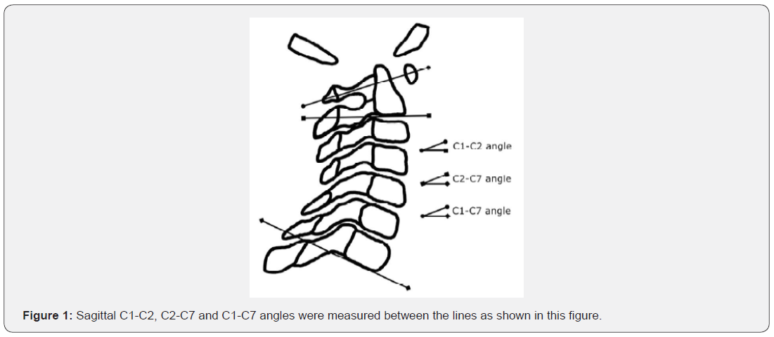
The mean follow-up period was 26 months (range 24 to 39 months). Pre- and post-operative sagittal measures of C1-C2, C2-C7, and C1-C7 angles (Figure 1), atlanto-dens interval (ADI), posterior atlanto-dens interval (PADI) and adjacent segment (i.e., C2-C3) anterior disc height (ADH) were recorded from lateral X-ray imaging. We adopted the measurement parameters of sagittal cervical alignment previously defined by Yoshimoto et al. [23]. ADI and PADI were measured along the line drawn from the craniocaudal midpoint of (and parallel to) the atlas [31]. Absolute measurements of C2-C3 ADH were recorded from X-rays taken in the neutral position (based on the hand-marked corners of the C2 and C3 vertebrae), in a method similar to that described by Frobin et al. [32]. The presence or absence of superior migration of the odontoid (SMO) and cervical sub-axial subluxation was noted on preoperative imaging, and these features were also considered along with C1-C2 bony fusion, screw pull-out and screw breakage on post-operative imaging. SMO was deemed to be present if there was encroachment of the dens beyond McRae’s line on static or dynamic imaging [33]. We judged cervical sub-axial subluxation if the distance along the superior endplate line of the lower vertebra was greater than 2.5 mm with respect to the vertebra above. C1-C2 bony fusion was inferred from plain film X-ray if there was evidence of new bone trabeculation at the AA interfaces without any evidence of periprosthetic lucency or intervertebral motion on dynamic imaging. All data measures were assessed and recorded by a single observer (JN). Statistical analysis was carried out using SAS® software (SAS Institute, Cary, NC).
Results
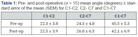
In relation to the pre-operative mean measures, there were no remarkable differences noted in the postoperative fixation, sub axial, and whole C-spine angles (i.e., the C1-C2, C2-C7, and C1- C7 angles, respectively) (Table 1). Following C1-C2 TAS fixation, we found that the mean distance for ADI was shorter and the mean distance for PADI was longer. The mean C2-C3 ADH was unchanged following C1-C2 TAS fixation. No patient had evidence pre- or post-operatively of SMO. We noted no significant evidence of cervical sub-axial subluxation on pre-operative imaging in any case. Two patients progressed to develop anterior subluxation at C5-C6 (this was noted in one case at 27 months, and in the other, at 36 months; in the second case, we also noted an associated anterior subluxation at C2-C3). Immediate corrective surgical intervention was not required in any case, however, once any late sub-axial subluxation was detected, clinical and imaging surveillance was intensified. C1-C2 bony fusion rate following fixation was 100%. We did not observe any evidence of screw pull-out or breakage.
Discussion
The AA segment is the most mobile region of the spine, and its stability can therefore be significantly affected by pathological changes which commonly occur as part of the natural disease process in RA. Given that AA instability has the potential to cause significant morbidity (and even mortality) if left untreated, serious consideration must be given to C1-C2 surgical stabilization. There have, however, been reports of sagittal alignment change following various posterior C1-C2 arthrodesis procedures [23,24], and subaxial subluxation has also been reported following TAS fixation in RA patients [25-27]. As well, the use of sublaminar wiring (to form a posterior point of fixation) is of course not without risk. Nonetheless, satisfactory clinical outcomes have been achieved with C1-C2 TAS fixation [20-22], and it is therefore not surprising that many spinal surgeons view the technique as the current gold standard in posterior AA arthrodesis [14-16].
Naderi et al. [34] have suggested, based on in vitro biomechanical data, and as seems intuitively obvious, that the overall stability of the AA segment can be increased by using as many fixation points as possible. Indeed, and though it may be that C1-C2 movement is better limited by a three- versus a two-point Magerl’s operation, whether one approach over the other confers a clear advantage, clinically, and in specific scenarios, remains unclear. In general, by not undertaking to supplement C1-C2 TAS fixations with any form of posterior construct (such as sublaminar wiring), some immediate and late risks of fusion surgery can be eliminated (e.g., supplementary posterior construct infection or failure causing morbidity). At the same time, there may also be other mutual benefits to be realized for patients, surgeons, and service providers, if it could be demonstrated that a twoversus three-point operative procedure was just simply quicker. Beyond the observation-based approach adopted in the present work, future case-controlled investigations comparing rates of infection, iatrogenic injury, operative time etc, may prove useful in establishing whether conclusive evidence that an overall treatment advantage does indeed exist.
Yoshimoto et al. [23] have reported that, in a variety of pathologies, and for various posterior AA fixation techniques (all of which involved posterior bone-wire or clamp constructs), the C1-C2 angle tends towards hyper-lordosis following fusion and this in turn causally leads the sub-axial cervical spine to kyphotic alignment. However, as described in the present study, our experience (exclusive to RA patients and C1-C2 TAS arthrodesis without block bone-wire, clamp, cable or claw construct) suggests that there is little if any difference to be expected between the mean pre- and post-operative C1-C2 angles. This may explain then why we also did not consequently observe any remarkable mean changes in sub-axial and whole C-spine angles at late follow-up. Mukai et al. [26] have suggested that biomechanical compensatory changes after C1-C2 TAS fixation in RA can occur over time (e.g., increase in C1-C2 lordosis, anterior shift of C2, decrease in C2-C7 lordosis and a tendency of C1 inclination to return to pre-operative inclination). However, we surmise that any supplementary posterior construct depends upon, to some extent, forces which have a tendency to fix the C1-C2 complex in a hyperlordotic position, and therefore, such effects may be mitigated by modification of the Magerl technique as we have described.
AA instability in RA is routinely assessed by measurements of ADI and/or PADI from lateral X-ray imaging. An ADI of more than 3 mm in an adult is generally considered to be abnormal. In terms of clinical usefulness, PADI has been shown to have a high negative predictive value, and as such, is felt to be the better screening tool in RA [35,36]. In the present study, the mean results obtained for both, ADI (which shortened) and PADI (which lengthened) appear to reflect our overall technical success in horizontal stabilization of the AA segment in our patients.
Caudal to the AA segment, cervical motion is distributed equally among the sub-axial elements. It is well recognized that fusion at any one spinal segment can cause increased loads at adjacent segments, and this may in turn lead to adjacent segment degenerative disease. One of the principal roentgenographic features of spinal degenerative disease is disc space narrowing/ loss of disc height. In the present study, we sought to know whether there was evidence of degenerative change in the adjacent segment to fusion, and for uniformity of measurement and ease of comparison, we limited our analysis to C2-C3 ADH. We did not find any notable difference between mean C2-C3 ADH pre- and post-operatively. Thus, adjacent segment change leading to disc space narrowing, if at all present at late follow-up in the present series, was not overt on lateral X-ray imaging.
SMO is not an uncommon finding in advanced RA, and it is not inconceivable that horizontal stabilization of the AA segment could diminish the risk of SMO onset/progression. In the present study, SMO was not observed pre-operatively. The absence of SMO post-operatively following C1-C2 TAS fixation, despite the lack of a supplementary posterior construct, would indicate that there is no added risk of vertical subluxation of the odontoid in RA patients. Our results may also provide some support to the view that AA arthrodesis may be a prophylactic measure in preventing the onset/progression of SMO in RA [24].
We did observe a total of three sub-axial subluxations/ kyphosis (i.e., at C5-C6 and C2-C3 and C5-C6, in two patients, at 27 and 36 months, respectively). We cannot precisely state the reason(s) for this, but it may simply be that the sub-axial subluxations we observed were manifestations of the natural disease process in RA. We note that the proportion of RA patients who develop sub-axial subluxations during the course of the disease has been reported to be at least one-fifth [37]. Ito et al have recently reported that sub-axial subluxation following C1- C2 TAS fixation (supplemented either with posterior wire, cable or claw construct), is more likely to occur in RA versus non-RA patients, but only in the immediate post-operative period (i.e., 3 months) [27]. In the present series, sub-axial subluxations were only seen relatively late in follow-up. Clarke et al have reported that sub-axial subluxation in RA patients who have undergone various forms of posterior AA arthrodesis (though not including TAS fixation) is most common at the C3-C4 segment. They reasoned that wiring the C2 lamina, followed by a period of rigid external bracing, may lead to C2-C3 auto-fusion, and this in turn may lead to increased biomechanical stresses which predispose the C3-C4 level to subsequent instability [24]. Clearly, this scenario would not apply to patients in the present series, and this may explain why we did not observe C3-C4 subluxation. It has recently been reported that Modic (particularly, Type 2) change in the cervical spine as observed from MRI scanning, is predictive of future degenerative change [38]. Although beyond the scope of the present study, we did note anecdotally and in retrospect, that for each of the late sub-axial subluxations we observed, there was pre-operative MRI evidence of Modic Type 2 change. MRIbased studies may perhaps provide further insight with respect to fusion surgery and sub-axial disease in RA.
Bahadur et al recently reported their clinical experience of 38 cases using a two-point, modified Magerl technique (bilateral TAS fixation with block bone graft, without supplementary posterior construct); most of their patients had AA instability secondary to trauma, and they attained a comparable fusion rate in relation to the traditional Magerl operation [16]. Similarly, Wang et al have reported their clinical experience of 57 cases of C1-C2 TAS fixation with morselized autologous bone graft without supplementary posterior construct; most of their patients had AA instability resulting from congenital malformation or trauma, and they achieved a 100% C1-C2 fusion rate [35]. For C1-C2 TAS without posterior construct in RA patients, the 100% fusion rate reported in the present technical report compares well with our previously published clinical experience [22]. In terms of screw pull-out, Bahadur et al in their series, reported only one case of pull-out and this was in a patient with RA [16]. Wang et al observed no cases of screw pull-out or screw breakage [35], and our technical results in that regard (albeit exclusively in RA patients), using a similar technique of anatomic reduction, does mirror the experience of our colleagues. It therefore appears that screw pull-out or screw breakage, whatever the altered biomechanics may be in twoversus three-point C1-C2 TAS fixation, and even in RA patients, is not to be expected.
Conclusion
Despite the limitations of the present study, namely small sample size and the lack of comparative cohort, it appears that technically satisfactory outcomes of AA segment arthrodesis in RA patients can be achieved by TAS fixation and morselized bone graft only, even without supplementary posterior construct. We did note at long-term follow-up a total of three sub-axial subluxations. However, these results are in keeping with the expected rate of such occurrences in the natural disease process of RA in the sub-axial cervical spine. Regular follow-up imaging for all patients who have undergone a modified Magerl C1-C2 TAS fixation is advised.
Acknowledgment
No funds were received in support of this work. No benefits in any form have been or will be received from a commercial company related directly or indirectly to the subject of this manuscript.
References
- Fielding JW (1957) Cineroentgenography of the normal cervical spine. J Bone Joint Surg Am 39-A(6): 1280-1288.
- Dvorak J, Hayek J, Zehnder R (1987) CT-functional diagnostics of the rotatory instability of the upper cervical spine. Part 2. An evaluation on healthy adults and patients with suspected instability. Spine 12(8): 726 –731.
- Ranawat CS, O'Leary P, Pellicci P, Tsairis P, Marchisello P, et al. (1979) Cervical spine fusion in rheumatoid arthritis. J Bone Joint Surg Am 61(7): 1003-1010.
- Pellicci PM, Ranawat CS, Tsairis P, Bryan WJ (1981) A prospective study of the progression of rheumatoid arthritis of the cervical spine. J Bone Joint Surg Am 63(3): 342-350.
- Isu T, Miyasaka K, Abe H, Ito T, Iwasaki Y, et al. (1983|) Atlantoaxial dislocation associated with neurofibromatosis. Report of three cases. J Neurosurg 58(3): 451-453.
- Crockard HA, Stevens JM (1995) Craniovertebral junction anomalies in inherited disorders: Part of the syndrome or caused by the disorder? Eur J Pediatr 154(7): 504-512.
- Bouchaud-Chabot A, Lioté F (2002) Cervical spine involvement in rheumatoid arthritis. A review. Joint Bone Spine 69(2): 141-154.
- Wolfs JF, Peul WC, Boers M, van Tulder MW, Brand R, et al. (2006) Rationale and design of The Delphi Trial – I(RCT)2: international randomized clinical trial of rheumatoid craniocervical treatment, an intervention-prognostic trial comparing 'early' surgery with conservative treatment. BMC Musculoskelet Disord 7: 14.
- Matsunaga S, Sakou T, Onishi T, Hayashi K, Taketomi E, et al. (2003) Prognosis of patients with upper cervical lesions caused by rheumatoid arthritis: comparison of occipitocervical fusion between c1 laminectomy and nonsurgical management. Spine 28(14): 1581-1587.
- Dreyer SJ, Boden SD (1999) Natural history of rheumatoid arthritis of the cervical spine. Clin Orthop Relat Res 366: 98-106.
- Gallie WE (1939) Fractures and dislocations of the cervical spine. Am J Surg 46(3): 495-499.
- Tucker HH (1975) Technical report: method of fixation of subluxed or dislocated cervical spine below C1-C2. Can J Neurol Sci 2(4): 381-382.
- Brooks AL, Jenkins EB (1978) Atlanto-axial arthrodesis by the wedge compression method. J Bone Joint Surg Am 60(3): 279-284.
- Mummaneni PV, Haid RW (2005) Atlantoaxial fixation: Overview of all techniques. Neurol India 53(4): 408-415.
- Härtl R, Chamberlain RH, Fifield MS, Chou D, Sonntag VK, et al. (2006) Biomechanical comparison of two new atlantoaxial fixation techniques with C1-2 trans-articular screw-graft fixation. J Neurosurg Spine 5(4): 336-342.
- Bahadur R, Goyal T, Dhatt SS, Tripathy SK (2010) Transarticular screw fixation for atlantoaxial instability - modified Magerl's technique in 38 patients. J Orthop Surg Res 5: 87.
- Magerl F, Seemann PS (1987) Stable posterior fusion of the atlas and axis by trans-articular screw fixation. In: Kehr P, Weidner A, eds. Cervical Spine, Vol 1. Vienna, Springer-Verlag, pp. 322-327.
- Grob D, Crisco JJ 3rd, Panjabi MM, Wang P, Dvorak J (1992) Biomechanical evaluation of four different posterior atlantoaxial fixation techniques. Spine 17(5): 480-490.
- Richter M, Schmidt R, Claes L, Puhl W, Wilke HJ (2002) Posterior atlantoaxial fixation: biomechanical in vitro comparison of six different techniques. Spine 27(16): 1724-1732.
- Haid RW Jr, Subach BR, McLaughlin MR, Rodts GE Jr, Wahlig JB Jr (2001) C1-C2 trans-articular screw fixation for atlantoaxial instability: a 6-year experience. Neurosurgery 49(1): 65-70.
- Dickman CA, Sonntag VK (1998) Posterior C1-C2 trans-articular screw fixation for atlantoaxial arthrodesis. Neurosurgery 43(2): 275-281.
- Nagaria J, Kelleher MO, McEvoy L, Edwards R, Kamel MH, et al. (2009) C1-C2 trans-articular screw fixation for atlantoaxial instability due to rheumatoid arthritis: a seven-year analysis of outcome. Spine 34(26): 2880-2885.
- Yoshimoto H, Ito M, Abumi K, Kotani Y, Shono Y, et al. (2004) A retrospective radiographic analysis of sub-axial sagittal alignment after posterior C1-C2 fusion. Spine 29(2): 175-181.
- Clarke MJ, Cohen-Gadol AA, Ebersold MJ, Cabanela ME (2006) Long-term incidence of sub-axial cervical spine instability following cervical arthrodesis surgery in patients with rheumatoid arthritis. Surg Neurol 66(2): 136-140.
- Kato Y, Itoh T, Kanaya K, Kubota M, Ito S (2006) Relation between atlantoaxial (C1/2) and cervical alignment (C2-C7) angles with Magerl and Brooks techniques for atlantoaxial subluxation in rheumatoid arthritis. J Orthop Sci 11(4): 347-352.
- Mukai Y, Hosono N, Sakaura H, Fujii R, Iwasaki M, et al. (2007) Sagittal alignment of the sub-axial cervical spine after C1-C2 trans-articular screw fixation in rheumatoid arthritis. J Spinal Disord Tech 20(6): 436-441.
- Ito H, Neo M, Sakamoto T, Fujibayashi S, Yoshitomi H, et al. (2009) Subaxial subluxation after atlantoaxial trans-articular screw fixation in rheumatoid patients. Eur Spine J 18(6): 869-876.
- Bolger C, Wigfield C, Melkent T, Smith K (1999) Frameless stereotaxy and anterior cervical surgery. Comput Aided Surg 4(6): 322-327.
- Bolger C, Wigfield C (2000) Image-guided surgery: applications to the cervical and thoracic spine and a review of the first 120 procedures. J Neurosurg 92(2 Suppl): 175-180.
- Wigfield C, Bolger C (2001) A technique for frameless stereotaxy and placement of trans-articular screws for atlanto-axial instability in rheumatoid arthritis. Eur Spine J 10(3): 264-268.
- Bono CM, Vaccaro AR, Fehlings M, Fisher C, Dvorak M, et al. (2007) Measurement Techniques for Upper Cervical Spine Injuries: Consensus Statement of the Spine Trauma Study Group. Spine 32(5): 593-600.
- Frobin W, Leivseth G, Biggemann M, Brinckmann P (2002) Vertebral height, disc height, posteroanterior displacement and dens-atlas gap in the cervical spine: precision measurement protocol and normal data. Clin Biomech 17(6): 423-431.
- McRae DL, Barnum AS (1953) Occipitalization of the atlas. Am J Roentgenol Radium Ther Nucl Med 70(1): 23-46.
- Naderi S, Crawford NR, Song GS, Sonntag VK, Dickman CA (1998) Biomechanical comparison of C1-C2 posterior fixations. Cable, graft, and screw combinations. Spine 23(18): 1946-1955.
- Wang C, Yan M, Zhou H, Wang S, Dang G (2007) Atlantoaxial trans-articular screw fixation with morselized autograft and without additional internal fixation: technical description and report of 57 cases. Spine 32(6): 643-646.
- Boden SD, Dodge LD, Bohlman HH, Rechtine GR (1993) Rheumatoid arthritis of the cervical spine. A long-term analysis with predictors of paralysis and recovery. J Bone Joint Surg 75(9): 1282-1297.
- Oda T, Fujiwara K, Yonenobu K, Azuma B, Ochi T (1995) Natural course of cervical spine lesions in rheumatoid arthritis. Spine 20(10): 1128-1135.
- Mann E, Peterson CK, Hodler J (2011) Degenerative Marrow (Modic) Changes on Cervical Spine MRI Scans: Prevalence, Inter- and Intra-examiner Reliability and Link to Disc Herniation. Spine 36(14): 1081-1085.

























