Metastatic Adenocarcinoma of the Breast as a cause of Pathological Hip Fracture: About a case
Lázaro Robaina Ruíz1* and Alan Robaina Machado2
1Specialist in Traumatology and Orthopedics of the Specialty Hospital of Guayaquil, Spain
2Integral General Medicine Specialist, Spain
Submission: May 06, 2019;;Published: June 25, 2019
*Corresponding author: Lázaro Robaina Ruíz, Specialist in Traumatology and Orthopedics of the Specialty, Spain
How to cite this article: Lázaro Robaina Ruíz, Alan Robaina Machado. Metastatic Adenocarcinoma of the Breast as a cause of Pathological Hip Fracture: About a case. Ortho & Rheum Open Access J 2019; 14(2): 555884. DOI: 10.19080/OROAJ.2019.14.555884
Abstract
E.E.J.M, 57 years old, HC: XX. ID: Pathological fracture of the right hip referred to the social insurance center, Clinic of the Pacific where he remained hospitalized for two weeks, for non-coverage of social insurance derive it to the public health system coordinated by Zonal eight and refer it to the Specialty Hospital of Guayaquil where it is entered for study and treatment. A protocol with tumor biopsy was performed in the first time, in a second time we performed wide surgical resection of eleven cm of proximal femur, including metastatic tumor of the right hip and placement of a cemented tumor of the hip, subsequently associated with a neoadjuvant chemotherapy protocol by oncology. With immediate incorporation and ambulation after the implantation of the hip prosthesis, we follow up by ambulatory trauma consultation confirming satisfactory evolution. One year and six months after tumor resection due to metastatic fracture and placement of the right hip prosthesis, consult for contralateral pain in the hip and spine, we performed new imaging studies and found new multiple metastases. APP of right breast adenocarcinoma with total mastectomy year 2013, Stage IA (T0N1M0).
Keywords: Metastatic breast adenocarcinoma; Hip fracture
Introduction
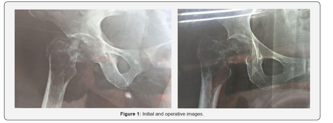
General notes on metastatic breast cancer. Breast cancer is the most frequent type in women (skin cancer is excluded), it is the second most frequent cause of cancer death in women. Metastatic breast cancer will generate most of such deaths. About 5% of women have metastatic breast cancer when they are first diagnosed. More research is needed to determine how many people with non-metastatic breast cancer subsequently have metastatic breast cancer. The 5-year survival rate indicates the percentage of people who survive at least 5 years after the cancer is detected [1]. The 5-year survival rate of women with metastatic breast cancer is 27%. The 5-year survival rate of men with metastatic breast cancer is 20%. Breast cancer may be limited to the breast and / or nearby lymph node regions, which we call early stage or locally advanced cancer (Figures 1 & 2).
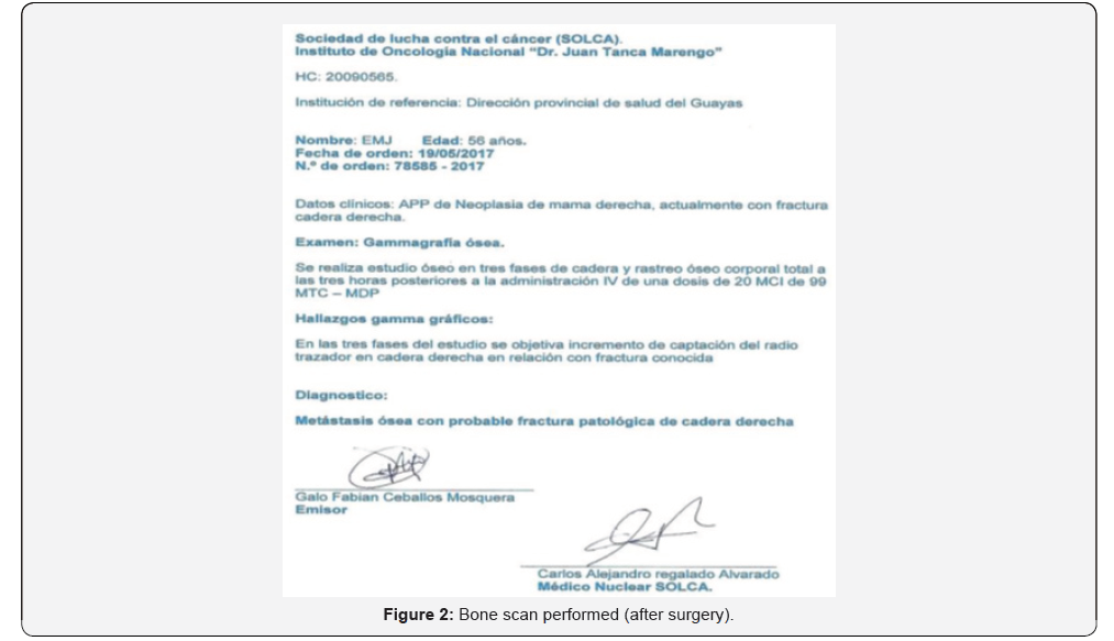
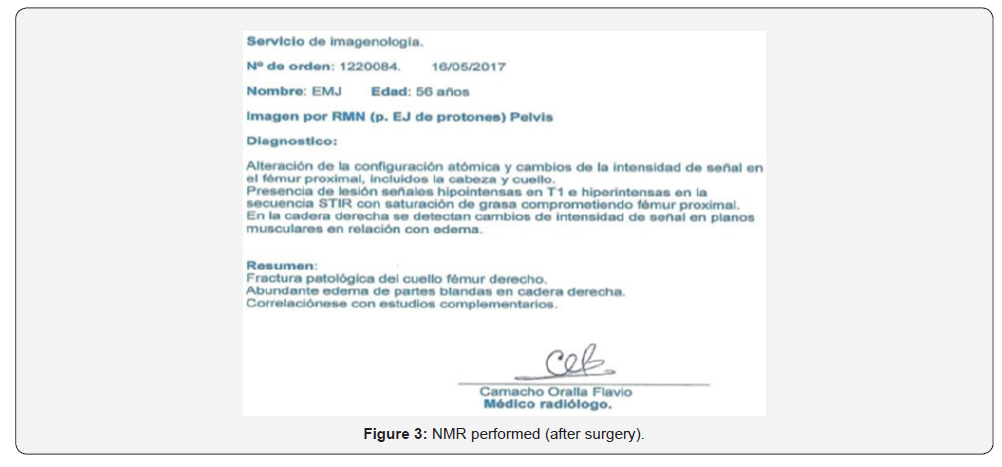
Conversely, when breast cancer spreads to a distant area from which it originates, it is defined as „metastatic“ cancer [2]. Denominating to the area of dissemination „metastasis“, either one or several areas; It most often spreads to bones, liver, lungs, or the brain. By criterion of Prof Martin (president of SEOM), which considers the term „metastasis“ when the physical continuity between the primary tumor and the new outbreaks of advanced pathology is lost, for this reason it also includes the extension of cancer to the drainage ganglia. In the same sense it considers that the lethality of the metastasis is not immediate and in multiple occasions the metastasis takes time in releasing the most harmful substance If the cancer has spread to several places (Figures 3-5). The disease is called stage IV metastatic breast cancer [3]. Even after breast cancer spreads, it is still called the area that gave rise to it (breast), „primary tumor.“
Metastatic breast cancer can develop when cancer cells in the breast separate from the primary tumor and enter the bloodstream or lymphatic system. Both systems transport bodily fluids, by which cancer cells can move away from the original tumor, settle and grow in a different part of the body and form new tumors. Sometimes, the first diagnosis of breast cancer occurs when it has already spread, called „de novo“ metastatic breast cancer. Most often, metastatic breast cancer is diagnosed after a person has previously received treatment for breast cancer at a more initial (non-metastatic) stage. Which they call „distant recurrence“ or „metastatic recurrence.“


Types
Several types are known, any of them can present metastasis, most of them start in the ducts or lobes,
being called ductal carcinomas or lobular carcinomas:
i. Ductal carcinoma: they originate in the cells that internally cover the milk ducts and make up most breast cancers.
ii. Lobular carcinoma: originates in the lobes.
The less common types of breast cancers include: Spinal, Mucinous, Tubular, Metaplastic, Papillary. Inflammatory breast cancer is a rapidly growing cancer that accounts for approximately 1% to 5% of all breast cancer cases. Paget‘s disease is a type of cancer that starts in the nipple ducts. Breast cancer can develop in men and women. However, breast cancer in men is rare and accounts for less than 1% of all breast cancers.
Other Current Types
a. Negative tripe, represents 15% of the cases, is hereditary, very aggressive and proliferative, with a level of survival not exceeding one year, for which unfortunately there are currently no therapeutic targets and in which only the chemotherapy.
b. Hormono dependent or ER positive, represents 67% of cases, The HER-2 expresses different prognoses, for which hormone therapy is used to delay the use of chemotherapy.
c. -HER 2 - positive, represents 18% of the cases and thanks to advances in molecular biology offers greater current therapeutic possibilities, modifying its course or almost certain death sentence for those affected by this type of cancer.
Treatment Approaches
The comprehensive treatment of the patient is carried out by a multidisciplinary team and within its therapeutic options we include: Surgery and systemic therapies (hormonal, chemotherapy, targeted therapy) Surgery: Surgical treatment is usually recommended to remove a metastatic tumor that generates
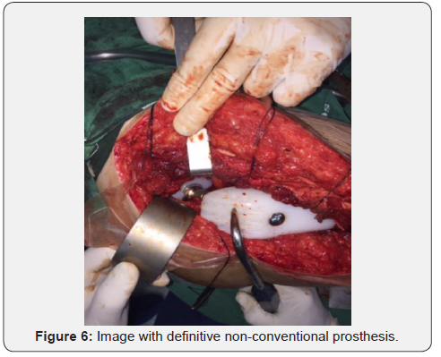
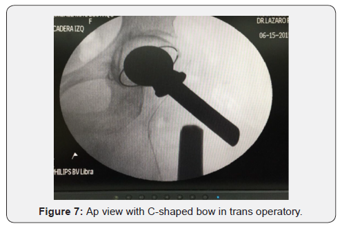
Symptoms or discomfort, including pathological fractures for stabilization and improve the quality of life of the patient, however, complementary examinations continue their clinical course. Surgery can be used alone or associated with radiation therapy (Figures 6 & 7).
The types of systemic therapies that are used for metastatic breast cancer include: Hormone therapy known as endocrine therapy, it is an effective treatment for tumors positive for estrogen or progesterone. Its goal is to reduce the levels of estrogen and progesterone in the body or prevent these hormones from reaching the cancer cells by preventing their use for their growth. They include: Tamoxifen, aromatase inhibitors, ovarian suppression, Fulvestrant, other therapies.
Chemotherapy
Using medications (one or combinations) in various programs, on a weekly, biweekly, 21 days or monthly basis, with the aim of destroying cancer cells and preventing their growth and division. There are several drugs used with uses and doses according to programs, including: Capecitabine, Carboplatin, Cisplatin, Cyclophosphamide, Docetaxel, Doxorubicin, Epirubicin, Eribulin, Fluoracil, Gemcabene, Iritecan, Ixabepilone, Metrotexate, Paclitaxel, Vinorelbine. Chemotherapy can be combined with therapies directed to the recipient.
Targeted Therapy
Treatment is targeted to cancer-specific genes or proteins, or tissue conditions that contribute to cancer growth and survival [4]. These treatments are very focused, and their function is different from chemotherapy or hormone therapy. This type of treatment blocks the growth and spread of cancer cells and, at the same time, limits damage to healthy cells.
Different types of targeted therapies vary in how they attack Cancer Cells
Monoclonal antibodies recognize a specific protein and, through their binding, act on cancer cells, without affecting cells that do not have that protein. Monoclonal antibodies used for breast cancer include trastuzumab, pertuzumab, and TDM- 1. Small molecule inhibitors are drugs made to attack specific parts of cancerous tissue (receptors or enzymes) or to different parts of a cell chasing multiple targets. The most used for breast cancer include; Lapatinib, Palbociclib, Ribociclib and Everolimus, Trastuzumab, Pertuzumab, Ado trazumab, Lapatinib, others [5].
Radiotherapy: Using X-rays (external beam radiation therapy) or other particles with high potency to eliminate cancer cells, to reduce the size of the tumor or to slow down its growth, as well as to treat the symptoms of the tumor.
Types: Total brain radiotherapy; Stereotactic radiotherapy with a single high dose of radiation applied directly to the tumor site and fractionated stereotactic radiotherapy.
About A Case
E.E.J.M, 57 years old, HC: XX. ID: Pathological fracture of the right hip referred to the social insurance center, Clinic of the Pacific where he remained hospitalized for two weeks, for noncoverage of social insurance derive it to the public health system coordinated by Zone eight and send it to the hospital of specialties of Guayaquil where you enter for your study and treatment. APP: Right breast adenocarcinoma with total mastectomy year 2013, Stage IA (T0N1M0) (Figures 8 & 9).


Discussion
Upon physical examination of the patient, we observed external rotation with shortening 1.8 cm of the right lower extremity, predominance of the right hip and thigh, functional impotence due to pain on palpating and moving the right hip, increase in local volume in the trochanteric region of the femur straight. We indicate preoperative protocol to perform tumor biopsy, in addition to general hematology, biochemistry, ESR, and PCR tests. LDH, Phosphatases, calcium, phosphorus, tumor markers, imaging tests, simple hip Rx, bone pelvis, thorax, bone scintigraphy; T.A.C of thorax and abdomen for tumor staging.
In the anteroposterior Rx of the right hip at a real distance, we observed an extracapsular pathological fracture, with a main intertrochanteric line, with a wide osteolytic image that extends from the femoral neck to the whole trochanter mass, and extends 2.5 cm sub trochanteric , with 6 cm of proximal femur with tumor image. The complements showed haemoglobin at 7.3 g/L, phosphatase at 286 U/L, other tests within normal ranges, negative chest and abdomen T.A.C, bone scintigraphy, and hip CT showed evidence of metastatic fracture. Three units of red blood cell concentrate compatible with group and Rh factor and subsequent control are transfused, with a result of haemoglobin at 12 g/l; We prepare the patient for a surgical first time by tumor biopsy.
We perform incisional tumor bone biopsy; simultaneous samples are sent to pathological anatomy laboratories of Guayaquil hospital and SOLCA hospital. (Society to fight against cancer). Ten days later we received a report by pathologists diagnosed with metastatic adenocarcinoma, histology with fibrous stroma, cordons and large cell masses. The case is discussed in Medical Staff integrated by Internal Medicine, Anesthesiology, Clinical Oncology, Traumatology and by consensus of the Medical Staff after clinical discussion, considering all the aspects inherent to the patient, their age, current physical state and their metastatic tumor disease. decided to perform a wide surgical resection that included ten to eleven cm of proximal femur, due to the magnitude of the tumor that covered the whole area of the hip, including trochanters in about seven - eight cm of proximal femur, associated with extracapsular pathological fracture with displacement in varus and with shortening of the lower limb. The patient and her family are informed about everything inherent to the procedure of wide tumor resection and the placement of a total non-conventional prosthesis of the cemented hip. The patient and family members accept the surgical proposal and sign informed consent of surgery Control and preoperative valuations of the patient by anesthesiology, internal medicine and cardiology with report of apt for the surgery posed.
We programmed and performed surgery with wide surgical resection, using the Smith Petersen approach
for the right hip, we found intra-operatively that the entire proximal femur was fractured in several fragments of different sizes and invaded by bone tumor, eleven cm of proximal femur were resected and sent to pathological anatomy laboratory; Unconventional total cemented hip prosthesis, control rx by intensifier of images in satisfactory trans operative, mobility and stability of the satisfactory prosthesis with recovery of the length of the right lower extremity compared with left extremity. She remains hospitalized for six days after surgery. 24 hours after surgery, early ambulation begins with the support of forearm crutches, removal of drains at 72 hours with good postsurgical evolution; Hospital discharge is given with treatment and interconsultation with hospital oncologists for neoadjuvant chemotherapy and follow-up by external consultation of oncology and traumatology [6-9].
The patient and her family express their well-being and gratitude for the care and treatment received from the pathological fracture of the hip, as well as their prompt ambulation and post-operative physical autonomy; We follow up by traumatology at 15 days, at month, three months, six months and eleven months after surgery, with favorable evolution, radiographic control and complementary tests in normal ranges. The patient consults us at one year and six months after surgery for pain in the left hip and in the spine. Prior to clinical examination, we performed rx of pain areas and verified new scattered tumor images in shoulders, thoracolumbar spine, pelvis, left hip, among others, all related to stage IV metastasis. The patient is evaluated by a team of oncologists, oncoradiology and a new cycle of chemotherapy and symptom control is agreed upon [10-12].
Conclusion
We performed extensive surgical resection, based on Enneking 1980 and in the Capanacci classification for surgical treatment the reference patient was in a stage III of Capanacci; Whose author proposes the wide resection associated with the reconstruction of the affected area. It was found intraoperatively that the entire proximal femur was fractured with several fragments of different sizes and completely invaded by tumor, eleven cm of proximal femur were resected with tumor safety margin and total non-conventional prosthesis of cemented hip was placed, interconsultation was performed with hospital oncologists for post-surgical neoadjuvant chemotherapy and follow-up by trauma and oncology external consultation. With immediate incorporation and ambulation after the implantation of the hip prosthesis, we follow up by ambulatory trauma consultation confirming favorable evolution.
One year and six months after tumor resection due to metastatic fracture and placement of the right hip prosthesis, consult for contralateral pain in the hip and spine, we performed new imaging studies and found new multiple metastases.
Conflicts of Interest
In this article there are no conflicts of interest.
References
- American Society of Clinical Oncology (ASCO). Editorial board for cancer.
- Barnadas A, Algara M, Cordoba O, Casas A, Gonzalez M, et al. (2018) Recommendations for the follow-up care of female breast cancer survivors: a guideline of the Spanish Society of Medical Oncology (SEOM), Spanish Society of General Medicine (SEMERGEN), Spanish Society for Family and Community Medicine (SEMFYC), Spanish Society for General and Family Physicians (SEMG), Spanish Society of Obstetrics and Gynecology (SEGO), Spanish Society of Radiation Oncology (SEOR), Spanish Society of Senology and Breast Pathology (SESPM), and Spanish Society of Cardiology (SEC). Clin Transl Oncol 20(6): 687-694.
- Breast metastasis, EFE Health (2017).
- Agency for Research on Cancer (IARC). Globocan International 2008.
- Agudelo BM (2011) Levels, trends and impact of mortality from breast cancer in Costa Rica by provinces, 2000-2009. Population and Health in Mesoamerica. 9(1): 1-14.
- American Cancer Society (2018) Breast Cancer Facts & figures 2011-2012. Atlanta: American Cancer Society, Inc. p. 1-8.
- American Cancer Society. Data and Statistics on Cancer among Hispanics/Latinos 2009-2011.
- Atlanta: American Cancer Society, Inc (2009) Cancer Facts and Statistics. American Cancer Society, US.
- Aparicio LI A, Morera SM (2009) Geographic patterns of incidence and avoidable mortality due to breast cancer in Costa Rica. Rev Costarr Salud Pública.
- Ortiz BA, Vargas AR, Muñoz LG (2005) Incidence and Mortality of Cancer in Costa Rica 1990-2003. Ministry of Health, San José, Costa Rica.
- Parker JS, Mullins M, Cheang MC, Leung S, Voduc D, et al. (2009) Supervised risk predictor of breast cancer based on intrinsic subtypes. J Clin Oncol 27(8): 1160-1167.
- Perou CM, Sørlie T, Eisen MB, van de Rijn M, Jeffrey SS, et al. (2000) Molecular portraits of human breast tumors. Nature 406 (6797): 747-752.


