Correlation between Morfologic and Functional Status in Patients with Rheumatoid Arthritis
Boris Prodanovic1,2*, Snjezana Novakovic-Bursac¹, Goran Talic¹,2, Sandra Trivunovic¹, Daliborka Golic¹ and Nenad Prodanovic2,3
1Institute for Physical Medicine and Rehabilitation, Dr Miroslav Zotović Hospital, Bosnia and Herzegovina
2Faculty of Medicine, University of Banja Luka, Save Mrkalja 14, Bosnia and Herzegovina
2University Clinical Center, Republic of Srpska, Dvanaest beba, Bosnia and Herzegovina
Submission: February 25, 2019;Published: March 07, 2019
*Corresponding author: Boris Prodanovic, Institute for Physical Medicine and Rehabilitation, Dr Miroslav Zotović Hospital, Slatinka 11, Banja Luka, Bosnia and Herzegovina
How to cite this article: Boris P, Nenad P, Goran T, Sandra T, Daliborka G, et al. Correlation between Morfologic and Functional Status in Patients with Rheumatoid Arthritis. Ortho & Rheum Open Access J 2019; 13(5): 555873. DOI: 10.19080/OROAJ.2019.13.555873
Abstract
Objective: To determine the correlation between clinical tests for disease activity assessment and MSS (radiological activity), and to establish the difference in functional status of patients on admission and discharge from stationary rehabilitation compared to the radiological stadium of the disease.
Patients and Methods: The study was carried out as a prospective study during the period from May to July 2017, which included 50 RA patients who have spent a stationary physical therapy at the Rheumatology Division of the Institute for Physical Medicine and Rehabilitation “Dr Miroslav Zotovic” Banjaluka. Testing was performed by the method HAQ questionnaire, DAS28 ESR/CRP, CDAI, SDAI tests and modified Sharp score (MSS).
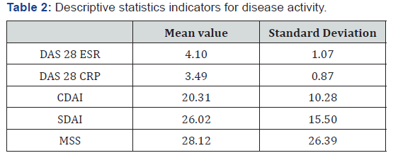
Results: In the population there were 92% women, and 8% of men, the average age was 62.54 +9,44 and the duration of disease 9.41 +7,10 years. The mean values of the tests for the evaluation of the disease activity were: DAS28 ESR 4,10±1,07, DAS28 CRP 3.49±0,87 , CDAI 20.31±10,28, SDAI 26.02±15,51 . MSS average was 28.12±26,39. The average HAQ score on admission was 1.12±0,49 , and on discharge it was 0.94±0,49. No significant correlation between radiological and clinical tests for assessment disease activity, was found. Significant positive correlation of radiological stages with HAQ on admission and at discharge did not result in significant correlation with their divergence.
Conclusion: Clinical tests for RA activity assessment are not necessarily in correlation with radiological activity, and functional recovery at the admission and discharge from stationary rehabilitation were not accompanied by radiological stadiums.
Keywords: Rheumatoid arthtitis; Clinical disease activity; Radiological activity; Functional recovery
Introduction
Rheumatoid arthritis (RA) is an autoimmune disease of unknown etiology characterized by symmetrical peripheral polyarthritis. Inflammatory process leads to bone and cartilage destruction, deformity and functional deficiency [1,2]. Evolution of disease is hardly predictable, with periods of remission and exacerbation. In approximately 80% of the patient’s disease starts gradually, and most commonly clinically occurs after infection, obstetric delivery, physical or psychological trauma [3,4]. The expected consequence of longer duration of RA is the creation of a functional deficit. More than 80% of patients after 10 to 12 years of RA duration, have functional deficiencies and signs of joint injury. About 15% of patients have a short-term inflammatory process that ends with no residual significant functional deficit [3,5-6]. The results of epidemiological studies suggest that RA prevalence ranges from 0.4% to 1.9% worldwide, and it is most commonly around 1% [7-8].
Key symptoms and signs of RA are intense pain and functional disability of different degree, which have significant influence on the life quality of the diseased [9]. Main goal of the treatment of patients with RA is improvement of life quality. The treatment consists of prevention and control of articular lesions, prevention and decrease of functional deficit and treatment of pain [2]. In Bosnia and Herzegovina, in pharmacological therapy, the most common in use of the so called “basic antirheumatic therapy”, which implies the use of non-steroid, anti-inflammatory drugs (NSAIDs), drugs that modify the course of the disease – disease-modifying anti-rheumatic drugs (DMARDs), cytostatics and corticosteroids in different combinations.
Nowadays, in clinical practice, tests most frequently used for disease activity assessment, are: Disease Activity Score 28 (DAS 28); Clinical Disease Activity Index (CDAI); Simplified Disease Activity Index (SDAI); Health Assessment Questionnaire (HAQ). Besides listed tests for disease activity assessment, radiological disease activity assessment is widely used in clinical studies, which is calculated using “modified Sharp score” (MSS) [10]. Goal of this research was to determine the correlation between clinical tests for disease activity assessment and MSS (radiological activity), and to establish the existence of difference in functional status of patients on admission and discharge from stationary rehabilitation compared to the radiological stadium of the disease. Expected results are: Clinical tests for disease activity assessment are in positive correlation with radiological activity of RA analysed by MSS [10]; Radiological stadium of the RA has significant influence on functional recovery of the patients after the rehabilitation [10].
Patients and Methods
The research was conducted as a prospective study in period from May to July 2017, and it was approved by Ethics Committee of the Institute for Physical Medicine and Rehabilitation “Dr Miroslav Zotovic” in Banjaluka. Research was conducted at the Department for Rehabilitation of Patients with Rheumatological, Post-operative and Post-traumatic conditions of the Institute for Physical Medicine and Rehabilitation “Dr Miroslav Zotovic” in Banjaluka. The sample comprised patients of both genders, of different age range, regardless of presence or absence of comorbidity.
Inclusion criteria were:
RA diagnosed based on American Rheumatological Society criteria revised in 1987;
Basic anti-rheumatic therapy as foundation of medicamentous therapy;
Adequate stationary physical treatment (≥15 therapy days)
Non-inclusion criteria was:
Biological therapy as basis of the treatment Exclusion criteria was:
Interrupted stationary physical treatment, because of any reasons.
In order to collect as much important data for research as possible, Anamnestic sheet and review form were used, which were made based on earlier relevant research and information from the literature. The research has involved basic anamnestic data (first name and last name, age, sex), data about comorbidities, duration of RA, as well as the way of its treatment. Patients have assessed their condition on visual analogue scale (VAS), in order to calculate the value of the tests for disease activity assessment. Main researcher has assessed the condition of the patient based on clinical examination, as well as the number of painful and swollen joints. After that, values of the tests for disease activity assessment have been calculated by using the following formulas:
DAS 28 ESR= 0.56 x √(TJC28) + 0.28 x √(SJC28) + 0.70 x log (ESR) + 0.014 x GH – where the TJC stands for tender joint count, SJC for swollen joint count, ESR for erythrocyte sedimentation rate and GH for the patient´s global health estimated by using VAS;
DAS 28 CRP= 0.56 x √(TJC28) + 0.28 x √(SJC28) + 0.36 x log (CRP + 1) + 0.014 x GH where CRP stands for C reactive protein and GH for global health;
CDAI= SJC28 + TJC28 + PtGA + EGA + CRP – where PtGA stands for patient´s global health estimated by using VAS and the EGA for estimator global assessment
SDAI= SJC28 + TJC28 + PtGA + EGA [10].
Examination of patients’ functional abilities was performed by using the HAQ questionnaire, which has 20 questions divided in 8 functional categories. Answers to the questions about possibilities of doing certain functions are rated with: 0 – without difficulties; 1 – with small difficulties; 2 – with large difficulties; 3 – inability to perform. The total mark obtained is divided by 20 and HAQ category index is obtained, which represents a twodigit decimal number. HAQ index has 3 gradations: 0-1: lightly decreased functions in everyday life; 1-2: shows more serious damage in all segments; 2-3: complete disability with necessity of other person’s help. The HAQ score is calculated during at admittance to stationary physical treatment and after the last conducted physical therapy [10].
Biochemical blood analysis, which means measuring of the value of CRP, was conducted by using standardised methods on Roche Cobas C11 apparatus. Besides the mentioned, erythrocyte sedimentation value reading has also been done within the first hour. For the needs of determining the radiological stadium and radiological activity of RA using MSS, roentgengraphies of fist and hand joints in anteroposterior projection and machine profile have been done. MSS has been calculated by assessing involvement of joint spaces on 13 characteristic joints of both fists. If the joint wasn’t affected by the process, value 0 has been assigned; value 0,5 if it’s unclear if the joint is constricted; value 1 if the joint crack is mildly focally narrowed; 1,5 if it is still light joint involvement, but slightly bigger than focal; value 2 if the joint is moderately affected, but 50% of the joint’s crack is still intact; value 2.5 if it is moderate joint affection, but slightly “worse radiological manifestation”; value 3 if the process has spread to more than 50% of joint’s crack, while the value 3.5 has been given if the whole joint’s crack has been narrowed and modified; value 4 has been added in cases of joint ankylosis [10].
Radiological (morphological) disease stadium was determined by using Steinbrocker’s classification in 4 stages. First stage indicated early radiological changes like periarticular swelling of the spindle shaped soft tissue, most commonly localised on the third and fourth proximal interphalangeal hand joint. In the joint ends, juxta articular hypomineralisation of the bones and light constriction of the joint’s spaces can be seen in the first stage. In this stage, small subcortical cysts can also be seen, most commonly on distal ends of the first phalanges and in ulna’s styloid process. In the second stage, constrictions of articular spaces are much more expressed, shallow dents appear as well. In the third stage, dents with subluxation or joint luxation are present. In the fourth stage, fibrotic changes and later bone ankylosis, appear as well [10]. Statistical data interpretation was performed by using the licensed SPSS 22 software package. Statistical significance was set at p≤0. 05. Normality of data distribution was assessed by means of the using Kolmogorov-Smirnov test. Since only age range and HAQ score values on admittance and release from hospital had normal data distribution, nonparametric tests were used for further analysis. Correlation degree was estimated by calculating the Spearman rho coefficient.
Results


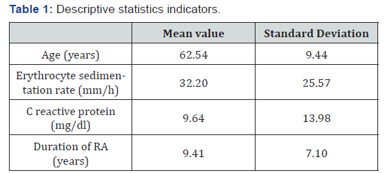
A total of 50 patients out of whom 46 or 92% were women, and 4 or 8% were men, were included in this clinical trial. Table 1 shows arithmetic middles (AM) and standard deviations of age range, SE value, CRP, as well as duration of RA. Descriptive statistics indicators, Table 2 shows arithmetic means and standard deviations of the values for RA activity assessment: DAS 28 SE, DAS 28 CRP, CDAI, SDAI and MSS. Descriptive statistics indicators for disease activity. In Table 3, values of the test for HAQ functional ability assessment with arithmetic means and standard deviations at the beginning and at the end of the stationary physical treatment and rehabilitation are shown. Descriptive statistics indicators for HAQ.

Negative correlation between the tests for disease activity assessment (DAS28 SE, DAS28 CRP, CDAI and SDAI) and MSS was found, but it wasn’t statistically significant. At the same time, value of listed clinical parameters for disease activity assessment (DAS28 SE, DAS28 CRP, CDAI and SDAI) are in significant mutual positive correlation.

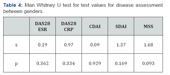
Values of SE and CRP are in strong positive correlation which is statistically significant (rho=0.645, p=0.000), and none of these parameters is in correlation with disease duration. By using t-test for independent samples, it was found that the male patients were significantly older (t=2.619, p=0.012). By using Mann Whitney U test, it has been found that the values of CRP are significantly higher in male patients (z=2,54, p=0,011), while the disease duration values and SE, statistically significant differences were not found Table 4. Mann Whitney U test for test values for disease assessment between genders Statistically significant differences has not been found for HAQ values on admission and discharge between genders (HAQ reception z=1,11, p=0,267; HAQ release z=0,412, p=0,681). Kruskal Wallis test showed that there was no statistically significant difference in functional recovery after stationary rehabilitation with patients belonging to different radiological stadiums (χ=2.056, p=0.561).
By correlating radiological stadiums of RA with gender, age range, duration of the disease, and values of SE and CRP, positive statistically significant correlation with age range (rho=0,344, p=0,015) and borderline positive correlation with duration of rheumatoid arthritis (rho=0.266, p=0.059) was obtained Table 5. shows correlations between radiological stadium with tests for disease activity assessment. Correlation of radiological stadium with RA activity assessment tests Significant positive connection of radiological stadiums with HAQ on admission and discharge (HAQ reception rho=0.3‚ p=0.03; HAQ release rho=0.295, p=0.04) did not result in significant connection with their difference (DeltaHAQ rho=0,125; p=0,388). Using ANOVA test, statistically significant difference in age range with patients belonging to different radiological stadiums of RA (F=4.44, p=0.008) has been established.

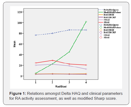
Statistically significant difference in SE values (χ=4.873, p=0.181) i CRP-a (χ=1.491, p=0.684) has not been established. Kruskal Wallis test results for difference comparison among disease activity assessment test values among patients belonging to different radiological stadiums of the RA, are shown in Table 6. Differences in values of disease activity assessment among patients belonging to different radiological stadiums of RA. Figure 1 shows relations amongst Delta HAQ and clinical parameters for RA activity assessment, as well as modified Sharp score. Values of radiological stadium are located on abscisse, while test value is found on the ordinate (for Delta HAQ procentually).


Discussion
Rheumatoid arthritis is systemical, auto-immune disease that leads to deformation and destruction of joints (mostly tiny hand and foot joints), therefore compromising hand functions and patient‘s walk, as well as patient‘s work ability. According to data from the literature [4], women have RA three to five times more often than men, with most common incidence of disease appearing between third and seventh decade of life. Gender distribution in this study was like that, and women were considerably more represented in comparison to men (92%: 8%), while the average age range of patients was 62.54, which is in accordance with literature data. Average duration of RA was 9.41 years.
Rheumatoid arthritis is systemical, auto-immune disease that leads to deformation and destruction of joints (mostly tiny hand and foot joints), therefore compromising hand functions and patient‘s walk, as well as patient‘s work ability. According to data from the literature [4], women have RA three to five times more often than men, with most common incidence of disease appearing between third and seventh decade of life. Gender distribution in this study was like that, and women were considerably more represented in comparison to men (92%: 8%), while the average age range of patients was 62.54, which is in accordance with literature data. Average duration of RA was 9.41 years. was used as significant minimal difference of HAQ score on the admission on stationary physical treatment and after the discharge from the treatment, obtained results would imply that 14 patients or 28%, have established the mentioned minimal difference of HAQ score, which would furthermore rate the applied physical treatment as successful [12].
By analysing the anatomic stadium, according to Steinbrocker, the majority of patients were in II stadium (24 or 48%), while the number of patients in the I stadium (12 or 24%) or in the III stadium (11 or 22%) was almost equal. No significant differences were found in the values of HAQ score on admission and release from stationary physical treatment among the patients who belonged to different radiological stadiums of RA. In the study of cases published by Todorovic-Tomasevic [13,14], majority of patients who were examined belonged to II and III stadium of the disease according to Steinbrocker (80%), which our study did not confirm. These sorts of differences could be related to total duration of the main disease of our patients. In the mentioned study, no statistically important correlation of anatomical stadium according to Steinbrocker, and the value of HAQ score was found, which was also confirmed in our studies. Functional ability examination results using the HAQ questionnaire survey in the mentioned research, showed that the average values of HAQ score are 1,25±0,7. Weak monotone direct correlation bond between the duration of disease and functional status assessed by HAQ score (r= -0.088, p= 0.501) was obtained.
In our research, we included large number of patients and obtained an average value of HAQ score on reception (HAQ score on reception is compared because the patients of the mentioned group of authors did not underwent stationary physical treatment), which is the same gradation level as the one in Tomasevic-Todorovic and co-workers’ research. As for the significance of the correlation between HAQ reception score and disease duration, our research obtained similar results as the study mentioned before. Results of our study imply that higher radiological stadium accompanies higher age range and longer RA duration, which was established by Todorovic-Tomasevic and co-workers’ survey.
MSS is in negative correlation with clinical parameters for RA activity assessment. Our results are in accordance with Kunihiro Yamaoka results [15], who obtained results which implied the non-existence of correlation between MSS and clinical parameters for RA activity assessment (DAS28 ESR, SDAI, CDAI). The study lasted for a year and was conducted on 37 patients. These results can be related to a certain degree of subjectivity of RA activity assessment clinical tests. Difference between our study and Kunihiro Yamaoka study is in patients’ main therapy. Even though the patients were treated with more potent drugs (JAK inhibitors) in the mentioned study, it did not result in a better correlation between clinical and radiological disease activity.
Research that was conducted by a group of authors from USA and Norway [16] who were evaluating measuring instruments used in everyday rheumatological practice, and which refer to functional status and disease activity, and degree of radiological progress, also did not found any correlation between MSS and disease activity assessment clinical tests. Their sample had many similar features to ours. The RA duration and radiological disease activity measured with MSS, are in weak positive correlation, which is not statistically significant, while it is in positive statistically significant correlation with HAQ scores on admission and at discharge. These sorts of results speak in favour of the fact that the longer RA duration is followed by higher radiological disease activity, which leads to poor functional status measured by HAQ score, as well as poor postrehabilitation recovery [17]. Higher radiological stadium relates to higher values of ModSharp and HAQ reception score, which did not result in HAQ score’s positive connection between admission and release [18].
Conclusion
Clinical tests for RA activity assessment are not necessarily in correlation with RA radiological activity degrees, measured by MSS. No statistically significant differences in functional recovery of the patients with RA who belong to different radiological stadiums according to Steinbrocker, after stationary rehabilitation was conducted, were found.
References
- Fries GF, Ramey DR (1997) Arthritis specific” global health analogue scale assess “generic” health related quality of life in patient with rheumatoid arthritis. J Rheumatol 24(9): 1697-1702.
- Ward MM, Leigh JP (1993) The relative importance of pain and functional disability to patient with rheumatoid arthritis. J Rheumatol 20: 1494-1499.
- Yelin E, Meenan RF, Nevitt M, Epstein WV (1980) Work disability in rheumatoid arthritis: effects of disease, social and work factors. Ann Intern Med 93(4): 551-556.
- Wolfe F, Mitchell DM, Sibley JT, Fries JF, Bloch DA, et al. (1994) The mortality of rheumatoid arthritis. Arthritis Rheum 37(4): 481-494.
- Lipsky PE (2001) Rheumatoid arthritis. In: Braunwald E, Fauci AS, Kasper DL, Hauser SL, Longo DL, Jameson JL, eds. Harrison's Principles of Internal Medicine, (15th edn), McGraw-Hill, New York, USA, pp. 1928-1937.
- Furu M, Hashimoto M, Ito H, Fuji T, Terao C, et al. (2014) Discordance and accordance between patient's and physician's assessments in rheumatoid arthritis. Scand J Rheumatol 43(4): 291-295.
- Studenic P, Radner H, Smolen JS, Aletaha D (2012) Discrepancies between patients and physicians in their perception of rheumatoid arthritis disease activity. Arthritis Rheum 64(9): 2814-2823.
- Markenson JA, Koenig AS, Feng JY, Chaudhari S, Zack DJ, et al. (2013) Comparison of physicians ant patient global assessments over time in patients with rheumatoid arthritis. A retrospective analysis from the RADIUS cohort. J Clin Rheumatol 19(6): 317-323.
- Cordingley L, Prajapati R, Plant D, Maskell D, Morgan C, et al. (2014) Impact of psychological factors on subjective disease activity assessment in patients with severe rheumatoid arthritis. Arthritis Care Res 66(6): 861-868.
- Firestein G, Budd R, Gabriel S, McInnes I, O’Dell-Kelley R, et al. (2017) Textbook of Rheumatology. Elsevier, Amsterdam, Netherlands.
- Keystone CE, Pope EJ, Carter Thorne J, Poulin-Costell M, Phan-Chronis K, et al. (2016) Two-year radiographic and clinical outcomes from the Canadian Methotrexate and Etanercept Outcome study in patients with rheumatoid arthritis. Rheumatology 55(2): 327-334.
- Ward MM, Guthrie LC, Dasgupta A (2017) Direct and indirect determinants of the patients global assessment in rheumatoid arthritis: differences by level of disease activity. Arthritis Care Res (Hoboken) 69(3): 323-329.
- Tomašević-Todorović S, Branković S, Bošković K (2009) Procena funkcijskog stanja bolesnika sa reumatoidnim artritisom. Med Pregl 62(5-6): 273-277.
- Tomašević-Todorović S, Branković S Pridružena (2008) oboljenja i reumatoidni artritis. Praxis Medica 36: 65-67.
- Yamaoka K, Kubo S, Sonomoto K, Tanaka Y, Maeshima K (2012) Antirheumatic effect of JAK-inhibitors. Japanese Journal of Clinical Immunology 35(2): 112-117.
- Sonali PD, Chih-Chin L, Heather T, Tabatha N, Fritsa M, et al. (2014) Rheumatoid arthritis quality measures and radiographic progression. Semin Arthritis Reum 44(1): 9-13.
- Tanaka Y, Yamanaka H, Saito K, Iwata S, Miyagawa I, et al. (2012) Structural damages disturb functional improvement in patients with rheumatoid arthritis treated with etanercept. Mod Rheumatol 22(2): 186-194.
- Keystone EC, Haraoui B, Guérette B, Mozaffarian N, Liu S, et al. (2014) Clinical, Functional, and Radiographic Implications of Time to Treatment Response in Patients With Early Rheumatoid Arthritis: a Posthoc Analysis of the PREMIER Study. J Rheumatol 41(2): 235-243.


