A Novel Intramedullary Telescopic Rod for Management of Fracture in Osteogenesis Imperfecta Child
Mohamed Waheeb*1 and Mohamed Almojtaba2
1Orthopedic consultant, Curative governmental organization, Egypt
2Orthopedic department, Saudi German hospital group-Jeddah, Saudi Arabia
Submission: December 18, 2019;Published: January 11, 2019
*Corresponding author: Mohamed Waheeb, Orthopedic consultant, curative governmental organization, Egypt
How to cite this article: Mohamed Waheeb. A Novel Intramedullary Telescopic Rod for Management of Fracture in Osteogenesis Imperfecta Child. Ortho & Rheum Open Access J 2019; 13(3): 555865864.DOI: 10.19080/OROAJ.2019.13.555864
Abstract
Introduction: Manipulating a technique to innovate a new model of instrumentations was the challenge we faced in Osteogenesis Imperfecta case (OI), as until 2018 we did not have in Egypt a standard telescoping nail which makes it possible for us to facilitate the telescoping technique by utilizing innovative rod components that will give us excellent results over two consecutive years.
Patient and methods: I used the humeral nail and took advantage of it as a female rod which was introduced in the antegrade technique through the piriformis fossa. I also used an Ender nail as a male rod which was introduced in retrograde technique via knee arthrotomy, and finally, a new tactic was used to prevent rods dislodgement by using proximal and distal locking of the component’s extremities. Humeral and Ender Nails (HEN) were used to treat fractures in an 8-year-old girl with an OI, suffering from both bilateral femoral fractures and the lack of either physical activity or walking ability. The condition developed into complications of fragile bone in the form of sequential femoral fractures at the age of 4 years.
Results: The girl achieved excellent fractures union, the bones acquired the appropriate length, and a stable highly efficient internal splint for the long bones was successively done. The girl began a good range of physical activities.
Introduction
Osteogenesis imperfecta (OI) is the clinical manifestation of genetic disorder, which often modifies the production of type I collagen. Patients of OI suffer from critical structural skeletal modifications that often progress into fractures and / or deformities involving long bones and axial skeletons deformities. Medical treatment includes the use of bisphosphonates compound which improves bone density and reduces the risk of fracture, however, fracture defects often require surgical correction where intramedullary rods are the most common treatment in these cases. The first intramedullary rod technique developed to treat patients with OI was created by Scofield and Millar [1]. Because of growth, fixed-length rods require frequent [2] revision and this prompted the introduction of the telescopic intramedullary rod (TIMR), by Bailey and Dubow [3]. Here I present a novel telescoping technique utilizing a novel orthopedic instrument cooperation.
Patient and Methods
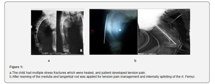
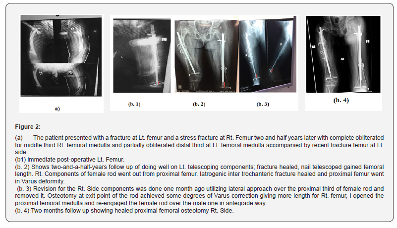
An 8-year-old female child suffers from a previously condition known as OI and has undergone several surgical procedures since she was 4 years old in form of medullary reaming as management of tension pain of closed medulla and tangential rod as management of fracture Rt. Femur which was united Figure 1. Later in 2016, the girl had fracture Lt. Femur on partially obliterated medulla and complete obliterated Rt. Femoral medulla. The standard management for her is the telescoping rod which was not available at the market in Egypt till 2018. The telescoping rod was very expensive and patients cannot financially afford, so we challenged the situation and introduced novel telescoping technique with another available instruments already exist at market. Based on male and female rods for telescoping nail, I decided to use a humeral interlocking nail as a female rod and an Ender nail as a male rod, the proximal screw at humeral nail (female rod) was locked and the ender nail (male rod) was introduced to humeral nail medulla through knee arthrotomy and was locked and secured with trans-hole k-wire. Under general anesthesia, lateral decubitus positions Rt. LL. Up then Lt. LL were adopted. I made use of the lateral approach multiple level osteotomies at Rt. Femur which had deformed and stress fractures, then I opened the medulla with drill bolt 4mm, I used retrograde and antegrade reaming via sequential rigid reamers, Lt. Femur was fractured with no deformity, medulla of Lt. femur was partially obliterated, I opened femoral medulla with the same technique described before for Rt. Femur through lateral approach at the fracture site.While reaming of Rt. Femoral medulla inter-trochanteric fracture iatrogenic occurred, which was fixed with multiple K-wires, multiple bone cracking occurred at bone segments between the osteotomy levels, which were fixed with cerclage wires around the nail Figure 2.


Proximal locking screw at humeral nail (female rod) was inserted. Retrograde insertion of Enders nail (male rod) into medulla of female rod was done through snip arthrotomy of both knees via the intercondylar notches after opening the near cortex using drill bolt 2.5 mm. Distal locking for male rod was done by K-wire passing through the hole of Enders nail (male rod). Patient receive one unit of packed RBCs intraoperative and two units of them post-operative. High above Lt. knee cast and hip Spica for Rt. Femur was made. Patient was discharged from the hospital after two days post-operative in a good health, cast and K-wires were removed from Rt. and Lt. LL. After one and half months, wounds were healed by 1ry intension. Walking exercise and physiotherapy started after three months and fractures healed.
After 6 months, nail at Rt. Femur went out proximally with no fractures and revision for the telescoping components adjustment was done one month ago. Under general anesthesia, patient positioned lateral decubitus Rt. LL. Up, utilizing direct lateral approach, female rod was extracted, osteotomy at proximal 3rd Rt. Femur was done for some Varus correction and retrograde reaming was operated for proximal segment of femur, antegrade re-insertion of female rod over male rod and proximal locking screw was done. Patient recovery from anesthesia was smooth and she discharged from the hospital in a good health with no cast and no blood transfusion.
Results
I had introduced my novel technique for telescoping intramedullary rods at bilateral femoral fractures in a patient complaining of OI. Type IV. Main follow up time is two years. The patient generally did some activities that she could not do before, like starting, walking, standing up depending upon herself and setting at ease. Fracture healing was achieved in average time 3 months and no wound healing complications were observed. Revision for one side resulted due to intra-operative sequelae inter-trochanteric Rt. Femoral fracture which was fixed by K-wires and shortened the surgical procedure time and decreased the blood loss. Revision was done utilizing the same implants which were previously introduced.
Discussion
OI must be clinically treated, using bisphosphonates, to increase bone resistance and mineral density [4]. In moderate and severe cases where children present with excessive fractures or deformities, surgical treatment using intramedullary rods is recommended [5], which aids in correcting these problems and facilitates the possibility of walking in severely affected patients, thus improving their quality of life [6]. Telescopic rods have evident advantages, as they require fewer surgical interventions in response to children’s growth compared with non-telescopic rods because of their ability to adapt to bone growth, serve as an internal template and prevent deformities [7].
Various telescopic rods have been designed. Recent literature recommends the FD rod since studies have reported lower complication rates and a higher permanence period [8]. A distinct characteristic of my Humeral Ender Nail (HEN) components in relation to other described rods is their lower price, less destructive for distal femoral growth plate, proximal and distal locking technique using screw and wire makes lower incidence of dislodgement of its components.
Conclusion
The HEN telescoping components with distal and proximal locking technique using screw and wire are suitable and affordable safe components for surgical management of OI patients older than 5 years old.
References
- Sofield HA, Millar EA (1959) Fragmentation, realignment, and intramedullary rod xation of deformities of the long bones in children. J Bone Joint Surg Am 41(8): 1371-1391.
- Bailey RW (1981) Further clinical experience with the extensible nail. Clin Orthop 159: 171-176.
- Bailey RW, Dubow HI (1963) Studies of longitudinal bone growth resulting in an extensi- ble nail. Surg Forum 14: 455-458.
- Van Dijk FS, Sillence DO (2014) Osteogenesis imperfecta: clinical diagnosis, nomenclature and severity assessment. Am J Med Genet A 164A(6): 1470-1481.
- Lee K, Park MS, Yoo WJ, Chung CY, Choi IH, et al. (2015) Proximal migration of femoral telescopic rod in children with osteogenesis imperfecta. J Pediatr Orthop 35(2): 178-184.
- Escribano-Rey RJ, Duart-Clemente J, Martínez de la Llana O, Beguiristáin-Gúrpide JL (2014) Osteogénesis imperfecta: tratamiento y resultado de una serie de casos. Rev Esp Cir Ortop Traumatol 58(2): 114-119.
- Boutaud B, Laville J (2004) L’embrochage centro-médullaire coulissant dans l’ostéogenèse imparfaite. Rev Chir Orthop Reparatrice Appar Mot 90(4): 304-311.
- Kaiser MM (2012) 31th Meeting of the Pediatric Section of the German Society of Trauma Surgeons (DGU). Eur J Trauma Emerg Surg 38(3): 327-345.


