Bone Density versus Bone Quality as a Predictor of Bone Strength
Jindal M1*, Lakhwani OP2, Kaur Omkar3, Agarwal S2 and Garg Keerty4
1Department of Orthopaedics, Kalpana Chawla Govt Medical College, India
2Department of Orthopaedics, ESI – PGIMSR, India
3Department of Pathology, ESI – PGIMSR, India
4Department of Anaesthesia, Guru Nanak Medical College, India
Submission: April 11, 2018; Published: June 18, 2018
*Corresponding author: Mohit Jindal, Assistant Professor, Department of Orthopaedics, Kalpana Chawla Govt Medical College, Tel: +91 9891904545; Email: mohitjidal2006@gmail.com
How to cite this article: Jindal M, Lakhwani OP, Kaur O, Agarwal S, Garg K. Bone Density versus Bone Quality as a Predictor of Bone Strength. Ortho & Rheum Open Access J 2018; 12(1): 555830. DOI: 10.19080/OROAJ.2018.12.555830.
Abstract
Introduction: Bone mineral density (BMD) is considered to be the standard method to evaluate bone strength and stratify risk of fracture Kanis et al. [1,2]. However, while easy to administer, the limitation of relying on BMD measurements is that they are uniplanar measurements in terms that the three-dimensional structure of bone is reduced to two dimensions and then the BMD values are computed. Also BMD measures only the mineral component of bone and does not give any information regarding its microstructure. Materials engineering has demonstrated that all the material properties including mechanical properties are a function of material microstructure understood as spatial properties and arrangement of constituent basic structural elements; in the case of bone these being the trabeculae. Thus the evaluation of bone microstructure plays important role in defining material properties of bone including the bone Strength.
Materials and Methods: In this study we have used cubical bone specimens obtained from patients undergoing hip replacement procedures and compared their BMD values with those obtained using static Histomorphometry (Trabecular No., Trabecular Seperation, Trabecular Thickness and Bone Volume) to their axial compression strength ( using Yield point at failure measured in Megapascals).
Results: We found a better correlation was found between microstructural parameters and Bone strength {Bone Volume (Spearman correlation 0.9652), Trabecular Number (Spearman correlation 0.7646), Trabecular Seperation (Spearman correlation 0.7541) Trabecular Thickness ( Spearman correlation 0.6726)} as compared to BMD and Bone strength (Spearman correlation 0.3746).
Discussion and Conclusion: While using Bone Mineral Density (BMD) as a sole predictor of bone strength, Bone Quality (microstructural indices) also needs to be considered. Bone Quality appears to be a better predictor of bone strength as compared to Bone density. However Histomorphometric Bone Quality parameters will require invasive procedures and no other in vivo imaging modality has been available to quantitatively measure Bone Microstructure. Bone Density measurement on the contrary is still a rather more available in vivo imaging modality to determine bone strength and still finds its place as a surrogate marker of Bone strength. There is a need of development of newer noninvasive in vivo imaging methods (microstructural computerized tomography) that assess bone strength independent of BMD and utilize measures for Bone Quality and these methods should improve the assessment of fracture risk and response to treatment.
Keywords: Histomorphometry; Bone Quality; Bone Density; BMD
Introduction
Bone mineral density (BMD) is considered to be the standard method to evaluate bone strength and stratify risk of fracture Kanis et al. [1,2]. However, while easy to administer, the limitation of relying on BMD measurements is that BMD measures only the mineral component of bone and does not give any information regarding its microstructure. Furthermore, studies have shown that higher BMD measurements do not directly translate to mechanical properties of bone and fracture risk. Schuit SC, van der Klift M, Weel AE, de Laet CE, Burger H et al. [3] evaluated the fracture incidence and association with bone mineral density in elderly men and women and found that only 34% women and 21% men with non vertebral fracture had BMD in osteoporotic range.
Stacey A Wainwright [4] also in their study only half of elderly women with incidental hip fracture had Bone Mineral Density in osteoporotic range at base line. This single BMD value, while easy for means of comparison between individuals, may not provide sufficient evaluation of bone trabecular architecture. Hence additional data is needed for predicting bone strength. Our study aims at assessing BMD Versus Histomorphometry so as to assess a better predictor of Bone Strength and hence propensity to fracture.
Material and Methods
The structural and mechanical characteristics of Bone have been extensively studied to understand the functional adaptations associated with the properties of biological material. It is known that morphology varies between bones and even within a bone Pugh et al. [5], Raux, et al. [6], Williams & Lewis [7], Whitehouse et al. [8] as do the mechanical properties Brown & Ferguson [9], Ciarelli et al. [10]. While measures of apparent density used as an estimate of mechanical properties does not explain all the variance nor does it account for the structural organization of the trabecular bone, thus rendering this scalar measure inadequate for predicting the material properties Goldstein [11]. Materials engineering has demonstrated that all the material properties including mechanical properties are a function of material microstructure understood as spatial properties and arrangement of constituent basic structural elements, in the case of bone these being the trabeculae. Thus the evaluation of bone microstructure plays important role in defining material properties of bone including the bone Strength.
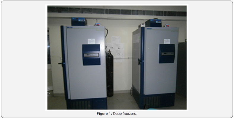
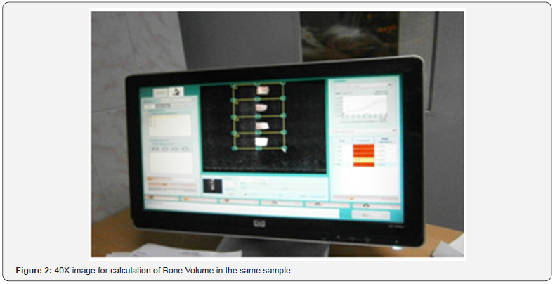
The relationship between trabecular micro-architectural pattern and bone strength has long been proposed. The importance of trabecular arrangement in bone and their significance in bone strength was first observed by Hermann Von Meyer an anatomist and Karl Culmann an engineer (1860). This arrangement of trabeculae ensures the maximum strength with the available bone material. Julius Wolff also based his theory on the similarity of trabecular pattern and maximum stress which formed the main idea in Wolff’s law. Currently, micro-architecture of trabeculae can be measured by several histomorphological parameters like the number of trabeculae defined in a given volume (Tb.N), their mean thickness (Tb. Th), their average separation distance (Tb.Sp) and their threedimensional volume (BV/TV). Histomorphometric evaluation of these microstructural indices has the potential of giving a better estimate of actual bone strength than BMD values Parfitt et al. [12]. Availability of the material to be used for simultaneously measuring the BMD and histomorphometric characterstics and its correlation with mechanical bone strength is a big challenge. Allogenic bone graft procured and processed from bone bank can serve as the substrate for this purpose. Hence this in vitro study is being undertaken using the retrieved femoral heads from living donors processed in bone bank (deep freezing and gamma irradiation) as a substrate to compare the bone mineral density (measured by dual energy x ray absorptiometry) and bone quality indices (measured using histomorphometry) as a better indicator of bone strength (Figures 1 & 2)..
In this Experimental in vitro study carried in 2012 -2014, femoral heads retrieved during surgical hip replacement procedures were made devoid of all soft tissue and cartilage and cut into equal cubical pieces. Exclusion criteria included femoral Head involved in - infection, Paget’s disease, Rheumatoid arthritis, osteoarthritis, neoplastic conditions, pathological fractures and other pathological conditions which were non representative of general population. Each piece was subjected to DEXA Scan, Compression tests and Histomorphometric analysis.
Bone Mineral Density
Areal Bone Mineral Density was measured using the dual energy x ray absorptiometry (DEXA) scan and analyzed.
Biomechanical Compression Testing
Each piece of the bone sample was tested for failure in Compression tests using (model 4482, Instron Corporation) at a constant unilateral deformation rate 0.5 mm/sec. The resultant deformation curve was analysed and yield point calculated (Figures 3-5).
Histomorphometric Analysis
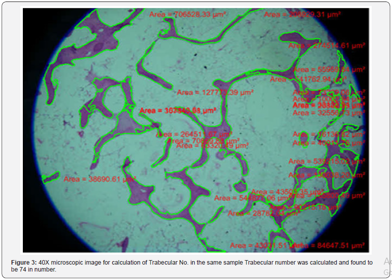
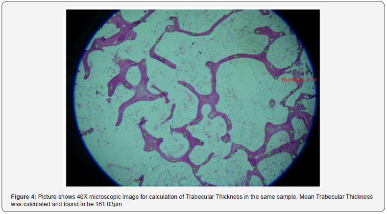
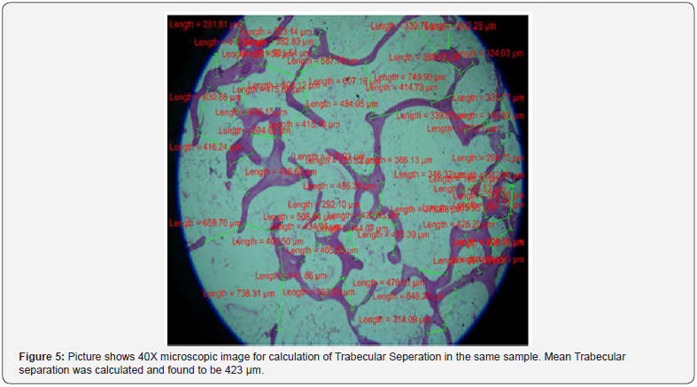
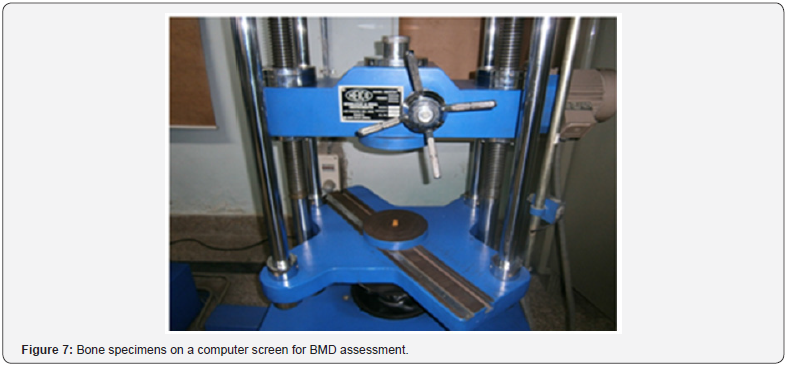
Bone histomorphometry is a quantitative histological examination of bone to access micro structure and predict the biomechanical strength. Preparation The Cancellous bone graft material decalcified using 5% Nitric acid for a period of 10 days (solution changed every 2 days) then dehydrated in ascending grades of alcohol, cleared in xylene and embedded in paraffin. Paraffin sections of 5 micrometre cut using microtome and stained with Hematoxylin & Eosin. The image was processed using NIKON based computerized softwares [40X Magnification] and histomorphometric parameters calculated (Figures 6 & 7).

Parameter undertaken to analyze the structure include
i. Bone volume (Trabecular bone volume/Tissue volume) - Represents the area occupied by the trabeculae Vs the total area of the field. Trabecular bone volume (volume of total cancellous bone) measured and expressed as percentage.
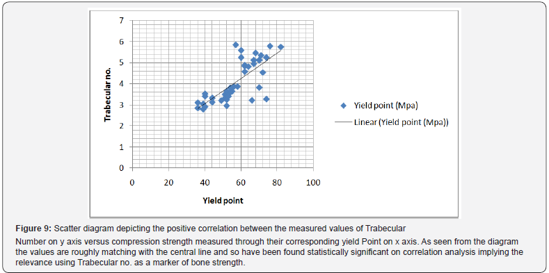
ii. Trabecular number - number of trabeculae per unit area. In this study single high power is taken as unit area.
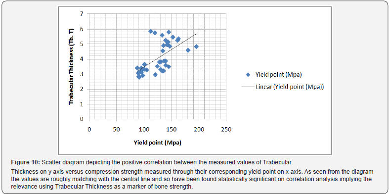
iii. Trabecular Thickness – mean trabecular thickness is measured and analysed.
iv. Trabecular separation –mean of average distance between the two trabeculae is measured and analysed.
Results
Bmd as A Measure Of Bone Strength
BMD of all calculated bone samples are compared to their corresponding compression strength at Yield Point
On comparison the spearman first rank correlation coefficient was 0.3746 corresponding to a p value of 0.0172(<0.05) indicating that a significant positive correlation is present between BMD and compression strength of cancellous bone (Figure 8).

Microstructural Parameters as a Measure of Bone Strength
Trabecular number(Tb.N) versus Compression strength
Analysis was done between all measured values of Trabecular Number and compared to their corresponding compression strength measured through their corresponding yield point. On performing statistical tests, Spearman first rank correlation coefficient was 0.7646 which was found statistically significant. A scatter diagram depicting the correlation is hereby drawn (Figure 9).
Analysis was done between all measured values of Trabecular Thickness and compared to their corresponding measured through their corresponding yield point obtained. On performing statistical tests, Spearman first rank correlation coefficient was 0.6726 which was found statistically significant. A scatter diagram depicting the correlation is hereby drawn (Figure 10).
Trabecular separation Vs Bone strength
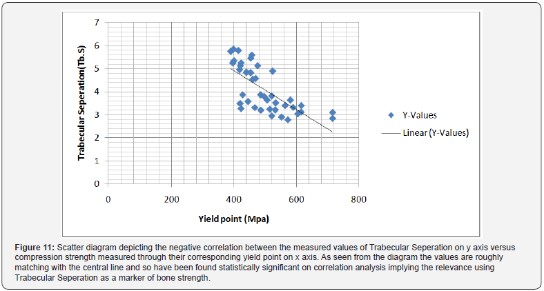
Analysis was done between all measured values of Trabecular Seperation and compared to their corresponding compression strength measured through their corresponding yield point obtained. On performing statistical tests, Spearman first rank correlation coefficient was 0.7541 which was found statistically very significant. A scatter diagram depicting the correlation is hereby drawn (Figure 11).
Bone Volume versus Bone strength
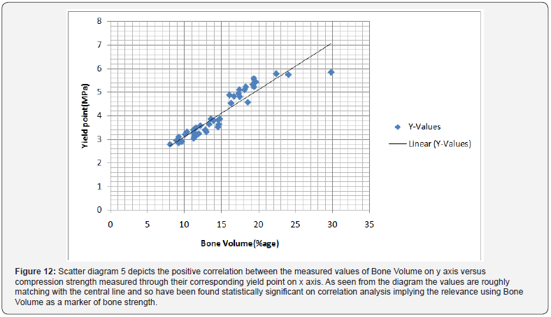
Analysis was done between all measured values of Bone Volume and compared to their corresponding compression strength measured through their corresponding yield point obtained. On performing statistical tests, Spearman first rank correlation coefficient was 0.9652 which was found statistically very significant. A scatter diagram depicting the correlation is hereby drawn (Figure 12).
Discussion
Osteoporosis is characterized by increased skeletal fragility as a result of reduced bone strength. The most severe consequences include bone fractures from unexpected and sudden increased in load (e.g. in accidents) which the bone is not usually accustomed to. The condition in osteoporosis where bone strength is compromised results from the imbalance where bone formation rate is reduced compared to resorption. For every 10% loss of bone mass, the fracture risk doubles. Osteoporosis is becoming a major health concern with the rapidly graying population in many countries. Bone fractures from falls and accidents could result in major costs in hospitalizations and surgery, especially for the elderly where the chances of complete recovery is low and permanent disability is likely.
Measures of Bone Strength
Bone Mineral Density (BMD) measured by Dual Energy X-ray Absorptiometry (DXA), has been traditionally used for the diagnosis of osteoporosis and thereby Bone Strength. However, recent research is indicating that the DXA test is not as reliable a measure of bone strength as originally thought. In fact, several studies have shown that many fractures occur in people with only moderately decreased Bone Mineral Density Wainwright et al. [4] Further, half of all postmenopausal fractures occur in women with Bone Mineral Density test results that indicate that their bone density levels are not low enough for them to be categorized as having osteoporosis Schuit et al. [13]. Quality of bone structure is more important than the density when determining the bone strength. The BMD measured by DXA does not provide information on the bone quality and it does not provide any insight into the architectural structure of bones.
Bone Quality
Bone quality is defined as the sum of the structural and material properties of the trabecular bone. The structural properties of bone refer to the trabecular microstructure. Trabecular or cancellous bone is made up of many interconnected trabeculae which may be composed of narrow rod like trabeculae or the flatter plate-like trabeculae. The more of the thicker plate-like trabeculae present in the structure as compared to the thinner, rod-like trabeculae, the better the quality and strength of the bone. Now, bone quality is measured by various microstructural indices i.e. Trabecular number, Trabecular Seperation, Trabecular Thickness and Bone volume which are measurable by Histomorphometry Parfitt et al. [14], Mc Donell et al. [15], Manal H Moussa et al. [16], Charles S Haworth [17].
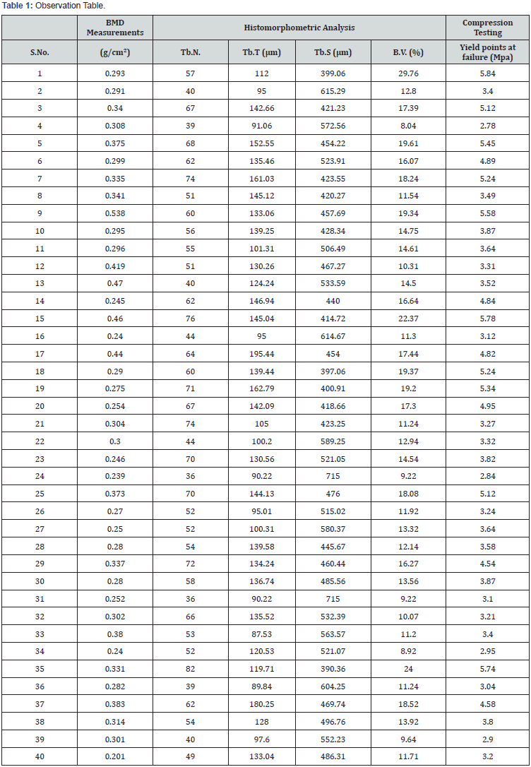
In this study BMD and Microarchitectural indices of Bone quality were used as measures of bone strength (Table 1). A significant correlation was found between bone strength and microstructural indices of bone quality. Although BMD measurements were also found to be significantly associated with Bone strength, the measure of its significance was much less when compared to Bone Quality indices. Among the Bone Quality indices, Bone volume was the most significantly associated with Bone strength followed by Trabecular Number.
The order of significance was; Bone Volume (Spearman correlation 0.9652)>Trabecular Number( Spearman correlation 0.7646)> Trabecular Seperation (Spearman correlation 0.7541)>Trabecular Thickness (Spearman correlation 0.6726)>>BMD(Spearman correlation 0.3746) As expected, microstructural parameters i.e. Bone Quality were found to be more correlated with Bone Strength as compared to Bone Density measurements.
Conclusion
In our study while comparing the Bone Strength with Bone Mineral Density and Histomorphometric parameters, a better correlation was found between microstructural parameters and Bone strength {Bone Volume (Spearman correlation 0.9652), Trabecular Number (Spearman correlation 0.7646), Trabecular Seperation (Spearman correlation 0.7541) Trabecular Thickness (Spearman correlation 0.6726)} as compared to BMD and Bone strength(Spearman correlation 0.3746). While using Bone Mineral Density (BMD) as a sole predictor of bone strength, Bone Quality (microstructural indices) also needs to be considered. Bone Quality appears to be a better predictor of bone strength as compared to Bone density. However Histomorphometric Bone Quality parameters will require invasive procedures and no other in vivo imaging modality has been available to quantitatively measure Bone Microstructure. Bone Density measurement on the contrary is still a rather more available in vivo imaging modality to determine bone strength and still finds its place as a surrogate marker of Bone strength. There is a need of development of newer noninvasive in vivo imaging methods (microstructural computerized tomography) that assess bone strength independent of BMD and utilize measures for Bone Quality and these methods should improve the assessment of fracture risk and response to treatment.
References
- Kanis JA, Melton LJ 3rd, Christiansen C, Johnston CC, Khaltaev N (1994) The diagnosis of osteoporosis. J Bone Miner Res 9(8): 1137-1141.
- Kanis JA, Johnell O, Oden A, Johnston B, de Lact C, et al. (2000) Risk of hip fracture according to the world health organization criteria for osteopenia and osteoporosis. Bone 27(5): 585-590.
- Schuit SC, van der Klift M, Weel AE, de Laet CE, Burger H, et al. (2000) Fracture incidence and association with bone mineral density in elderly men and women: the Rotterdam Study Bone 27(5): 585-590.
- Stacey A Wainwright, Lynn M Marshall, Kristine E Ensrud, Jane A Cauley, Dennis M Black, et al. (2005) Hip Fracture in Women without Osteoporosis. J Clin Endocrinol Metab 90(5): 2787–2793.
- Pugh JW, Rose RM, Radin EL (1973) Elastic and viscoelastic properties of trabecular bone: dependence on structure. J. Biomechanics 6(5): 475-485.
- Raux P, Townsend PR, Miegel R, Rose RM, Radin EL (1975) Trabecular architecture of the humanpatella. J Biomechanics 8(1): l-7.
- Williams JL, Lewis JL (1982) Properties and an anisotropic model of cancellous bone from the proximal tibia1 epiphysis. J biomed Eng 104(1): 50-56.
- Whitehouse WJ (1974) The quantitative morphology of anisotropic trabecular bone. J Microscopy 101(2): 153-168.
- Brown TD, Ferguson AB (1980) Mechanical property distributions in the cancellous bone of the humanproximal femur. Actu Orthop Stand 51(1-6): 429-437.
- Ciarelli MJ, Goldstein SA, Kuhn JL, Cody DD, Brown MB (1991) Evaluation of orthogonal mechanical properties and density of human trabecular bone from the major metaphyseal regions with materials testing andcomputed tomography. J orthop Res 9(5): 674-682.
- Goldstein SA (1987) The mechanical properties of trabecular bone: dependence on anatomical location and function. J Biomech 20(11- 12): 1055-1061.
- Parfitt AM (1987) Trabecular bone architecture in the pathogenesis and prevention of fracture, Am. J. Med 82(1B): 68-72.
- Yuehuei H An, Robert A (1999) Draughn Mechanical testing And The Bone Implant Interface. CRC Press, pp. 648.
- Parfitt AM, Mathews CH, Villanueva AR, Kleerekoper M, Frame B, et al. (1983) Relationship between surface, volume, and thickness of iliac trabecular bone in aging and in osteoporosis. Implications for the microanatomic and cellular mechanisms of bone loss. J Clin Invest 72(4): 1396-1409.
- McDonnell P, McHugh PE, O’ Mahoney D (2007) Vertebral osteoporosis and trabecular bone quality. Ann Biomed Eng 35(2): 170-189.
- Manal H Moussa (2008) A Comparative Study of the Static Histomorphometry and Marrow Content of Human Vertebral and Iliac Crest Trabecular Bone Egypt. J Histol 31(2): 290.
- Haworth CS, Webb AK, Egan JJ, Selby PL, Hasleton PS, et al. (2000) Bone Histomorphometry in adult Patients With Cystic Fibrosis. chest 118(2): 434–439.


